Joint Pathology Center
Veterinary Pathology Services
Wednesday Slide Conference
2019-2020
Conference 2
28 August, 2019
CASE III: Case #2 (JPC 4084695).
Signalment: 12-year-old, spayed female, Mix breed (Canis familiaris).
History: Patient was referred to a local veterinary hospital because of left exophthalmos. A very large orbital contrast-enhancing mass compressing the left eye was revealed by computerized tomography (CT). The mass along with the left eye was surgically resected.
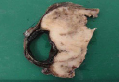
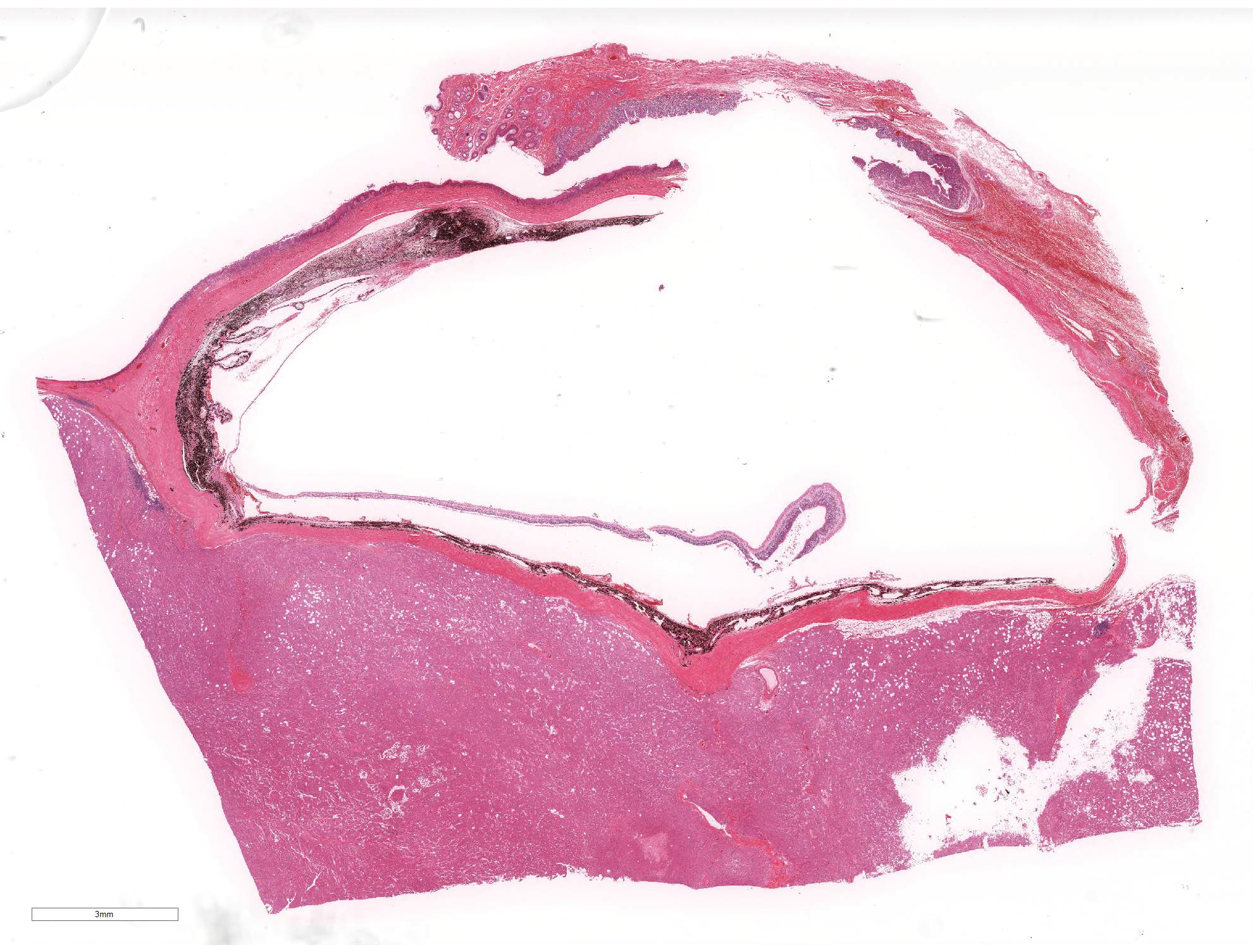
Gross Pathology: Grossly there was a whitish-gray, firm mass, approximately 5X4.3X3cm in diameter, located in the retrobulbar area. On cut surface, the mass was solid and whitish-marble, and completely involved the optic nerve.
Laboratory results: None
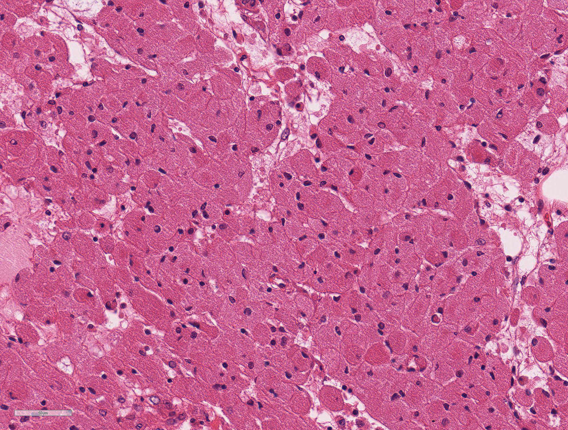
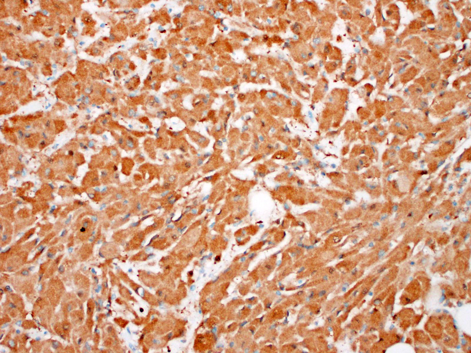
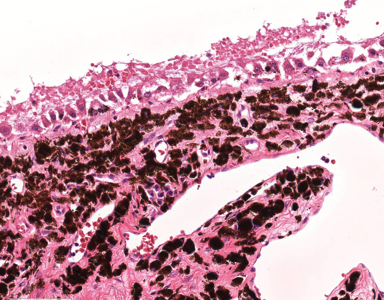
Microscopic Description: The tumor composed of sheets of closely packed large neoplastic cells supported by scant stroma harboring numerous small vessels. Neoplastic cells were large, mainly round to polygonal cells, with abundant eosinophilic granular cytoplasm. The nuclei showed minimal pleomorphism and hyperchromasia and tended to be situated towards the periphery of the cell. Mitoses were not observed. There were also few whorl-like structures. Abundant cytoplasmic eosinophilic granules were PAS-positive. Immunohistochemically, the neoplastic cells were diffusely positive for vimentin, but did not express cytokeratin AE1/AE3, neurofilament, GFAP and IBA-1.
Contributor Morphologic Diagnoses: Eye: Orbital meningioma, granular cell type.
Contributor Comment:
Orbital meningioma can derive from the optic nerve arachnoid cap cells that extend through the dura mater into the connective tissues of the orbit. In the dog, which has the higher incidence among the domestic species, 82% of the meningiomas are intracranial, 15% are intraspinal, and the remaining 3% are retrobulbar or orbital.5 Primary orbital meningiomas are generally thought to be slow growing and benign, but intraocular invasion and malignant variants with extracranial metastasis have been reported. Poodles, poodle crosses, Samoyeds, Samoyed crosses, German shepherds, and German shepherd crosses were the only breeds affected.7 In the same study, the mean age at the time of diagnosis was 9 years (range: 3 to 7 years of age), and a male sex predisposition with a male:female ratio of 2:1 was also evident.7 Clinically, they are frequently associated with unilateral protrusion of the ocular globe and blindness. Apparently there is no predilection for either side. Although not highly invasive, canine orbital meningiomas are difficult to remove, and local regrowth or extension through the optic foramen leading to blindness is a common complication.7
Orbital neoplasms are seen rarely in the dog, almost always present as slowly progressing, painless exophthalmos in older dogs, and are often malignant. A wide variety of tumor types can occur in the orbit, with fibroma, meningioma, osteosarcoma, malignant lymphoma, lipoma, rhabdomyomas, and lachrimal gland tumor.
In this case, histomorphological differentials are thought to be included rhabdomyomas, oncocytomas, and histiocytic sarcoma. Rhabdomyomas are benign skeletal muscle neoplasms that are characterized by glycogen-rich, eosinophilic, striated muscle cells with numerous mitochondria, and myofilaments. In oncocytomas, the abundant eosinophilic cytoplasmic granules correspond to large numbers of mitochondria. In histiocytic sarcoma, tumor cells sometimes contain many small vacuoles. Multinucleated giant cells with variably sized nuclei are common, and the mitotic rate is high in many areas.
The current classification of meningiomas in domestic animals describes nine histological types: meningothelial, fibrous, transitional, psammomatous, angiomatous, papillary, granular cell, myxoid and anaplastic.4 Among these types, granular cell meningioma is specific to domestic animals and does not exist in the WHO classification of human meningioma.6 Otherwise, there are some reports of canine granular cell tumors in the meninges.2,8,10 At present, the histopathological differences between granular cell meningiomas and granular cell tumors in the central nervous systems are obscure. Furthermore, the description of the histological characteristics of granular cell meningioma in the WHO classification for domestic animals is very similar to that of granular cell tumors. In the rat, there is convincing gross, histologic, and ultrastructural evidence suggesting that intracranial granular cell tumors originate from meningeal arachnoid cells and that some of these tumors may also contain mixtures of both cell types.2,12 The major meningeal involvement in both these canine tumors suggests that, as in the rat, they may be derived from a cell type forming the leptomeninges. Other support for this hypothesis is provided by a canine meningotheliomatous meningioma in which a substantial granular cell component has been described.2,9
JPC Diagnosis: 1. Retro-orbital fat: Granular cell tumor.
2. Eye:
Retinal detachment and atrophy with diffuse severe ganglion cell loss. .
3. Eyelid: Conjunctivitis, lymphoplasmacytic, chronic, diffuse, mild.
JPC Comment: While careful consideration was given to the contributorâs
diagnosis of "orbital meningioma, granular cell type", we prefer the diagnosis
of granular cell tumor in this case.
Much of the diagnostic confusion in this
particular case arises from changes in the current classification of
meningiomas. While present in the 1999 WHO Classification of Tumor of Domestic
Animals4 (referenced by the contributor), "granular cell meningioma"
has been dropped in more recent classifications.3 In addition, the
images of the âgranular cell meningioma" in the WHO Classification4
are quite similar to those ascribed to âgranular cell tumors" in other
publications, including the paper by Higgins et al.2,3
A number of WSC cases have dealt with the unique classification
of orbital meningioma in previous years, including WSC
2015-2016, Conference 20, Case 4 and WSC
2018-2019, Conference 7, case 4. A common
feature of orbital meningiomas is the presence of chondroid or osseous
metaplasia,1,7 which is absent in this case, as are other features
of meningioma. While a few granular cell meningiomas are present within the
collection of the Comparative Ocular Pathology Lab of Wisconsin (COPLOW) (and
one had been published)11, these tumors all had areas of osseous and
cartilaginous metaplasia, as well as areas of neoplastic cells that resemble
those seen in more classic meningiomas of the meningotheliomatous type (Personal
communication, Leandro Teixeira. ) Careful examination of the available
literature on canine orbital meningiomas1,7 failed to disclose
mention of a âgranular cell type"; in one reference of an orbital meningioma
with a âgranular cell component",9 the morphology is significantly
different that that seen in this case, with central nucleolus and homogenous
eosinophilic cytoplasm as compared to that seen here, in which the cytoplasm is
granular and the nuclei are peripheralized and hyperchromatic.
Granular cell tumors have been described in the central nervous system of the rat,
ferret, dog, and humans.2 The etiology of these neoplasm remains in
question, although prevailing theory in the rat, dog, and ferret is that they indeed
are of meningothelial origin. Granular cell tumors in humans may be of
astrocytic origin2 (A JPC-run GFAP stain was negative).
Additionally, they may have a similar appearance to oncocytic meningeal
variants in which the granules are numerous mitochondria rather than lysosomes,
as seen in animal species (Personal communication, Jey Koehler, Auburn
University).
The second morphologic diagnosis was suggested by Dr. Leandro Teixeira of
COPLOW upon slide review: If you look at the retina in the slide it is completely
devoid of ganglion cells. âThis is due to compression of the optic nerve by the
orbital neoplasm causing axonal (wallerian) degeneration and complete ganglion
cell loss. This is a very important lesion since it is the reason these dogs go
blind and thus I think it deserves a separate morphologic diagnosis." (Personal
communication, Leandro Teixeira).
The moderator reviewed differential diagnosis of retro-orbital meningioma, oncocytoma, rhabdomyosarcoma, and granular cell tumor. In addition, in this particular case, a section was floated off the section for ultrastructural analysis and the granules in the cells were demonstrated to be lysosomes further cementing the diagnosis of granular cell tumor. The moderator also presented data supporting her contention that the morphology of granular cell tumors may be the result of widespread cellular degeneration and lysosomal overload, which might explain how various cell types in various organs in various species may attain a similar morphologic appearance.
References:
1. Dubielzieg, RR, Ketring KL, McLellan GJ, Albert DM. The optic nerve. In: Veterinary Ocular Pathology: A comparative review. Edinburgh UK: Saunders/Elsevier; 2010; pp. 410-411.
2. Higgins RJ, LeCouteur RA, Vernau KM, Sturges BK, et al. Granular cell tumor of the canine central nervous system: Two cases. 2001. Vet Pathol. 38: 620-627.
3. Higgins RJ, Bollen AW, Dickinson PJ, Siso-Llonch S. Tumors of the Nervous System. In: Tumors in Domestic Animals, Fifth Ed, Meuten DJ, ed.; Ames IA: Wiley Blackwell 2016; pp. 864-872.
4. Koestner A, Bilzer T, Fatzer R, Schulman FY, Summers BA, Van Winkle TJ. Meningioma In: Histological Classification of Tumors of the Nervous System of Domestic Animals. 2nd series. Vol. V. Washington, DC: Armed Forces Institute of Pathology; 1999: 27-29.
5. Koestner A, Higgins RJ: Tumors of the nervous system. In: Tumors in Domestic Animals, 4ed. Meuten DJ, Iowa State Press, Ames, 2002: 697â738.
6. Louis DN, Perry A, Reifenberger G, von Deimling A, et al. The 2016 World Health Organization classification of tumors of the central nervous system: A summary. 2016. Acta Neuropathol. 131(6):803-820.
7. Mauldin EA, Deehr AJ, Hertzke D, Dubielzig RR. Canine orbital meningiomas: a review of 22 cases. Vet Ophthalmol. 2000; 3:11-16.
8. Mishra S, Kent M, Haley A, Platt S, et al. Atypical meningeal granular cell tumor in a dog. 2012. J Vet Diagn Invest. 24(1):192-197.
9. Perez V, Vidal E, Gonzalez N, Benavides J, et al. Orbital meningioma with a granular cell
component in a dog, with extracranial metastasis. J Comp Pathol. 2005; 133: 212- 217.
10. Sharkey LC, McDonnell JJ, Alroy J. Cytology of a mass on the meningeal surface of the left brain in a dog. 2004. Vet Clin Pathol. 33: 111-114.
11. Shaw GC, Miller SN, Dubielzig RR, Teixeira LBC (2015). Granular cell variant of a canine orbital meningioma: a case report. American College of Veterinary Pathologists, American Society for Veterinary Clinical Pathology, and Society of Toxicologic Pathology Combined Annual Meeting, Minneapolis, Minnesota.
12. Yoshida T, Mitsumori M, Harada T, Maita K. Mmorphological and ultrastructural study of the histogenesis of meningeal granular cell tumors in rats. 1997. Toxicol Pathol. 25(2):211-216.