Joint Pathology Center
Veterinary Pathology Services
Wednesday Slide Conference
2018-2019
Conference 7
October 17, 2018
CASE IV: AVC C5181-11 (JPC 4019356-00).
Signalment: 5 year old, castrated male Boxer, (Canis familiaris)
History: A mass was located behind the eye in the region of the optic nerve resulting in exophthalmia. It was noticed 4 months prior to presentation. There was no evidence of metastasis.
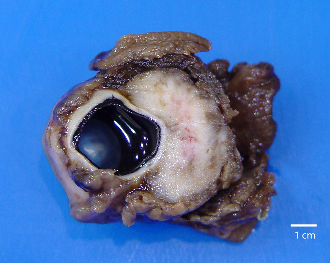
Gross Pathology: A globe with attached orbital soft tissue and eyelids was submitted. A firm to hard lobulated white mass which measured approximately 2 cm x 3 cm x 3 cm surrounded the optic nerve and infiltrated the orbital soft tissues on the posterior aspect of the globe.
Laboratory results: NA
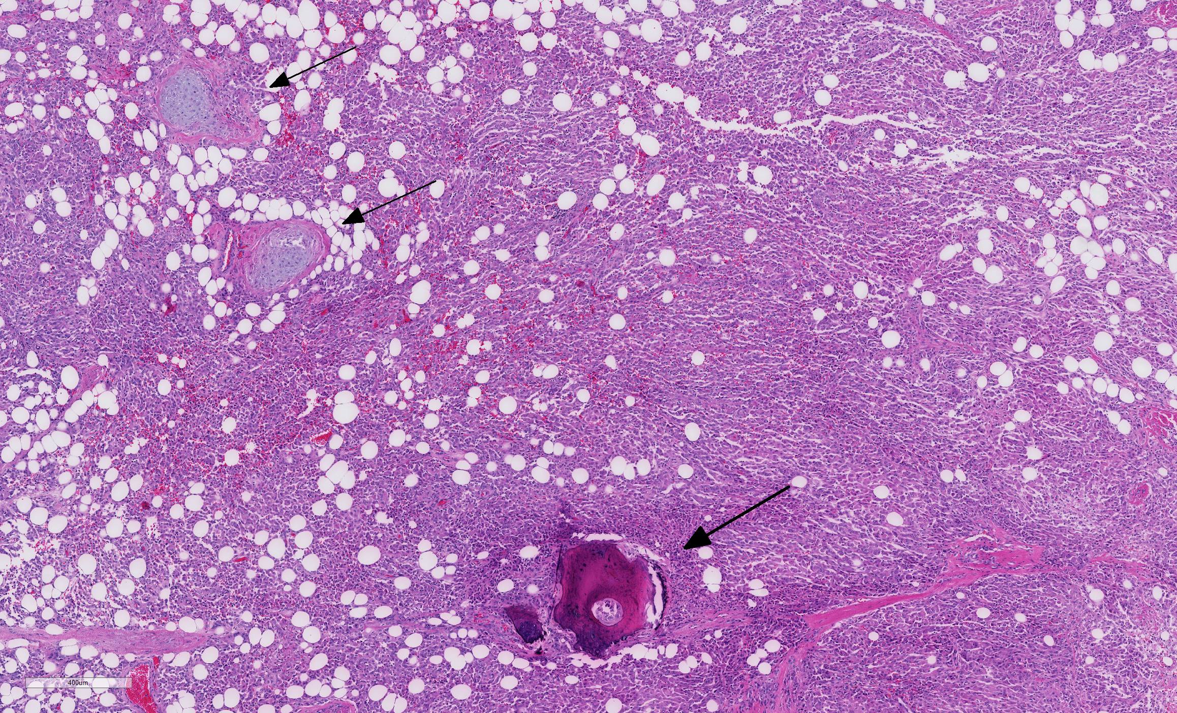
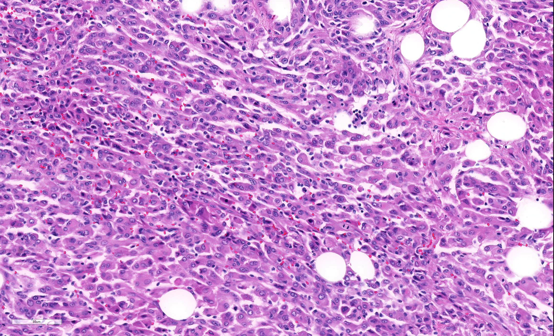
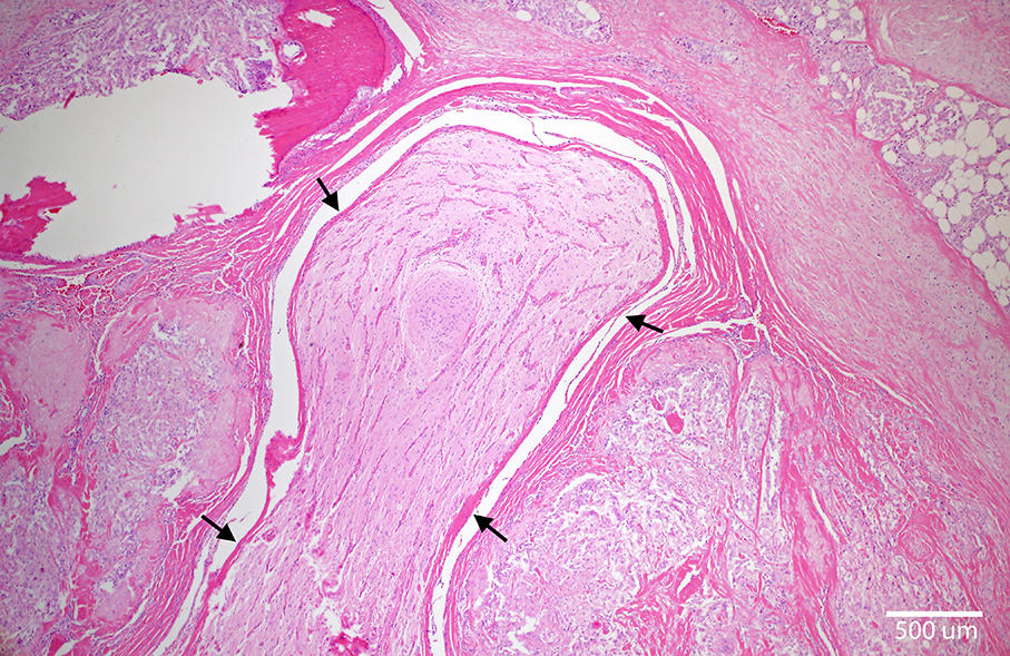
Microscopic Description: The nonencapsulated, well-delineated, moderately cellular mass surrounds the optic nerve, invades the orbital adipose and skeletal tissue, and extends slightly into the sclera. The mass is composed of nests and sheets of moderately densely packed neoplastic cells separated into lobules by fibrous connective tissue septa with scattered nodules of myxoid tissue and multifocal osseous and chondroid metaplasia. The majority of the neoplastic cells are large and polygonal with distinct cell margins, a moderate to abundant amount of glassy eosinophilic cytoplasm, and a large eccentric ovoid vesicular nucleus, occasionally with peripheralized chromatin, and one or two small distinct nucleoli. Scattered neoplastic cells contain intranuclear cytoplasmic invaginations. There is moderate to marked anisocytosis and anisokaryosis and scattered cells contain lobulated or multiple nuclei. Five mitotic figures are counted in 10 randomly selected HPF (40x) and rare mitotic atypia is noted. Small foci of necrosis, suppurative infiltration, and hemorrhage are present multifocally and small aggregates of lymphocytes are scattered amongst the neoplastic cells.
Contributor’s Morphologic Diagnoses:
Orbital (retrobulbar) meningioma
Contributor’s Comment: Along with the gross appearance and location (surrounding the optic nerve), the histomorphologic features, which include dense aggregates of large epithelioid cells and foci of cartilaginous and osseous metaplasia, are characteristic of orbital (retrobulbar) meningioma.8
Meningiomas are considered common tumors of dogs and Boxers may be one of several breeds with a predilection for developing these masses.9,10 Most meningiomas arise intracranially; tumors in the retrobulbar location are considered rare1,3,9,14 accounting for only 3% of meningiomas in the dog.7 Retrobulbar meningiomas may be primary (also called orbital meningioma), arising from the arachnoidal cap cells of optic nerve sheath, or secondary, representing extension of an intracranial neoplasm along the optic nerve.6,7 In addition to dogs1,2,4,8,9,11,12 and humans6, meningiomas arising from the optic nerve have been reported in a cat,5 F334 rats,16 and a Simmental cow.13
Grossly orbital meningiomas are tan solid conical masses that surround the optic nerve and are firmly adhered to the posterior aspect of the globe with tapering towards the optic canal.3 These tumors compress the optic nerve and circumferentially invade the adipose and muscular tissue of the orbit.3
Several histomorphologic subtypes of meningioma have been recognized in domestic species including: meningothelial, fibroblastic, transitional, psammomatous, papillary, microcystic, myxoid, angiomatous/angioblastic, and atypical/anaplastic.7,9,14 Histologically, orbital meningiomas of dogs (and humans) fit best within the meningothelial and/or transitional types.1,3,6,8,12 Granular cell differentiation, which has previously been described in meningothelial meningiomas,7 has recently been recognized within a retrobulbar meningioma in a Golden Retriever dog.11 More typically however, the neoplastic cells form lobules and dense aggregates and are epithelioid in appearance being polygonal with moderate to abundant eosinophilic cytoplasm and round to oval, often vesicular, nuclei.1,3,8,9,12,15 The presence of myxoid, cartilaginous and osseous metaplasia are common features1,2,4,8,9,11,15 and may be useful aids in both the histologic and radiographic or ultrasound diagnosis of these tumours.3,8
Immunohistochemistry is useful in ruling out carcinoma, an important differential diagnosis in these cases.3,14 Unlike carcinomas, the cells of orbital meningioma typically stain positively for vimentin and S-100 (although variably) and are usually negative for cytokeratin.8,11 In one reported case,12 tumor cells stained positively for vimentin (cytoplasmic), E-cadherin (membranous), and were negative for S100, pancytokeratin, and cytokeratins AE1 and AE3. Ultrastructurally, these masses have the typical features of meningioma14 with interdigitating processes containing intermediate filaments and occasional desmosomes present between the cell membranes.8
Dogs with orbital meningioma are usually aged (mean age of 9 years)8 and present with orbital swelling or exophthalmos with2,4,8,12 or without1,8 blindness. These tumors are generally thought to be slow-growing and benign, however tumor recurrence following extenoration,2,4,11 intraocular invasion,1,2 and malignant variants with extracranial metastasis (mainly to the lungs)2,4,11 have been reported. In one review paper8 evaluating orbital meningioma in 22 dogs, follow-up information was available in 17 cases: enucleation with excisional biopsy was therapeutically effective in 11 cases (median follow-up time 1.7 years) and local recurrence was reported in 6 cases. While none of the 22 dogs had evidence of metastases, 2 of the 6 dogs with tumor recurrence developed central blindness in the opposite eye, suggesting spread along the optic nerve to the level of the optic chiasm.8
Contributing Institution:
Atlantic Veterinary College, University of Prince Edward Island
http://avc.upei.ca
JPC Diagnosis: Eye: Orbital meningioma.
JPC Comment: The contributor has done an outstanding job in describing this unique classification of meningioma. There are few differentials for this fairly characteristic neoplasm of the orbit; the morphologic appearance of epithelioid to spindle cells infiltrating the periorbital tissues with multiple areas of osseous and chondroid differentiation is unique, even in the absence of immunohistochemical findings. In addition to being strongly immunopositive for vimentin, canine meningiomas may be multifocally positive for other neural markers, such as S-100 and NSE (WSC 2015-2016, Conference 20, Case 4). An S-100 immunostain was performed on this case at the JPC; neoplastic cells were strongly immunopositive.
Differential diagnosis would include malignant peripheral nerve sheath tumor (especially epithelioid variants), multilobular tumor of bone, and possibly orbital lobular adenoma. Of these three, only the multilobular tumor of bone would be expected to have any significant osseous or cartilaginous differentiation, and obviously far exceeding that which is seen in this slide. Histiocytic sarcoma commonly metastasizes to the eye and may also have a dimorphic appearance with both epithelioid and spindle cells, but lacks osseous and chondrous metaplasia and possesses a different immunohistochemical profile (immunopositive for vimentin and IBA-1 and other leukocyte markers, while negative for S-100 and NSE.)
The conical gross appearance of orbital meningiomas is a characteristic finding, tapering in proximity to the brain. Infiltration through the optic foramen may be life-threatening; metastasis of this tumor is extremely rare. 3
References:
- Barnett KC, Kelly DF, Singleton WB. Retrobulbar and chiasmal meningioma in a dog. J Small Anim Pract. 1967; 8: 391-394
- Buyukmihci N. Orbital meningioma with intraocular invasion in a dog: histology and ultrastructure. Vet Pathol. 1977; 14: 521-523.
- Dubielzig RR. Tumours of the eye. In Meuten DJ, ed. Tumors In Domestic Animals. 5th ed. Ames, IA: Blackwell Publishing; 2017: 892-922.
- Geib LW. Ossifying meningioma with extracranial metastasis in a dog. Vet Pathol. 1966; 3: 247-254
- Grahn BH, Stewart WA, Towner RA, Noseworthy MD. Magnetic resonance imaging of the canine and feline eye, orbit, and optic nerves and its clinical application. Can Vet J. 1993; 34: 418-424.
- Jain D, Ebrahimi KB, Miller NR, Eberhart CG. Intraorbital meningiomas: a pathologic review using the current World Health Organization criteria. Arch Pathol Lab Med. 2010; 134: 766-770
- Koester A, Higgins,RJ. Tumours of the nervous system. In Meuten DJ, ed. Tumors In Domestic Animals. 4th ed. Ames, IA: Blackwell Publishing; 2002: 697-738.
- Mauldin EA, Deehr AJ, Hertzke D, Dubielzig RR. Canine orbital meningiomas: a review of 22 cases. Vet Ophthal. 2000; 3: 11-16.
- Montoliu P, Anor S, Vidal E, Pumarola M. Histologic and immunohistochemical study of 30 cases of canine meningioma. J Comp Path. 2006; 135: 200-207
- Patnaik AK, Kay WJ, Hurvitz AI. Intracranial meningioma: a comparative pathologic study of 28 dogs. Vet Pathol. 1986; 23: 369-373.
- Perez V, Vidal E, Gonzalez N, Benavides J, Ferreras MC, Villagrasa M, Pumarola M. Orbital meningioma with a granular cell component in a dog, with extracranial metastasis. J Comp Path. 2005; 133: 212-217.
- Regan DP, Kent M, Mathes R, Almy FS, Moore PA, Howerth EW. Clinicopathologic findings in a dog with retrobulbar meningioma. J Vet Diagn Inves. 2011; 23: 857-862.
- Reis JL, Kanamura CT, Machado GM, Franca RO, Borges JRJ, Santos RL. Orbital (retrobulbar) meningioma in a Simmental cow. Vet Pathol. 2007; 44: 504 - 507.
- Summers BA, Cummings JF, de Lahunta A. Tumors of the central nervous system. In: Summers BA, Cummings JF, de Lahunta A, eds. Veterinary Neuropathology. St Louis, MO: Mosby; 1995: 351–401.
- Wilcock BP. Eye and ear. In: Maxie MG, ed. Jubb, Kennedy and Palmer's Pathology of Domestic Animals. 4th ed. Vol 1. St Louis, MO: Elsevier; 2007: 459-552.
- Yoshitomi K, Everitt JI, Boorman GA. Primary optic nerve meningioma in F344 rats. Vet Pathol. 1991; 28: 79-81.