Wednesday Slide Conference Online
DODVPR
Conference: 20 - 2015 Case: 04 Eye - dog
Signalment:
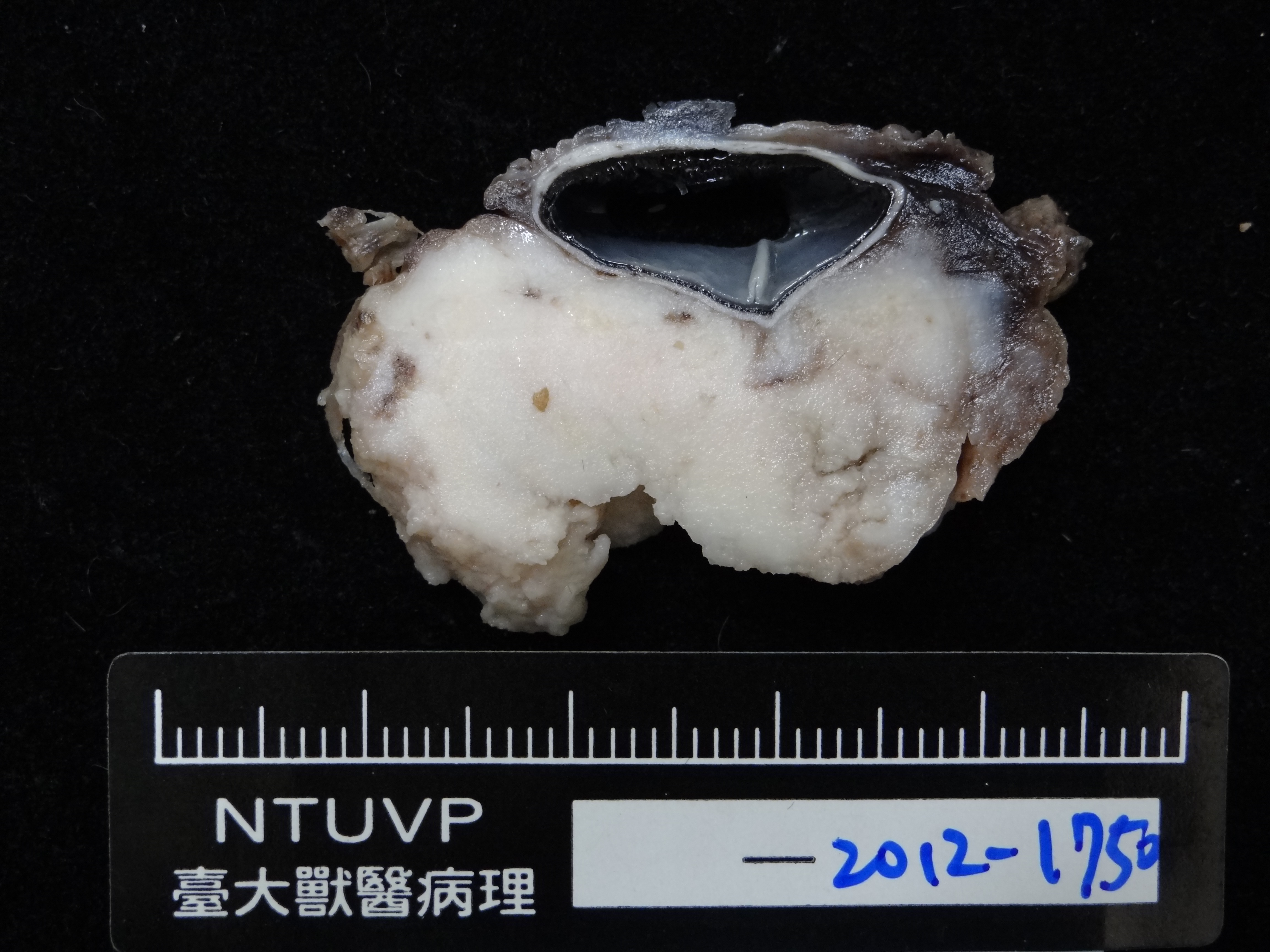
Gross Description:
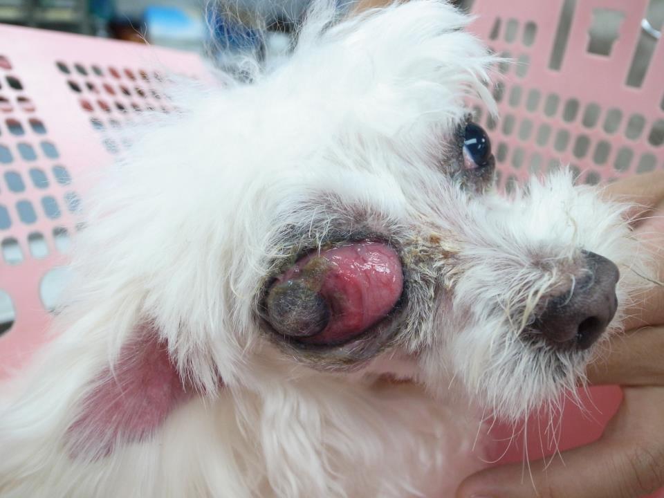
History:
Histopathologic Description:
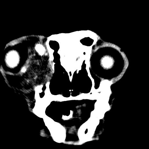
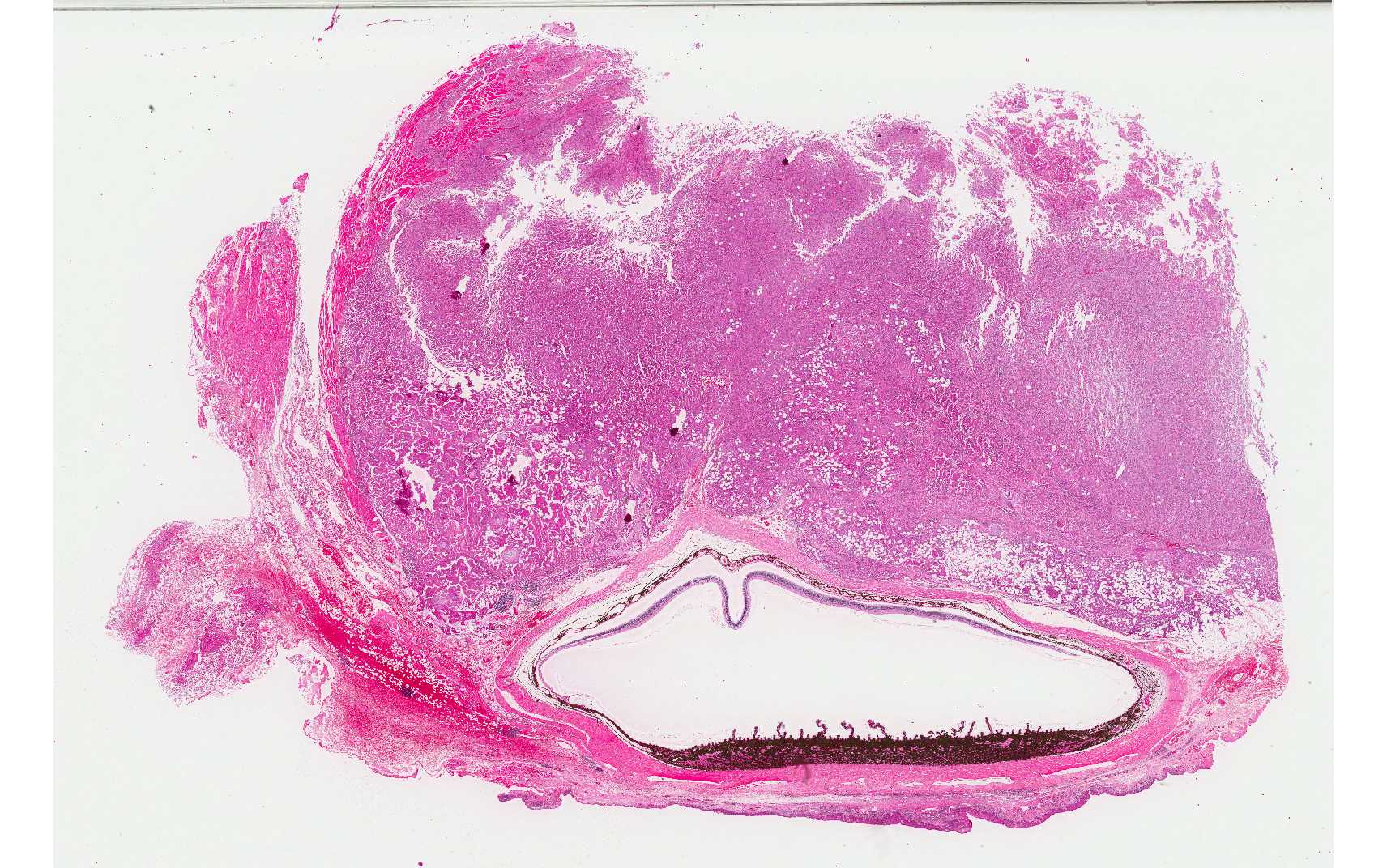
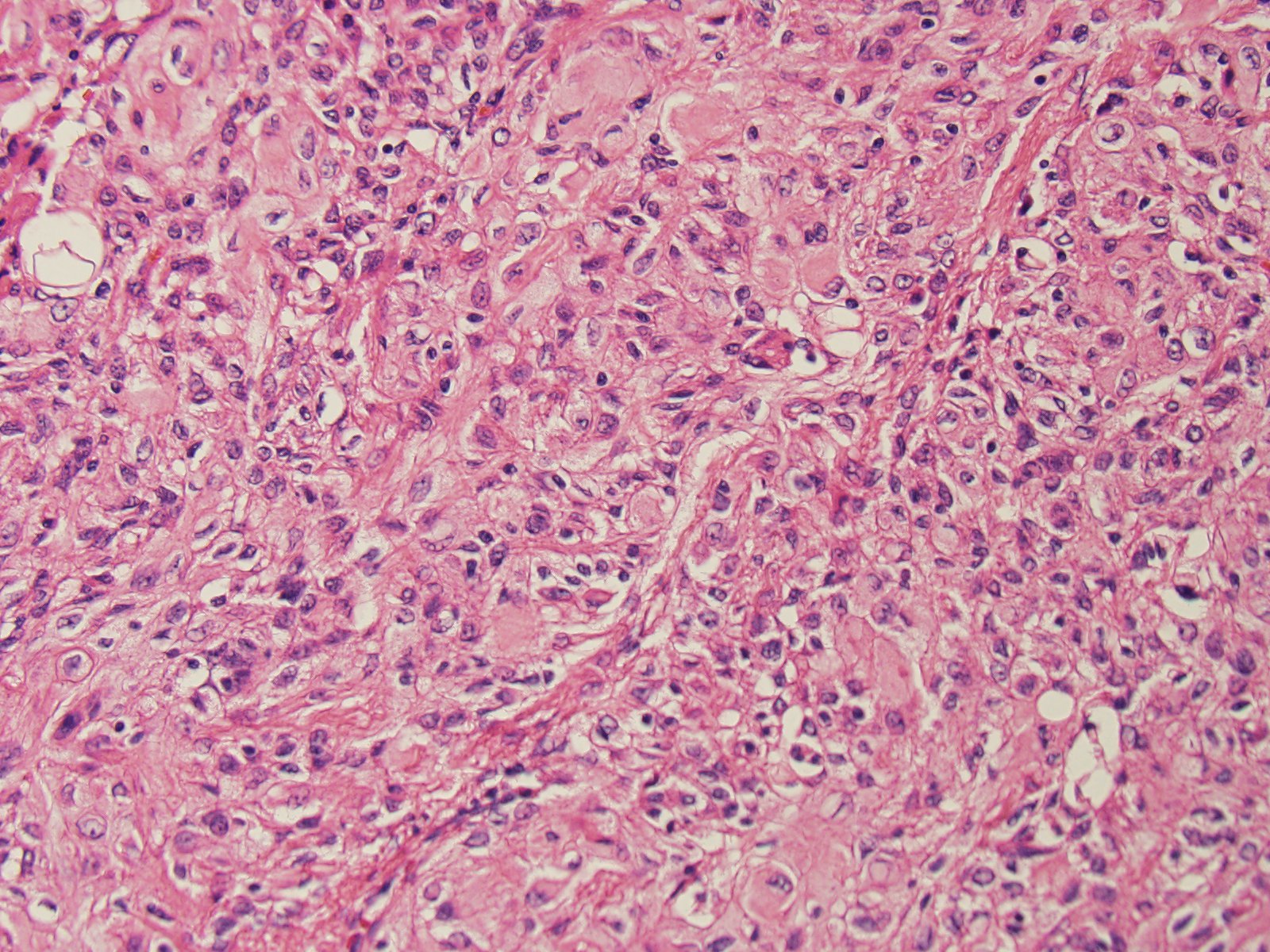
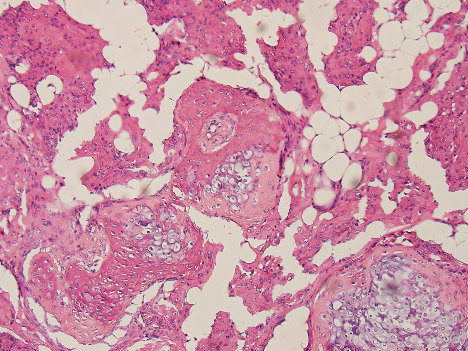
The periorbital mass, which is located behind the globe, is unencapsulated and poorly demarcated, and is causing the deformation of globe. The neoplastic cells are arranged in islands, small lobules and tight whorls or bundles, and are separated by a delicate fibrous stroma. These tumor cells have epithelioid appearance with ample eosinophilic cytoplasm, fairly distinct cell margins, and large open-faced nucleoli. Mitoses are rare. Islands of chondroid and osseous metaplasia, and varied size of vacuolation resembling adipose tissue, are present in the tumor area. The neoplasm invades the peripheral adipose tissues and extra-ocular muscles. Tumor cells infiltrate the lamina propria of the bulbar conjunctiva and partially replace the outer sclera. Multifocal necrosis is remarkable in the tumor area, but may be rare in the tissue of the submitted slide. Aggregations of lymphocytes and hemorrhage are also noted at the marginal area of the tumor. Retinal detachment, with remarkable hypertrophy of retinal pigment epithelium and degeneration of ganglion cells, is noted. Edema is present in the conjunctival epithelia. At the area behind the globe, the tumor cells encompassed an obscure nerve bundle with degenerative changes, suggestive of an orbital nerve.
By tissue immunohistochemistry tumor cells are immunopositive for vimentin, S-100 and neuron-specific enolase (NSE), and are immunonegative for cytokeratin (CK), desmin, glial fibrillary acidic protein (GFAP), Melan-A, and neurofilament (-).
The epithelia of eyelid and third eyelid demonstrate severe hyperplasia, and the lamina propria displays extensive plas-macytic infiltration and hemorrhage. The tissues from the deep mass display numerous aggregates of lymphocytes, edema, hemorrhage, and necrosis, with presence of suspected neoplastic cells as described.
Morphologic Diagnosis:
Lab Results:
Condition:
Contributor Comment:
The histological features of meningiomas can be classified as meningotheliomatous, fibromatous, psamommatous or transitional pattern. In a study from the Comparative Ocular Pathology Laboratory of Wisconsin,3 in 22 canine orbital meningiomas collected from 1981-1997, most of them were meningotheliomatous with rare fibromatous foci. The tumors characteristically have large epithelial-like neoplastic cells with nuclei containing disperse chromatin and prominent nucleoli. Psammoma bodies, which are commonly found in intracranial meningiomas, were not found in this study. In addition, islands of bone and cartilage metaplasia, which are very rare in the intracranial menin-giomas, are very commonly present.3 Metaplasia is very rare in human and canine meningiomas, but is more common in the orbital meningioma, which can be a diagnostic aid for both histologic and ultrasound analysis.3 In the present case, the histology findings are very similar to the Wisconsins study; nevertheless, prominent local invasion suggesting malignancy is noted in our case.
In
the present case, due to the overlapping expression of neural markers, the
diagnosis should rely on histologic findings of classic chondroid and osseous
metaplasia with myxomatous changes. It's reported that canine meningiomas
reveal variable positive results for neural markers, such as S-100
and NSE.2 Correlated to the location, morphology,
and IHC results, the differential diagnosis should include an epithelioid
variant of malignant peripheral nerve sheath tumor. The orbital meningioma classically
shows that epithelioid tumor cells envelope the optic nerve, expand into the
peripheral adipose and loose connective tissues with myxomatous stroma, and chondroid
and osseous metaplasia is present.6 In the veterinary
literature, epithelioid malignant PNST involving multiple organs with a mixture
of spindle and epithelioid cells histologically has been reported.4
JPC Diagnosis:
Conference Comment:
The moderator discussed the classic appearance of orbital meningioma in dogs and suggested that in an orbital neoplasm composed of large polygonal to spindle cells containing areas of cartilaginous and osseous differentiation, meningioma should be the first consideration.The differential diagnosis for orbital tumors in the dog includes multilobular tumor of bone, a mesenchymal neoplasm which also contains areas of cartilage and bone; these tumors have a characteristic lobulated pattern, and lobules are surrounded by spindle cells embedded in a rich collagenous matrix.Soft tissue sarcoma is an additional consideration for an orbital spindle cell neoplasm containing abundant collagen; these tumors typically lack bone and cartilage; however, and in dogs, morphologically low-grade but biologically high-grade fibrosarcoma would be the primary concern. -Other considerations for orbital neoplasia include lymphoma, osteosarcoma, lacrimal or sa-livary gland adenocarcinoma, hem-angiosarcoma, liposarcoma (including hibernoma) and canine lobular orbital adenoma. Microscopically, the presence of osteoid would be the discriminating feature for osteosarcoma; and the presence of strap cells, rowed nuclei, and cross striations are characteristic features of rhab-domyosarcoma.3 Macroscopically, canine orbital multilobular adenoma has a discriminating appearance and texture that may help differentiate it from other neoplasms, including the presence of friable, translucent lobules with a delicate capsule and often found to be challenging to manipulate and remove during surgery.Histologically, multilobular adenoma consists of well-differentiated lacrimal or salivary gland tissue with the absence of ducts, and may have PAS positive material within the cytoplasm.5
References:
3.Dubielzig RR, Ketring KL, McLellan GJ, Albert DM. Veterinary Ocular Pathology: A Comparative Review. Philadelphia, PA: Saunders Elsevier; 2010:126-141.
4.Garcia P, Sanchez B, Sanchez MA, Gonzalez M, Rollan E, Flores JM: Epithelioid malignant peripheral nerve sheath tumour in a dog. J Comp Pathol.2004;131: 87-91.
5.Headrick JF, Bentley E, Dubielzig RR.Canine lobular orbital adenoma: a report of 15 cases with distinctive features. Vet Ophthalmol. 2004; 7(1):47-51.
6.Mauldin EA, Deehr AJ, Hertzke D, Dubielzig RR. Canine orbital meningiomas: a review of 22 cases. Vet Ophthalmol. 2000; 3:11-16.
8. Regan DP, Kent M, Mathes R, Almy FS, et al.Clinicopathologic findings in a dog with a retrobulbar meningioma. J Vet Diag Invest. 2011; 23(4):857-862.
9. Reis Jr. JL, Kanamura CT, Machado GM, Franca RO, et al. Orbital (retrobulbar) meningioma in a Simmental cow. Vet Pathol. 2007; 44(4):504-507.
10. Tvedten H, Hillstrom A.Cytologic appearance of retinal cells included in a fine-needle aspirate of a meningioma around the optic nerve of a dog.Vet Clin Pathol. 2013; 42(2):234-237.