CASE III:
Signalment
Male Holstein fetus aborted at 234 days of gestation, Bos taurus, bovine.
History
An adult, female, second-gestation Holstein cow aborted a male fetus at 234 days of gestation.
Gross Pathology
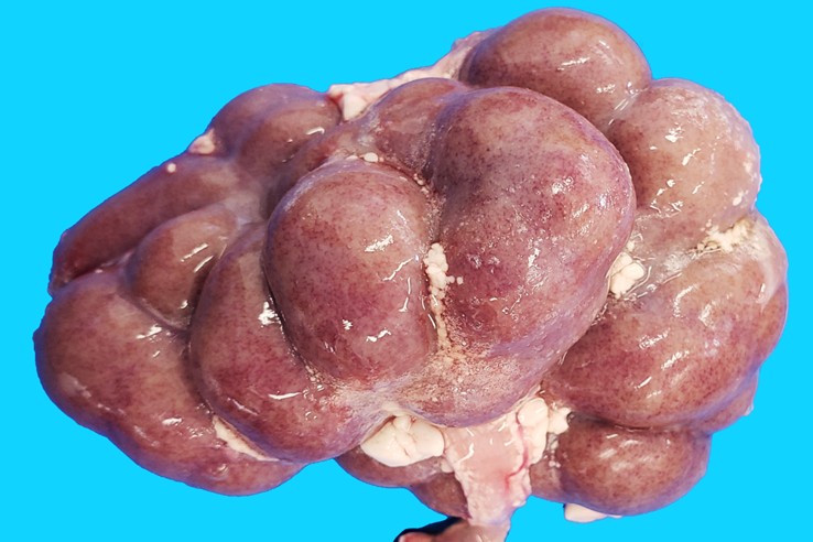
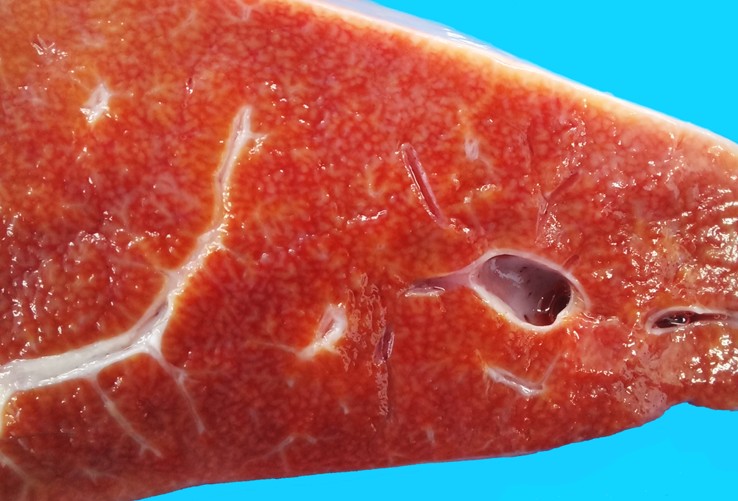
The fetus was in a good state of postmortem preservation, with a complete hair coat. No external abnormalities were noted. There was mild subcutaneous edema in the ventral neck, diffuse petechiae in the thymus, and paintbrush hemorrhages in the serosa/adventitia of the umbilical arteries. The abdominal, thoracic, and pericardial cavities contained a moderate amount of reddish serous fluid. In both kidneys there were numerous, discrete, pinpoint red foci with a generalized distribution alternating with areas of pallor visible from the capsular surface, which on cut section corresponded with enhanced cortical and medullary rays in the renal parenchyma. The liver had multifocally extensive areas of roughness and pale whitish discoloration visible from the diaphragmatic capsular surface; the underlying parenchyma had increased consistency and an enhanced reticular pattern, characterized by numerous pinpoint pale grayish foci, and indistinctly bordered interconnected linear areas ~1 mm wide alternating with reddish-orange areas of parenchyma.
Laboratory Results
None before the pathologic examination was conducted (see the "Contributor's Comment's" section below for results of laboratory tests conducted postmortem).
Microscopic Description
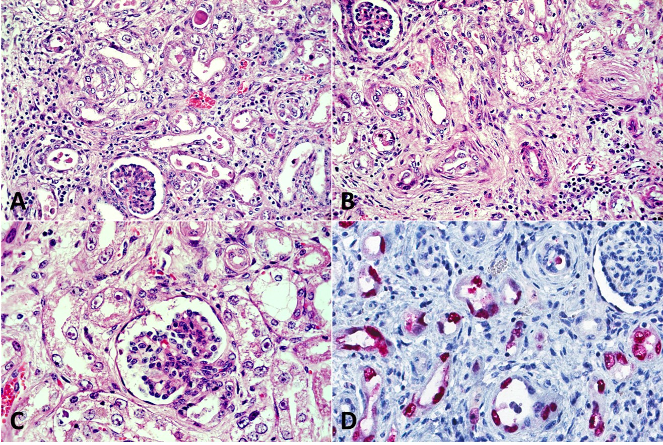
Kidney: with a multifocal disseminated distribution the cortical tubules are variably ectatic (luminal dilation), frequently contain hypereosinophilic and karyorrhectic (necrotic) cell debris in their lumina and are lined by attenuated epithelium. Multifocally tubular epithelial cells are either swollen and have vesicular nuclei (tumefaction) frequently containing one or more round or pleomorphic magenta viral inclusion bodies, surrounded by a clear halo (chromatin margination), or are shrunken with angular cell borders, hypereosinophilic cytoplasm and have pyknotic nuclei or karyorrhectic debris (necrosis). In affected areas, the interstitium is multifocally infiltrated by inflammatory cells, predominantly lymphocytes and histiocytes, with some plasma cells and occasional neutrophils, and/or expanded by spindle cells with an elongated nuclei (fibroblasts) embedded in a collagenous eosinophilic fibrillar extracellular matrix (fibrosis/fibroplasia).
Contributor's Morphologic Diagnoses:
Kidney: nephritis, tubulointerstitial, lymphocytic, and histiocytic, with intranuclear epithelial viral inclusion bodies, cortical tubular epithelial cell necrosis, and interstitial fibrosis, multifocally extensive, severe, bovine.
Contributor's Comment:
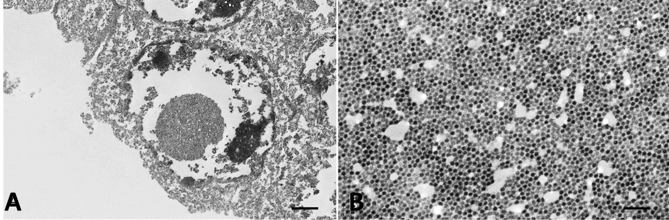
In this case, bovine polyomavirus 1 (BoPyV-1, Epsilonpolyomavirus bovis) was detected by qPCR, isolated from the kidney, and fully sequenced, while other viruses that can cause intranuclear inclusions (such as herpesviruses and adenoviruses) were ruled out. In addition, abundant polyomavirus large T antigen (LT Ag) was demonstrated intralesionally by immunohistochemistry in cortical tubular epithelial cells (Figure 3D), and large numbers of viral particles with typical polyomaviral morphology were found in the nuclei of renal tubular epithelial cells by transmission electron microscopy (Figure 4). Further details on the diagnostic investigation conducted in this case have been recently published.8
The pathological findings in the kidneys of the fetus were consistent with an active polyomavirus infection, with lytic replication of the virus, production of abundant intranuclear inclusion bodies composed of dense arrays of assembled virions, and LT Ag expression in the nuclei of renal tubular epithelial cells. In addition, severe degeneration (i.e., swelling, tumefaction, attenuation) and necrosis of renal tubular epithelial cells were indicative of a cytopathic viral effect, along with a prominent inflammatory response to the infection, and interstitial fibrosis suggestive of chronic active renal damage at the time of abortion.
Polyomaviruses are a diverse group of non-enveloped viruses with a small, circular, double-stranded DNA genome found in a wide variety of mammalian (including humans), avian, fish, amphibian, reptile, and invertebrate species.12 According to the International Committee on Taxonomy of Viruses (ICTV, https://talk.ictvonline.org/, accessed on June 20, 2023), the Polyomaviridae family contained 8 genera named Alpha-, Beta-, Delta-, Epsilon-, Eta-, Gamma-, Theta- and Zeta-polyomavirus, although many strains and species were awaiting genus assignation. While some polyomaviruses can cause acute disease and even death due to productive (lytic) replication in their hosts, most of the polyomaviruses infecting mammals establish persistent subclinical infections in healthy individuals, resulting in clinical disease only after reactivation of the infection in immunosuppressed hosts.11
The polyomaviruses currently known as bovine polyomavirus (BoPyV) were initially discovered in uninoculated cultures of kidney cell lines of stump-tailed macaques, rhesus monkeys, and cynomolgus macaques, and were referred to as Stump-Tailed Macaque Virus (STMV),18 cynomolgus kidney strain (CK-strain) of STMV,20 and Fetal Rhesus Kidney Virus (FRKV).15 Because of their growth in uninoculated cell cultures, it was initially thought that they were endogenous viruses of non-human primates. However, it has since been determined that they were polyomaviruses of bovine origin contaminating the bovine fetal serum used to supplement cell growth media.14 The first polyomavirus isolated in a bovine kidney cell line was obtained from a healthy newborn calf,2 and was designated Wokalup Research Station Virus (WRSV). It was suggested shortly after their discovery that STMV, FRKV, and WRSV were all isolates of an identical BoPyV,13 which was later supported by viral whole-genome sequencing.19
Species of polyomaviruses known to infect live cattle to date include Bos taurus polyomavirus-1 (syn. Bovine polyomavirus-1 —BoPyV-1—, Epsilonpolyomavirus bovis), and Bos taurus polyomavirus-2 (syn. Bovine polyomavirus-2 —BoPyV-2—, awaiting genus assignation). A third species named Bovine polyomavirus-3 (BoPyV-3, awaiting genus assignation) was originally detected in ground beef samples collected at a supermarket in the USA.16 To the best of our knowledge, BoPyV-1 and BoPyV-3 have never been causally associated with pathology or disease, while BoPyV-2 has been recently proposed as a probable cause of nonsuppurative encephalitis in cattle.10
Although the pathogenic mechanisms by which most polyomaviruses induce tissue damage are not yet fully understood, five patterns of polyomavirus-induced pathology have been proposed.4 These include: 1- cytopathic polyomavirus pathology pattern, 2- cytopathic-inflammatory pathology pattern, 3- immune-reconstitution inflammatory syndrome, 4- autoimmune polyomavirus pathology pattern, and 5- oncogenic polyomavirus pathology pattern. The cytopathic polyomavirus pathology pattern is characterized by uncontrolled viral replication in the infected cells without significant inflammation; PML caused by JCPyV replication in the oligodendrocytes is the prototype disease for this pattern. The cytopathic-inflammatory pathology pattern is characterized by high-level virus replication with cytopathic lysis of infected cells, along with necrosis and a significant inflammatory response. PyVAN in kidney allografts is the prototype disease for this pattern.4 The lesions in the kidneys of the aborted fetus described herein best fit with this latter pattern of polyomavirus-induced pathology.
Histologically, PyVAN in humans is characterized by varying degrees of multifocal random interstitial nephritis, tubulitis, cytopathic changes and basophilic intranuclear inclusions in renal epithelial cells, interstitial fibrosis, and tubulo-interstitial atrophy, depending on the stage of disease progression.17 Interestingly, these same lesions were observed in the kidneys of the aborted bovine fetus infected with BoPyV-1.8
Immunohistochemistry using antibodies against Simian Virus 40 (SV-40, Macaca mulatta polyomavirus 1, Betapolyomavirus macacae) LT Ag, as in this case, has been shown to cross-react with other polyomaviruses, such as BKPyV and JCPyV, in cases of PyVAN in humans.3 In concert with this, the antibody also cross-reacted with BoPyV-1, indicating that this test can be used for viral identification in formalin-fixed paraffin-embedded bovine tissues. This is not unexpected, considering that LT antigen has domains that are conserved among the polyomaviruses.12
Generally, occurrence and progression of diseases caused by polyomaviruses in mammals seem to be largely dependent on host immunosuppression.11,12 Pregnant females can experience modulation in their immune system making them more prone to pathogenic infections.7 While a real-time PCR performed on serum of the cow indicated that she was viremic at the time of the abortion,8 whether she or the aborted fetus were immunosuppressed was unknown. There was no evidence of lymphoid depletion on histologic examination of lymphoid tissues including thymus, spleen, and lymph nodes in the aborted fetus. Bovine viral diarrhea virus, a common immunosuppressive virus of cattle, was ruled out by reverse transcriptase qPCR in the fetal tissues.
This case suggests that BoPyV-1 is pathogenic to the bovine fetus and, thus, a probable cause of abortion in cattle.8 Pathogenicity seems to involve cytopathic viral effects with tissue damage including necrosis, inflammation, and fibrosis, with lesions resembling PyVAN in humans. Factors of the virus and host that would determine lesion and disease development, as well as the epidemiology and transmission routes, need further investigation.
Contributing Institution
Institucion Nacional de Investigacion Agropecuaria (INIA) Colonia, Uruguay www.inia.uy
JPC Diagnoses
Kidney: Tubular degeneration, necrosis, and regeneration, chronic, diffuse, moderate, with cellular and granular casts, interstitial nephritis and fibrosis, and tubular epithelial intranuclear viral inclusions.
JPC Comment
The contributor has provided an outstandingly thorough review of this entity. Polyomaviruses are also not uncommon visitors to the Wednesday Slide Conference with 18 cases dating back to 1994 (and likely quite a few before that).
Half of these cases are the various manifestations of primate polyomavirus (of which SV-40 is the prototypical NHP polyomavirus. This polyomavirus manifests in the brain, kidney, and in one case in the WSC Archive (WSC 1997, Conference 14, Case 1), in the lung (as well as in the kidney), representing well-known syndromes associated with polyomavirus infection in NHPs. In each of these cases, the affected non-human primate was immunosuppressed following experimental infection with similar lentivirus. In immunosuppressed non-human primates, polyomavirus infection results in Simian virus 40 (SV40) is an endemic polyomavirus in captive and free-ranging macaques. Infection does not cause lesions in immunocompetent animals, but immune suppression, such as SIV infection or immunosuppressive drug regimens, allows reactivation of latent infections or primary infections to cause disease.
There are two forms of nervous system disease in immunosuppressed macaques associated with polyomavirus infection. Progressive multifocal leukoencephalopathy (PML) results in demyelinating disease due to infection of oligodendroglia and is thought to occur in animals with underlying SV40 infection that subsequently are immunosuppressed.22, 23 A second nervous system manifestation of SV40 infection (in younger animals, but not necessarily immunosuppressed animals, is meningoencephalitis (ME), which is characterized by inflammatory infiltrates within the cortical grey matter and leptomeningeal edema.22,23 Renal infections in immunosuppressed macaques provide a model mirroring polyomavirus-associated renal transplant rejections in humans, and the pneumonia seen in immunosuppressed macaques mirrors a severe, rapidly fatal pneumonia seen in humans receiving hematopoietic stem cell transplants.
Several distinct syndromes associated with polyomavirus in birds have been seen in the WSC over the years. Budgerigar fledgling disease is a disease of young hand-reared psittacines which results in necrosis and intranuclear inclusions within tubular epithelium somewhat reminiscent of today's case in an aborted calf. Affected budgerigars that do not rapidly succumb to this disease may develop a range of feather abnormalities known as "French molt". Viral-associated hepatic necrosis is seen in affected birds as well. A single case of goose hemorrhagic polyomavirus (one of the four known avian polyomaviruses) appeared in the WSC in 2002 (Conference 7, Case 4), and is the only one of the avian polyomaviruses in which intranuclear viral inclusions are not seen.24
One final polyomavirus that has appeared several times in the WSC over the years is a unique polyomavirus infecting hamsters. This polyomavirus possesses the ability to infect both lymphocytes and undifferentiated keratinocytes. In young naive animals, epizootic polyomavirus infection causes lymphoma, resulting in mortality between 4 and 30 weeks. In older animals, enzootic polyomavirus infection of undifferentiated keratinocytes results in the generation of multiple beign follicular tumors similar to trichoepitheliomas. 25
Note: Images 3-1 through 3-4, and their respective captions were recently published8 in the journal Viruses (please see citation below), in an open access article distributed under the terms and conditions of the Creative Commons Attribution 4.0 International license (CC BY 4.0, https://creativecommons.org/licenses/by/4.0/). The contents of the article can be shared, copied, and distributed freely in any medium or format.
References
- Axthelm MK, Koralnik IJ, Dang X, et al. Meningoencephalitis and demyelination are pathologic manifestations of primary polyomavirus infection in immunosuppressed rhesus monkeys. J Neuropath Exp Neurol. 2004. 63 (7): 750-758.
- JPC Wednesday Slide Conference, Conference 7, Case 4, 2002.
- JPC Wednesday Slide Conference, Conference 19, Case 2, 2021.