Signalment:
Adult male garter snake, (
Thamnophis sirtalis parietalis).Recurring skin masses after surgical excision
Gross Description:
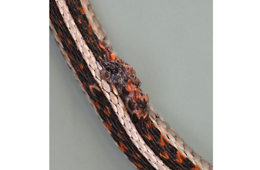
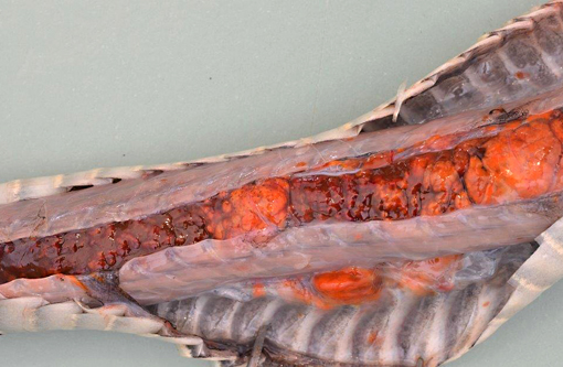
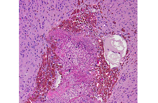
Located caudo-dorsally on the body there were two skin masses measuring 2 x 1 x 1cm and 0.5 x 0.5 x 0.5 cm in size, covered by crusts. The masses were orange and firm and the larger mass infiltrated the underlying muscle. Located on the ventral side of the body there were two smaller nodules. The kidneys and liver were moderately enlarged and both organs showed multifocal to coalescing, poorly demarcated and infiltrative growing orange nodules of 0.1 x 0.1 x 0.1cm up to 2 x 2 x 2 cm size. The coelomic cavity and hemipenis were also infiltrated by small multifocal orange masses.
Histopathologic Description:
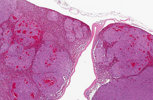
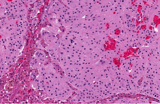
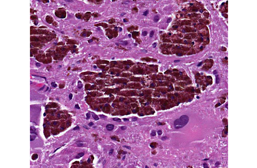
Liver: 90 - 95% of the tissue is replaced by a poorly demarcated, non encapsulated, infiltrative, multilobulated and densely cellular mass with small groups of remaining hepatocytes. Neoplastic cells are closely packed, supported by scant amount of fibrovascular stroma and arranged in anastomosing cords, islands and sheets. The neoplastic cells are polygonal to spindleoid, 15 to 30 μm in size with indistinct cell borders and have a high amount of granular eosinophilic cytoplasm and often contain birefringent olive-green to golden-brown granular pigment within the cytoplasm. Nuclei are round to oval to irregular with finely stippled chromatin and have up to three prominent nucleoli. Many cells contain up to five oval to irregular nuclei or have a huge nucleus (megakaryosis). Anisocytosis and anisokaryosis are high and there is anaplasia of neoplastic cells. In 10 HPF there are nine mitotic figures, some of which are bizarre. There are multifocal randomly distributed areas of necrosis, characterized by hypereosinophilia of the cytoplasm, pyknotic nuclei, karyolysis and karyorhexis mixed with hemorrhage. Multifocally there are small groups and cords of remaining hepatocytes often showing hypereosinophilic, vacuolated and pyknotic nuclei (degeneration).
Morphologic Diagnosis:
Liver: Iridophoroma, malignant, metastatic, garter snake (
Thamnophis sirtalis parietalis), reptile.
Condition:
Iridophoroma
Contributor Comment:
Fish, amphibians and reptiles contain pigment cells in their skin that are called dermochromatophores.(1) These cells are derived from the neural crest and are characterized by the pigment they contain within their cytoplasm.(1) There are four types of dermochromatophores: melanophores, which contain melanin pigment, iridophores containing purines (colorless pigment), xanthophores containing carotenoids (yellow pigments) and erythrophores containing carotenoids and pteridines (red pigments).(1) Chromatophoromas are tumors arising from the cutaneous pigment cells that have been rarely reported(2,3) and are subclassified into three types (melanophoromas or melanomas, xanthophoromas and iridophoromas) by the type of pigment they contain.(1,2) Chromatophoromas with combined features of all cell types have also been described.(4) The occurrence of these tumors in reptiles is higher than assumed and metastases are common.(1)
Iridophoromas occur as intraepidermal white masses and metastases have been observed in several organs.(2) Diagnosis of an iridophoroma is based on birefringent anisotropic granules within the neoplastic cells, distinguishing the iridophoroma from a melanophoroma and xanthophoroma. Based on the olive-green to golden-brown birefringent pigment within neoplastic cells we diagnosed a chromatophoroma of the subtype iridophoroma. Both benign and malignant iridophoromas have been reported in snakes, lizards, and fish.(1-3) Malignancy criteria of chromatophoromas in reptiles encompass the presence of intravascular (lymphatic/blood vessel) neoplastic cells, visceral metastasis, high pleomorphism and the presence of mitotic figures.(2) Both Melan-A and S-100 protein can be detected by immunohistochemistry in iridophoromas.(2)
JPC Diagnosis:
Liver: Iridophoroma.
Conference Comment:
The contributor provides an excellent summary of chromatophores and chromatophoromas in non-mammalian species. Chromatophores are contractile pigment cells that originate from embryonic neural crest cells and migrate to numerous tissues. Intracellular aggregation and dispersion of pigment granules engenders an ability to display rapid color changes in the skin, which can be important in camouflage, mating, and protection in many non-mammalian species.(4) Based on the presence of birefringent material within neoplastic iridophores, conference participants on the whole concurred with the diagnosis of iridophoroma; however, there was also a lively debate concerning the histogenesis of a collection of morphologically distinct cells within the neoplasm. These cells appear slightly more individualized and polygonal, with atypical nuclear morphology and abundant dark-brown to occasionally greenish intracellular pigment that is not as birefringent. Although a consensus was not reached, discussion centered upon whether these cells represent a unique clonal variant of the more spindled neoplastic cell population, or perhaps, as some participants suggested, these are simply aggregates of melanomacrophages, which are a normal component of many reptile and amphibian livers, trapped within the neoplasm.
References:
1. Okihiro MS. Chromatophoromas in two species of Hawaiian butterflyfish, Chaetodon multicinctus and C. miliaris. Vet Pathol. 1988;25:422-431.
2. Heckers KO, Aupperle H, Schmidt V, Pees M. Melanophoromas and iridophoromas in reptiles. J. Comp. Path. 2012;146:258-268.
3. Suedmeyer K, Bryan JN, Johnson G, Freeman A. Diagnosis and clinical management of multiple chromatophoromas in an eastern yellowbelly racer (Coluber constrictor flaviventris). J Zoo Wildl Med. 2007;38(1):127-130.
4. AFIP Wednesday Slide Conference 29, 3 May 2000, Case I e S-49-98 (AFIP 2681629) http://www.askjpc.org/wsco/wsc/wsc99/99wsc29.htm