Results
AFIP Wednesday Slide Conference - No. 29
3 May 2000
- Conference Moderator:
Dr. Don Nichols, Diplomate, ACVP
Department of Pathology
National Zoological Park
Washington, DC 20008
- Return to WSC Case Menu
- Case I - S-49-98 (AFIP 2681629)
-
- Signalment: Boa constrictor (Boa constrictor constrictor),
adult (est. 8 years), unknown sex.
-
- History: Snake presented with a 1 cm2 dorsal mass
in November 1993. At surgery, the mass was described as being
rubbery and white and did not appear to invade underlying dermis.
Biopsy was diagnosed as "granulation tissue". Local
recurrence of mass occurred in July 1995, at which time it was
again surgically removed.
-
- Gross Pathology: 3 x 2 cm2 white, firm, fibrous dermal
mass.
-
- Contributor's Diagnosis and Comments: Morphologic
Diagnosis: Iridophoroma, skin, snake.
-
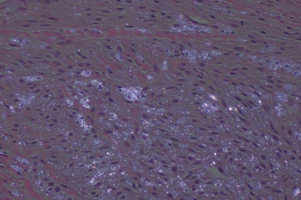
- Sections of skin containing an expansile, nonencapsulated,
multilobular, pigmented dermal mass which extends to both lateral
and ventral borders in some sections. The mass is composed of
whorls, interlacing bundles, nests and fascicles of a uniform
population of spindle cells on a fine fibrovascular stroma. The
cells are spindle to stellate with indistinct cell borders, moderate
amounts of eosinophilic fibrillar cytoplasm with varying amounts
of coarse to fine golden brown/olive green pigment granules.
The nuclei are ovoid to polyhedral with coarsely clumped chromatin
and a single, variably distinct, round, magenta nucleolus. The
mitotic index is less than l/HPF. Pigment granules are birefringent
on polarized light.
-
- Electron Microscopy: Cells had variable numbers of
empty elliptical cytoplasmic membrane lined spaces. The amount
of pigmentation seen on light microscopy related to the number
of spaces on EM. Membranes ranged in size, shape and arrangement
within the cytoplasm. The latticework of empty membranes is characteristic
and represents the loss of "reflecting platelets" during
routine uranyl acetate processing.
-
- While the histologic features of this mass were suggestive
of a melanoma, the presence of pigmented cells in a reptilian
skin tumor are not diagnostic of that tumor alone.
- In addition to melanophores, snake skin normally contains
3 other classes of pigment producing cells including iridophores,
erythrophores and xanthophores. All are derived from neural crest.
Neoplasms arising from all four classes have been described in
reptiles. Mosaic chromatophoromas with combined features of multiple
cell types have also been described.
-
- H&E light microscopic features of these tumors are indistinguishable
from those of a melanoma without the use of polarized light.
Ultrastructural features, however, are striking and diagnostic
and were pursued in this case.
Melanophoromas typically contain electron-dense melanosomes in
various stages of development with a characteristic internal
structure. Erythrophoromas contain characteristic lamellated
pterinosomes. Neither of these structures was identified in this
case. Instead, the cytoplasm was packed with a distinctive latticework
of empty membranes. These spaces represent the dissolved cytoplasmic
reflecting platelets of iridophores which are lost with routine
uranyl acetate and lead citrate EM staining.
-
 20x
obj
20x
obj
 20x
obj, polarized
20x
obj, polarized
- Case 29-1. Dermis. Variably pigmented spindle shaped
cells contain anisotropic crystalline material when viewed under
polarized light.
-
- AFIP Diagnosis: Skin: Iridophoroma, malignant, boa
constrictor, reptile.
-
- Conference Note: All conference participants agreed
with the contributor's morphologic diagnosis of iridophoroma.
The "malignant" designation was added because of infiltrative
growth pattern, a mitotic rate that ranged up to 7 per high power
field and nuclear atypia.
-
- Note: Communication with the contributor revealed
that this neoplasm has not recurred since it was removed in 1995.
The original excision from 1993 was reexamined and determined
to be the same neoplasm.
-
- Contributor: Department of Pathology, Wildlife Health
Center / WCS 185th St. and Southern Blvd., Bronx, New York, NY
10460.
-
- References:
- 1. Frye FL, Carney JD, Harshbarger JC, Zeigel FF: Malignant
chromatophoroma in a western terrestrial garter snake. J Am Vet
Med Assoc 167:557-558, 1975
- 2. Ghadially FN: Diagnostic Electron Microscopy of Tumors,
2nd ed., pp.106-121. Butterworths, London, England, 1985
- 3. Jacobson ER, Ferris W, Bagnara JT, Iverson WO: Chromatophoromas
in a Pine Snake. Pigment Cell Res 2:26-33, 1989
- 4. Leach MW, Nichols DK, Hartsell W, Torgerson RW: Radiation
therapy of a malignant chromatophoroma in a yellow rat snake
(Elaphe obsoleta quadrivittata). J Zoo and Wild Med 22(2):241-244,
1991
- 5. Okihiro MS: Chromatophoromas in two species of Hawaiian
butterfly fish, Chaetodon multicinctus and C. miliaris. Vet Pathol
25:422-43 1, 1988
- 6. Ryan MJ, Hill DL, Whitney GD: Malignant chromatophoroma
in a Gopher snake. Vet Pathol 18:827-829, 1981
-
-
- Case II - 40149 (AFIP 2679508)
-
- Signalment: Adult, female caiman lizard (Dracaena
guianensis)
-
- History: This lizard was one of two animals wild-caught
in Peru and spent approximately 1 year in at least 2 different
reptile dealerships in California. On arrival at the zoo, the
animals were in poor body condition and mildly dehydrated. Initial
treatment included subcutaneous fluid therapy and tube feeding.
Both lizards died suddenly within two weeks of arrival and had
similar necropsy findings.
-
- Gross Pathology: At necropsy, the lungs were diffusely
hyperemic and exuded copious amounts of clear fluid on section.
The lumen of each opened lung contained moderate amounts of a
yellow mucoid material. A single adult trematode parasite was
attached to the gastric mucosa.
-
- Laboratory Results:
- Aerobic bacterial culture of lung yielded 2+ Klebsiella pneumoniae,
3+ Proteus mirabilis, 3+ Pseudomonas sp., 2+ Citrobacter sp.,
and 2+ Aeromonas sp.
-
- Virus isolation from the lung on viper heart cells demonstrated
syncytial cell formation at 5 days post inoculation; negative
staining electron microscopy demonstrated virions consistent
with a paramyxovirus.
-
- Wet mounts of gastric fluid contained moderate numbers of
an unidentified trematode egg.
-
- Contributor's Diagnoses and Comments:
- 1. Lung: Pneumonia, proliferative and interstitial, diffuse,
moderate to marked with syncytial cells and rare intracytoplasmic
eosinophilic inclusion bodies.
- 2. Lung: Encapsulated trematode eggs (may not be present
in all sections).
-
- Histologically, there is expansion of faveolar septa by edema
and variable infiltrates of predominantly heterophils with fewer
mononuclear inflammatory cells. Multifocally, within the lumen
there are aggregates of similar inflammatory cells admixed with
fewer sloughed epithelial cells, erythrocytes, rare bacteria
and cellular debris. Diffusely, there is marked hyperplasia and
hypertrophy of faveolar lining epithelial cells (type-2 pneumocytes)
with formation of syncytial cells that have 2-3, and occasionally
up to 10, nuclei. Occasionally, epithelial cells contain discrete
variably sized eosinophilic cytoplasmic inclusion bodies. Rarely,
within faveolar septa, there are trematode eggs, which are surrounded
by a thin rim of fibrous connective tissue and/or low numbers
of macrophages, which are often multinucleate. In addition to
virus isolation (noted above), transmission electron microscopy
of formalin-fixed lung tissue showed moderate numbers of filamentous
and spheroidal virions, morphologically suggestive of paramyxovirus,
budding from pneumocyte cell membranes.
-
- The histologically observed proliferative pneumonia with
syncytial cell formation and the presence of cytoplasmic inclusion
bodies led to a presumptive diagnosis of a viral pneumonia in
this case. This was later confirmed by electron microscopy and
by isolation of a paramyxovirus from the lung. Although viral
pneumonias are well known in snakes, to our knowledge, this is
the first time that a viral pneumonia has been documented in
a lizard.
-
- Previous reports of paramyxoviruses in lizards have consisted
of a serosurvey of wild iguanas and of virus isolation without
correlative histopathology. In snakes, paramyxoviral infections
have been responsible for epizootics in private and zoological
collections and can be associated with high mortality. Lesions
in affected snakes are similar to those observed in this caiman
lizard and usually include proliferative pneumonia with or without
syncytial cell formation and rarely, intracytoplasmic inclusion
bodies.
-
- Another frequently reported lesion in affected snakes is
pancreatic necrosis with marked ductular hyperplasia and occasionally,
syncytial cell formation. Mild pancreatic lesions were also observed
in these affected lizards and consisted of mild interstitial
edema and ductular syncytial cell formation. In epizootics occurring
in snakes at our institution, pancreatic lesions have been more
prominent with the classical proliferative pneumonia being observed
only infrequently. Secondary bacterial pneumonia and septicemia
have been frequently described in snakes with paramyxovirus infections.
Consequently, when histologically evaluating what might seem
to be a clear-cut bacterial pneumonia in a reptile, care should
be taken not to overlook the sometimes-subtle proliferative changes
that could be suggestive of an underlying viral infection.
-
- The multiple bacterial isolates obtained from the lung of
this lizard were interpreted as representing secondary colonization
of the virally compromised respiratory tract. Trematode eggs
were observed in multiple tissues, including the lung, with minimal
associated host response and consequently were interpreted as
incidental findings. Because snails comprise the majority of
the diet in caiman lizards, the presence of trematode eggs in
tissues was not surprising.
-
 20x
obj
20x
obj
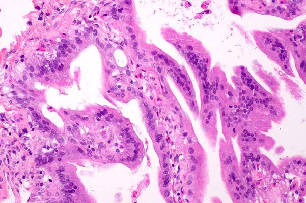
- Case 29-2. Lung. Multifocally air sac epithelium contains
syncytial cells.
 20x
obj
20x
obj
- Case 29-2. Lung. There are two deformed, collapsed,
empty, brown, trematode eggs expanding the interstitium.
-
- AFIP Diagnoses:
- 1. Lung: Pneumonia, interstitial, proliferative and exudative,
subacute, diffuse, moderate, with syncytial cells, caiman lizard
(Dracaena guianensis), reptile.
2. Lung, interstitium: Encapsulated trematode eggs (may not be
present in all sections).
Conference Note: Caiman lizards (members of the genus
Dracaena and the family Teiidae) are found in wet, forested areas
of South America. D. guianensis reaches a length of about 122
cm. Caiman lizards should not be confused with caimans, a group
which includes several species of Central and South American
reptiles related to alligators. The black caiman (Melanosuchus
niger) may reach 4.5 m; other species are generally in the 1.2
to 2.1 m range. The moderator, Dr. Nichols, noted that lizard
paramyxovirus has now been diagnosed at animal facilities in
San Diego, Baltimore, and Florida.
-
- Contributor: Department of Pathology, Zoological Society
of San Diego, PO Box 120551, San Diego, CA 92112-0551.
-
- References:
- 1. Gravendyck M, Ammermann P, Marschang RE, Kaleta EF: Paramyxoviral
and reoviral infections of iguanas on Honduran islands. J Wildl
Dis 34:33-38, 1998
- 2. Jacobsen ER, Adams HP, Geisbert TW, Tucker SJ, Hall BJ,
Homer BL: Pulmonary lesions in experimental ophidian paramyxovirus
pneumonia of Aruba Island rattlesnakes (Crotalus unicolor). Vet
Pathol 34:450-459,1997
- 3. Jacobsen ER, Flanagan JP, Rideout B, Ramsay EC, Morris
P: Ophidian paramyxovirus--roundtable discussion. Bull Assoc
Rept Amphib Vet 9:15-22, 1999
- 4. Jacobsen ER, Gaskin JM, Page D, Iverson WO, Johnson JW:
Illness associated with paramyxo-like virus infection in a zoologic
collection of snakes. JAVMA 179:1227-1230, 1981
- 5. Jacobsen ER, Gaskin JM, Wells S, Bowler K, Schumacher
J: Epizootic of ophidian paramyxovirus in a zoologic collection:
pathological, microbiological, and serological findings. J Zoo
Wildl Med 23:318-327, 1992
- 6. Schumacher J: Respiratory diseases of reptiles. Sem Av
Exot Pet Med 6 (4):209-215, 1997
-
-
- Case III - 1906/96 (AFIP 2694783)
-
- Signalment: Adult hawksbill turtle (Eretmochelys imbricata).
-
- History: The turtle had been in a zoo since 1970.
Following a shark bite in 1988, the animal had suffered from
recurrent episodes of cloacal obstipation and diphtheroid-necrotizing
cloacitis. In June 1996 it became anorectic and depressed. Early
in July, several well-circumscribed yellow nodules, up to 1 cm
in diameter, were noted in the skin of the ventral mandibular
and pericloacal area. The animal continued to deteriorate and
additional yellow nodules developed on neck and tail. It was
found dead one month later.
-

- Case 29-3. Gross tissue. There are multifocal irregularly
shaped pale zones (necrosis).
-
- Gross Pathology: Multiple yellowish firm nodules up
to 3 cm in diameter were present in the lung, liver, spleen,
kidney, esophagus, small and large intestine, pectoral and lumbar
muscles, and skin. Skin lesions were located in the mandibular
arch, ventral neck, the pericloacal region, and the ventral tail.
In addition to those lesions ulcerating the epidermis, several
nodules within the dermis were found. The liver revealed large
confluent and irregularly marginated areas of necrosis. The serosal
surface of liver, lung, spleen, kidney, small and large intestine
was ulcerated on multiple locations and covered with patchy fibrinous
layers. Furthermore, ulcerations of the intestinal mucosa were
present. On cut surface, nodules were friable and contained central
caseous material.
-
- Laboratory Results: The blood status showed high levels
of urea, uric acid, and CPK, whereas creatinine levels were considered
normal.
-
- Contributor's Diagnosis and Comments: Liver: granulomatous
hepatitis, multifocal, severe, with intralesional fungal hyphae
(Paecilomyces lilacinus)
-
- Paecilomyces spp. belongs to the section of hyaline Hyphomycetes
and is closely related to Penicillium spp. They are distinguished
from the latter often by their color and by their tapering phialides
and divergent conidial chains. P. lilacinus is characterized
by its vinaceus color, it's long and rough-walled, usually colored
conidiophores and the absence of chlamydospores. Paecilomyces
is a saprophytic organism, ubiquitous in soil and decaying material
and could be an airborne contaminant.
Several authors have described mycotic diseases in reptiles with
constant association of typical, mainly granulomatous lesions
to particular species of fungi like Paecilomyces spp., Aspergillus
spp. or Beauvaria sp. Most frequently affected organs are the
lung and the skin, but systemic disease is also reported. Systemic
infection with saprophytic molds is generally associated with
impaired immune defense, especially that of cellular immunity.
In our case, a possible site of entry for the fungus is the cloaca
or the pericloacal region, as repeated cloacitis was observed
in this turtle. Inside the body, the spread of fungi occurred
hematogenously and by direct implantation.
-
 40x
obj, PAS
40x
obj, PAS
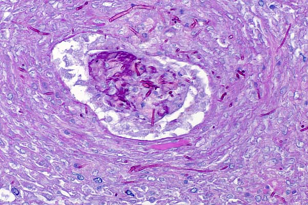
- Case 29-3. Liver. Multiple branching septate fungal
hyphae are present within the lumen and the wall of a blood vessel.
-
- AFIP Diagnosis: Liver: Hepatitis, necrotizing and
granulomatous, multifocally extensive, severe, with necrotizing
vasculitis, and fungal hyphae, hawksbill turtle (Eretmochelys
imbricata), reptile.
-
- Conference Note: Five species of Paecilomyces (P.
javanicus, P. lilacinus, P. marquandii, P. variotii and P. viridis)
have been reported to cause disease in humans and animals. In
animals, infection is generally characterized by chronic weight
loss. Lung is most often affected. The inflammatory reaction
is granulomatous. Histologically, budding yeast-like cells, septate
hyphae and conidia may be found.
-
- Contributor: Institut für Tierpathologie, Universität
Bern, Länggasse 122, CH-3012 Bern.
-
- References:
- 1. Heard DJ, Cantor GH, Jacobson ER, Purich B, Ajello L,
Padhye AA: Hyalohyphomycosis caused by Paecilomyces lilacinus
in an Aldabra tortoise. J Am Vet Med Assoc 9:1143-1145, 1986
- 2. Jacobson ER, Gaskin JM, Shields RP, White FH: Mycotic
pneumonia in mariculture-reared green sea turtles. J Am Vet Med
Assoc 175:929-933, 1979
- 3. Posthaus H, Krampe M, Pagan O, Guého E, Suter C,
Bacciarini L: Systemic Paecilomycosis in a Hawksbill Turtle (Eretmochelys
imbricata). J Mycol Med 7:223-226, 1997
-
-
- Case IV - 96-735 (AFIP 2701698)
-
- Signalment: 11+ -year-old, female, White's tree frog
(Litoria caerulea)
-
- History: This was an aged tree frog that had not been
eating well for several weeks. It was found dead in its cage.
-
- Gross Pathology: The carcass was in poor nutritional
condition; coelomic fat stores were markedly atrophied.
-
- Contributor's Diagnosis and Comments: Skin, hyperkeratosis
and acanthosis, diffuse, moderate, with multifocal epidermal
degeneration
- Etiologic Diagnosis: Mycotic (chytridiomycotic) dermatosis
- Etiology: Batrachochytrium dendrobatidis (Chytridiomycetes
fungus)
-
- Until recently, no species of fungi in the Phylum Chytridiomycota
was known to be a pathogen of vertebrate animals. However, fatal
cutaneous chytridiomycosis has now been reported in a wide variety
of wild and captive amphibians and has been associated with declines
of wild populations of frogs and toads in Australia, Panama,
and the United States.
-
- At the National Zoological Park (NZP), this disease has been
identified in more than 40 dead frogs of 4 different species.
The causative organism has been isolated in culture from blue
poison arrow frogs (Dendrobates azureus), green-and-black poison
arrow frogs (Dendrobates auratus), and a White's tree frog at
NZP. This organism is unlike any previously described fungal
genus or species and has been named Batrachochytrium dendrobatidis.
-
- The most characteristic lesions associated with cutaneous
chytridiomycete infection are hyperkeratosis and acanthosis.
Focal epidermal cell hypertrophy and/or degeneration are less
common. Inflammation is rare and, when it does occur, is usually
mild. Typically, low to moderate numbers of chytridiomycetes
are located in the superficial epidermis, primarily the keratinized
layers. In histologic sections, three stages of the organisms
are primarily seen: a uninucleated form, a multinucleated stage
containing internal septa, and a cyst-like form (zoosporangium)
containing multiple flagellated spores (zoospores). Each spore
has a single posterior flagellum that is very difficult to detect
in histologic sections.
-
- The entire life cycle of Batrachochytrium dendrobatidis has
not been determined. Spores are released through tubular projections
on the zoosporangia known as discharge papillae. The motile spores
are then thought to swim through water to infect other epidermal
cells.
-
- The mechanisms by which chytridiomycosis causes death is
not known. The skin of infected animals appears to be the only
organ to consistently have lesions. Normal skin functions in
amphibians include maintenance of hydration, osmoregulation,
thermoregulation, and respiration. The skin lesions probably
interfere with these functions and cause fatal metabolic alterations.
-
 40x
obj
40x
obj
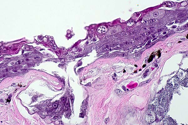
- Case 29-4. Skin. Multiple developmental stages of
fungal thalli and zoosporangia are present within the epidermis
and subepidermal glands.
-
- AFIP Diagnosis: Skin: Epidermal hyperplasia, diffuse,
moderate, with hyperkeratosis and numerous intracorneal fungi,
White's tree frog (Litoria caerulea), amphibian, etiology consistent
with Chytridiomycetes.
-
- Conference Note: Emerging infectious diseases are
believed to play important roles in the global declines in amphibian
populations. Various factors (host, pathogen, and environmental)
may be involved in disease emergence. Infectious diseases may
be only the proximate cause of death. Environmental factors such
as increased UV-B, chemical pollution, climate change and stress
have been hypothesized as possible underlying problems. In addition
to Chytridiomycetes, iridoviruses of the Ranavirus genus have
been implicated as contributing to the amphibian decline.
-
- Contributor: Department of Pathology, National Zoological
Park, Smithsonian Institution, Washington, DC, 20008.
-
- References:
- 1. Berger L, Speare R, Daszak P, Green DE, Cunningham AA,
Goggin CL, Slocombe R, Ragan MA, Hyatt AD, McDonald KR, Hines
HB, Lips KR, Marantelli G, Parkes H: Chytridiomycosis causes
amphibian mortality associated with population declines in the
rain forests of Australia and Central America. Proceedings of
the National Academy of Sciences 95:9031-9036, 1998
- 2. Longcore JE, Pessier AP, Nichols DK: Batrachochytrium
dendrobatidis gen. et sp. nov., a chytrid pathogenic to amphibians.
Mycologia 91(2): 219-227, 1999
- 3. Nichols DK, Pessier AP, Longcore JE: Cutaneous chytridiomycosis
in amphibians: an emerging disease? Proceedings, Annual Conference
of the American Association of Zoo Veterinarians, Omaha, NE,
pp. 269-271, 1998
- 4. Pessier AP, Nichols DK, Longcore JE, Fuller MS: Cutaneous
chytridiomycosis in poison dart frogs (Dendrobates spp.) and
White's tree frogs (Litoria caerulea). J Vet Diag Invest 11:194-199,
1999
- 5. Daszak P, Berger L, Cunningham AA, Hyatt AD, Green DE,
Speare R: Emerging infectious diseases and amphibian population
declines. Emerg Infect Dis 5(6):Nov-Dec,1999
-
-
- J Scot Estep, DVM
Captain, United States Army
Registry of Veterinary Pathology*
Department of Veterinary Pathology
Armed Forces Institute of Pathology
(202) 782-2615; DSN: 662-2615
Internet: estep@afip.osd.mil
-
- * The American Veterinary Medical Association and the American
College of Veterinary Pathologists are co-sponsors of the Registry
of Veterinary Pathology. The C.L. Davis Foundation also provides
substantial support for the Registry.
-
- Return to WSC Case Menu
 20x
obj
20x
obj
 20x
obj, polarized
20x
obj, polarized
 20x
obj
20x
obj
 20x
obj
20x
obj

 40x
obj, PAS
40x
obj, PAS
 40x
obj
40x
obj