Conference: 05 - 2010 Case: 03 Liver and cecum - Turkey
Signalment:
Young male intact commercial grower turkey.
Gross Description:
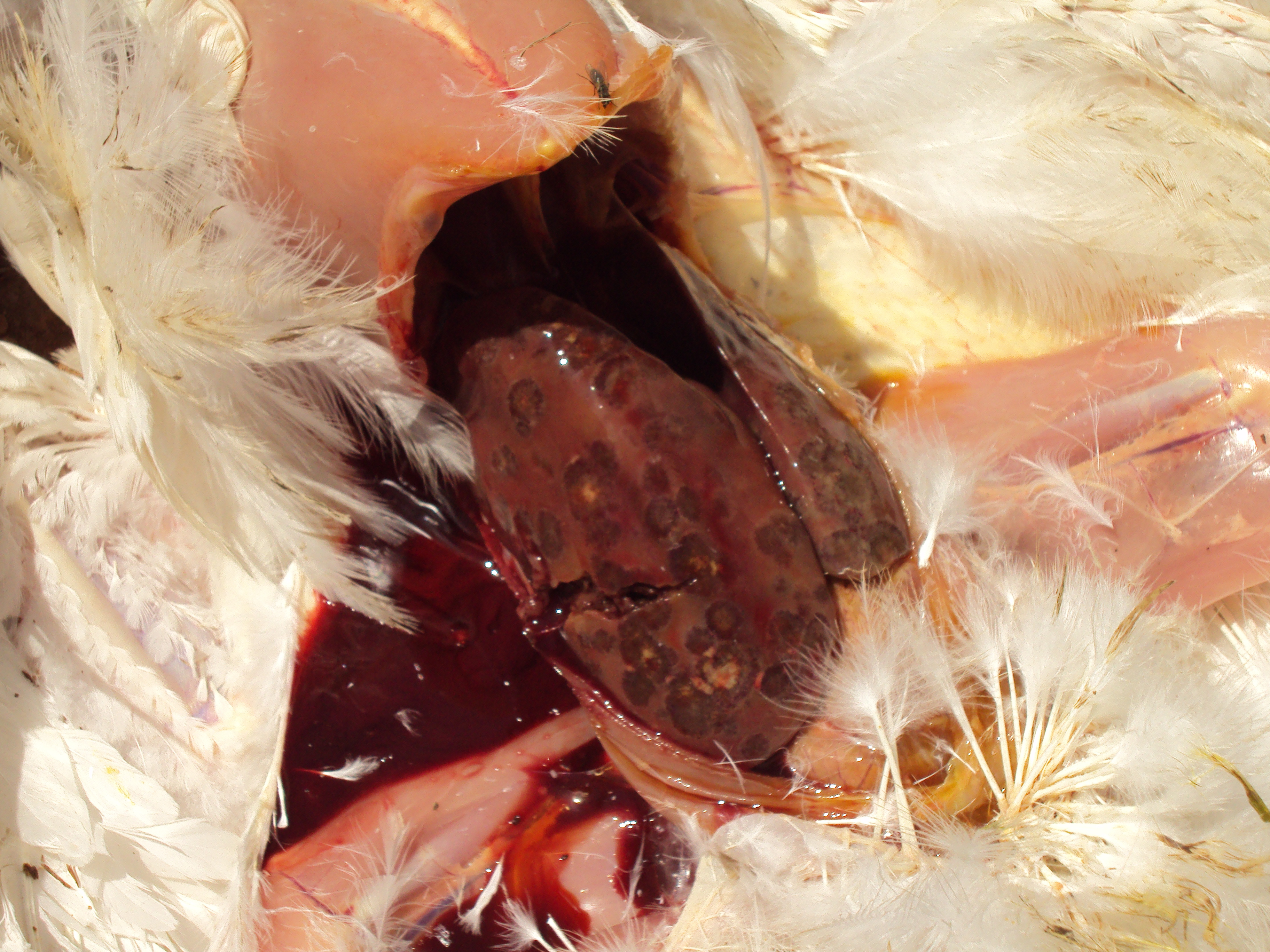
<br><u>Liver</u>: There are multifocal to coalescing circular regions of necrosis with depressed pale tan/yellow centers
surrounded by a slightly raised, often hemorrhagic rim. Lesions range in size from 0.5 to 3.0 cm in diameter and are
disseminated randomly throughout the parenchyma.
<br>
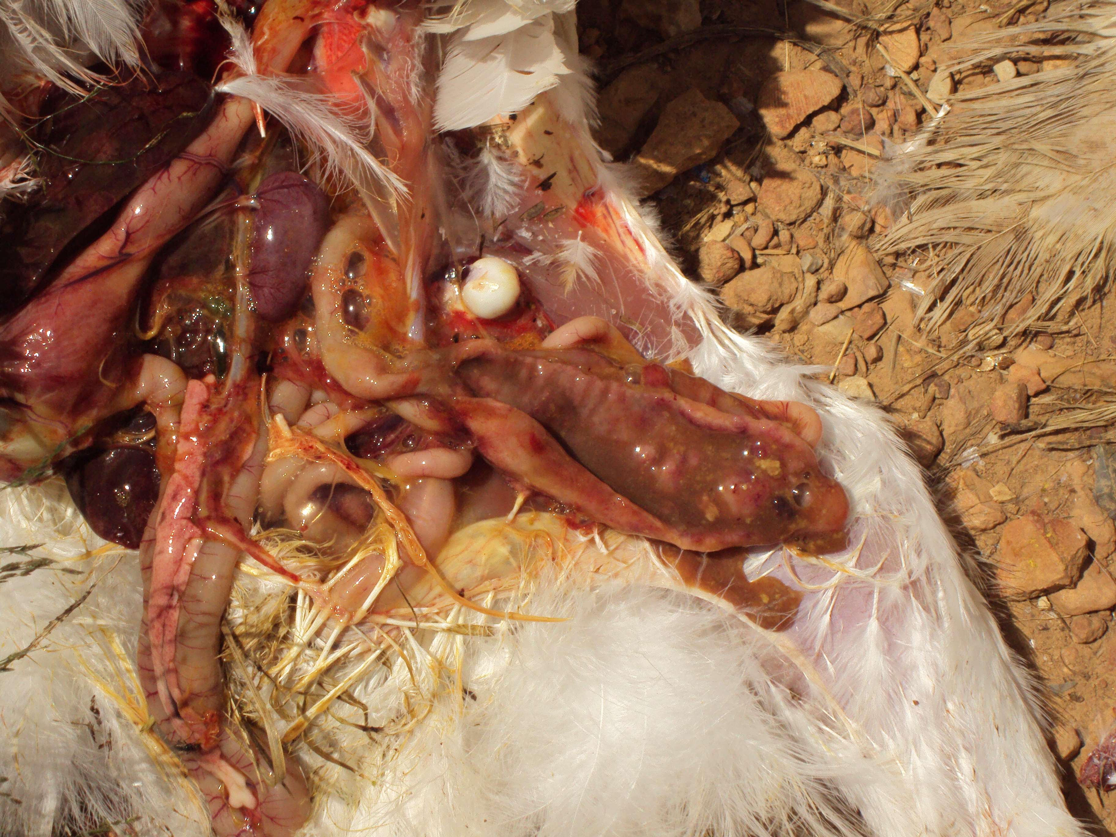
<u>Ceca</u>: Both ceca are moderately distended and fluid filled with thickened hyperemic walls mottled tan to dark red.
The mucosal surface is multifocally ulcerated with occasional partially adhered caseous debris amidst abundant
luminal hemorrhagic exudate.
History:
A male commercial production turkey was submitted for necropsy (donation) by a large commercial
production company. The company has multiple complexes in multiple states in the United States. This bird came
from a premise in Arkansas with a flock size of 40,000 birds. All birds in this flock are males and are sent to
slaughter around 20 weeks of age. This bird was found dead. The company did not report a higher than normal rate
of turkey mortality.
Histopathologic Description:
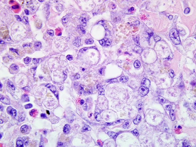
<u>Liver</u>: Approximately 50 to 80% of the parenchyma is largely obliterated by
multifocal, random, discrete and coalescing to regionally extensive areas of coagulative hepatic necrosis. Affected
areas are characterized by shrunken hepatocytes with intensely eosinophilic cytoplasm and pyknotic to lytic nuclei
or loss of hepatocytes, disruption of hepatic cords, and replacement with eosinophilic cellular and karyorrhectic
debris. Mainly within necrotic foci and variably expanding portal triads are pleocellular infiltrates composed of
moderate to large numbers of macrophages (many hemosiderin-laden), lymphocytes, heterophils, few plasma cells
and variable numbers of foreign body-type multinucleate giant cells. Phagocytized by multinucleated giant cells as
well as free and within the lumen of ectatic, hyperplastic bile ducts are individual to clustered 12-20 μm in diameter
eosinophilic to amphophilic protozoal trophozoites. Trophozoites may contain a 2-4μm diameter oval shaped
basophilic central to slightly eccentric nucleus. Pericholangial inflammation and fibrosis is prominent around large
sized bile ducts with remaining hepatic parenchyma interrupted by numerous variably sized newly formed bile
ductules. Numbers of multinucleate cells and organisms vary depending on the section received.
<br><br>
<u>Cecum</u>: Diffusely expanding the lamina propria and multifocally extending transmurally with effacement of up to
50% of the muscular coat is an inflammatory infiltrate composed predominantly of lymphocytes and macrophages
with fewer plasma cells and heterophils admixed with numerous 12-20μm diameter protozoal trophozoites similar to
those described within the liver. There is extensive loss of crypts and remnant crypts are hyperplastic, tortuous and
ectatic with piling of 2-3 epithelial cells layers, and luminal aggregates of detached epithelial cells and degenerate
leukocytes, mainly heterophils (crypt microabscesses). There is multifocal extensive mucosal ulceration, and in
some sections the cecal lumen contains a dense core of eosinophilic cellular and karyorrhectic debris, erythrocytes,
fibrin, degenerate protozoal trophozoites, macrophages and heterophils, and aggregates of rod shaped bacteria (cecal
core).
Morphologic Diagnosis:
<br><u>Liver</u>: Multifocal and coalescing, random, necrotizing, sub-acute-tochronic,
lymphohistiocytic and heterophilic hepatitis with biliary hyperplasia and intralesional protozoal
trophozoites, etiology consistent with <i>Histomonas meleagridis</i>.
<br>
<u>Cecum</u>: Transmural, marked, sub-acute-to-chronic lymphohistiocytic and heterophilic typhlitis with fibrinonecrotic
core (variable) and intralesional protozoal trophozoites consistent with <i>H. meleagridis</i>.
Condition:
Histomonas meleagridis
Contributor Comment:
Histomoniasis, also known as blackhead and infectious enterohepatitis, is a disease of
gallinaceous birds caused by <i>Histomonas meleagridis</i>, a flagellated amoeboid protozoan which remains an important
disease economically for the commercial poultry industry. Two aspects of this disease worthy of brief review are 1)
the unique life cycle of the organism and 2) differences in transmission and extent of disease in turkeys and
chickens.
<br><br>
The role of the cecal worm <i>Heterakis gallinarum</i> as in intermediate host has been well-described.(2,3) The exact
mechanism of infection of Heterakis eggs with histomonads remains unknown, but it has been suggested that the
protozoan may be transferred to the female worm during mating and are then incorporated into embryonated eggs.(2)
Infected eggs pass in feces where they may be ingested directly by birds or by earthworms who may serve as a
transport host. Ingestion of either egg-laden feces or earthworms by the bird results in transport to the cecum where
flagellated trophozoites are released from the nematode egg, multiply in the cecal lumen and penetrate the cecal
wall. The tissue stage loses the flagella becoming amoeboid. Eventually the histomonads gain entry into the
bloodstream and are carried to the liver via the hepatic-portal system. It is important to note that the fragile
trophozoite of <i>Histomonas</i>, which cannot survive long outside of any of its hosts, would be unable to survive
passage through the stomach if not within a nematode egg or an earthworm. Therefore, fecal oral transmission is not
thought to be an important route of transmission. Interestingly, some recent publications have shown the existence
of a cyst stage in some species of <i>Histomonas in vitro</i> sparking interest in the possibility of this happening under
certain conditions in the natural disease allowing for persistence in the environment and the possibility of oral
transmission.(4,7)
<br><br>
Extension to the liver as described above occurs at a higher rate in turkeys than chickens, with a much higher
associated mortality rate. Concurrent infection with <i>Eimeria tenella</i> results in increased liver lesions in chickens.
Histomonad virulence also requires the presence of cecal bacteria such as <i>Escherichia coli, Clostridium perfringens</i>,
and/or <i>Bacillus subtilis</i>, especially in turkeys.(2,3) As mentioned previously, turkeys suffer from a much higher
mortality rate than chickens, with the latter being better able to control the disease but still suffering decreased
productivity. A recent publication showed that chickens mount a more effective innate immune response to <i>H.
meleagridis</i> in the ceca than do turkeys resulting in better control of parasite numbers in chickens.(5) Furthermore,
unregulated cell-mediated immunity in the liver is more pronounced in the turkey than the chicken often leading to
lymphoid depletion of the spleen.(5) Another difference of histomoniasis in turkeys is that, in addition to transmission
via ingestion of <i>Heterakis</i> eggs, turkeys appear to be able to transmit histomonads directly via cloacal drinking
where cloacal droppings from an infected bird can be pulled retrograde into the ceca of a susceptible bird if the
droppings contact the vent of the uninfected bird.(2,3)
JPC Diagnosis:
<br>
1. Liver: Hepatitis, random, necrotizing, lymphohistiocytic and heterophilic, multifocal
coalescing, severe, with biliary hyperplasia and protozoal trophozoites, etiology consistent with <i>Histomonas
meleagridis</i>.
<br>
2. Cecum: Typhlitis, transmural, lymphohistiocytic and heterophilic, diffuse, severe, with fibrinonecrotic core and
protozoal trophozoites, etiology consistent with <i>Histomonas meleagridis</i>.
Conference Comment:
The contributor provides an excellent review of the pathology and life cycle of <i>Histomonas
meleagridis</i>. While attendees found this to be a relatively uncomplicated histologic diagnosis given the species and
abundance of trophozoites, all were interested and engaged in the discussion that the case precipitated.
<br><br>
Conference participants noted that blackhead is an imprecise colloquialism in that cyanosis of the head is neither a
constant feature nor a unique clinical sign of histomoniasis.(6) Other diseases, such as turkey enteric coronavirus
(bluecomb disease), avian influenza, and various respiratory pathogens may cause a cyanotic head in turkeys and
chickens. Gross digital images were shown during the conference to reinforce the point that the macroscopic lesions
of histomoniasis are very distinctive, and when classical lesions are present in both the liver and cecum
simultaneously, the findings are considered pathognomonic for the condition. When only cecal cores are present,
other etiologic considerations would include salmonellosis for both chickens and turkeys, and <i>Eimeria tenella</i> in
chickens.
<br><br>
In addition to histomoniasis, cecal heterakiasis was also discussed by participants. Although <i>H. gallinarum</i> can
cause thickened cecal walls, nodule formation, and inflammation in its own right in turkeys and chickens, its
practical and economic importance is the nematodes ability to serve as a carrier for <i>H. meleagridis</i>.(1) In pheasants,
however, <i>Heterakis isolonche</i> causes severe cecal disease with mortality rates that may exceed 50%.(2) The disease in
pheasants is characterized by a marked inflammatory response, fibrosis, and coalescing cecal wall nodules.
References:
1. Goodwin MA. Alimentary system. In: Riddell C, ed. <i>Avian Histopathology</i>. 2nd ed. Tallahassee, FL: American
Association of Avian Pathologists, Rose Printing; 1996: 127-129.
<br>
2. McDougald LR. Histomoniasis (Blackhead) and other protozoan diseases of the intestinal tract. In: Saif YM, ed.
<i>Disease of Poultry</i>, 12th ed. Ames, IA: Blackwell Publishing Professional; 2008:1095-1100.
<br>
3. McDougald LR. Blackhead disease (Histomoniasis) in poultry: a critical review. <i>Avian Dis.</i> 2005;49:462-476.
<br>
4. Munsch MH, Mehlhorn S, Al-Quraishy AR, Lotfi, Hafez HM. Molecular biological features of strains of
<i>Histomonas meleagridis. Parasitol Res.</i> 2009;104:1137-1140.
<br>
5. Powell FL, Rothwell L, Clarckson MJ, Kaiser P. The turkey, compared to the chicken, fails to mount an effective
early immune response to <i>Histomonas meleagridis</i> in the gut. <i>Parasite Immunol.</i> 2009;31:312-327.
<br>
6. Trees AJ. Parasitic diseases. In: McMullin PF, ed. <i>Poultry Diseases</i>, 6th ed., Edinburgh: Elsevier Limited;
2008:458-459.
<br>
7. Zaragatzki E, Hess M, Grabensteiner E, Abdel-Ghaffar F, Al-Rasheid K, Mehlhorn H. Light and transmission
electron microscopic studies of the encystation of <i>Histomonas meleagridis. Parasitol Res.</i> 2010;106:977-983.