Conference: 11 - 2019 Case: 01 Hiared skin -
History:
<html>
<head>
<meta http-equiv=Content-Type content="text/html; charset=windows-1252">
<meta name=Generator content="Microsoft Word 15 (filtered)">
<style>
<!--
/* Font Definitions */
@font-face
{font-family:Helvetica;
panose-1:2 11 6 4 2 2 2 2 2 4;}
@font-face
{font-family:SimSun;
panose-1:2 1 6 0 3 1 1 1 1 1;}
@font-face
{font-family:"Cambria Math";
panose-1:2 4 5 3 5 4 6 3 2 4;}
@font-face
{font-family:Calibri;
panose-1:2 15 5 2 2 2 4 3 2 4;}
@font-face
{font-family:Cambria;
panose-1:2 4 5 3 5 4 6 3 2 4;}
@font-face
{font-family:"\30D2\30E9\30AE\30CE\89D2\30B4 Pro W3";}
@font-face
{font-family:"Lucida Grande";}
@font-face
{font-family:"\@SimSun";
panose-1:2 1 6 0 3 1 1 1 1 1;}
@font-face
{font-family:"\@\30D2\30E9\30AE\30CE\89D2\30B4 Pro W3";}
/* Style Definitions */
p.MsoNormal, li.MsoNormal, div.MsoNormal
{margin:0in;
margin-bottom:.0001pt;
line-height:normal;
font-size:12.0pt;
font-family:"Times New Roman",serif;
color:windowtext;}
h1
{mso-style-link:"Heading 1 Char";
margin-right:0in;
margin-left:0in;
line-height:normal;
font-size:24.0pt;
font-family:"Times New Roman",serif;
color:windowtext;
font-weight:bold;}
h2
{mso-style-link:"Heading 2 Char";
margin-top:2.0pt;
margin-right:0in;
margin-bottom:0in;
margin-left:0in;
margin-bottom:.0001pt;
line-height:normal;
page-break-after:avoid;
font-size:13.0pt;
font-family:"Cambria",serif;
color:#365F91;
font-weight:normal;}
h4
{mso-style-link:"Heading 4 Char";
margin-top:2.0pt;
margin-right:0in;
margin-bottom:0in;
margin-left:0in;
margin-bottom:.0001pt;
line-height:normal;
page-break-after:avoid;
font-size:12.0pt;
font-family:"Cambria",serif;
color:#365F91;
font-weight:normal;
font-style:italic;}
p.MsoCommentText, li.MsoCommentText, div.MsoCommentText
{mso-style-link:"Comment Text Char";
margin-top:0in;
margin-right:0in;
margin-bottom:10.0pt;
margin-left:0in;
line-height:normal;
font-size:10.0pt;
font-family:"Lucida Grande";
color:black;}
p.MsoBodyText, li.MsoBodyText, div.MsoBodyText
{mso-style-link:"Body Text Char";
margin-top:0in;
margin-right:0in;
margin-bottom:12.0pt;
margin-left:0in;
line-height:125%;
font-size:12.0pt;
font-family:"Times New Roman",serif;
color:windowtext;}
a:link, span.MsoHyperlink
{color:blue;
text-decoration:underline;}
a:visited, span.MsoHyperlinkFollowed
{color:purple;
text-decoration:underline;}
p.MsoDocumentMap, li.MsoDocumentMap, div.MsoDocumentMap
{mso-style-link:"Document Map Char";
margin:0in;
margin-bottom:.0001pt;
line-height:normal;
font-size:12.0pt;
font-family:"Times New Roman",serif;
color:black;}
p.MsoPlainText, li.MsoPlainText, div.MsoPlainText
{mso-style-link:"Plain Text Char";
margin:0in;
margin-bottom:.0001pt;
line-height:normal;
font-size:11.0pt;
font-family:"Calibri",sans-serif;
color:windowtext;}
p
{margin-top:0in;
margin-right:0in;
margin-bottom:10.0pt;
margin-left:0in;
line-height:115%;
font-size:12.0pt;
font-family:"Times New Roman",serif;
color:black;}
pre
{mso-style-link:"HTML Preformatted Char";
margin:0in;
margin-bottom:.0001pt;
line-height:normal;
font-size:10.0pt;
font-family:"Courier New";
color:windowtext;}
p.MsoCommentSubject, li.MsoCommentSubject, div.MsoCommentSubject
{mso-style-link:"Comment Subject Char";
margin-top:0in;
margin-right:0in;
margin-bottom:10.0pt;
margin-left:0in;
line-height:normal;
font-size:10.0pt;
font-family:"Lucida Grande";
color:black;
font-weight:bold;}
p.MsoAcetate, li.MsoAcetate, div.MsoAcetate
{mso-style-link:"Balloon Text Char";
margin:0in;
margin-bottom:.0001pt;
line-height:normal;
font-size:9.0pt;
font-family:"Lucida Grande";
color:black;}
p.MsoNoSpacing, li.MsoNoSpacing, div.MsoNoSpacing
{margin:0in;
margin-bottom:.0001pt;
line-height:normal;
font-size:11.0pt;
font-family:"Calibri",sans-serif;
color:windowtext;}
p.MsoRMPane, li.MsoRMPane, div.MsoRMPane
{margin:0in;
margin-bottom:.0001pt;
line-height:normal;
font-size:11.0pt;
font-family:"Lucida Grande";
color:black;}
p.MsoListParagraph, li.MsoListParagraph, div.MsoListParagraph
{margin-top:0in;
margin-right:0in;
margin-bottom:10.0pt;
margin-left:.5in;
line-height:115%;
font-size:11.0pt;
font-family:"Lucida Grande";
color:black;}
p.MsoListParagraphCxSpFirst, li.MsoListParagraphCxSpFirst, div.MsoListParagraphCxSpFirst
{margin-top:0in;
margin-right:0in;
margin-bottom:0in;
margin-left:.5in;
margin-bottom:.0001pt;
line-height:115%;
font-size:11.0pt;
font-family:"Lucida Grande";
color:black;}
p.MsoListParagraphCxSpMiddle, li.MsoListParagraphCxSpMiddle, div.MsoListParagraphCxSpMiddle
{margin-top:0in;
margin-right:0in;
margin-bottom:0in;
margin-left:.5in;
margin-bottom:.0001pt;
line-height:115%;
font-size:11.0pt;
font-family:"Lucida Grande";
color:black;}
p.MsoListParagraphCxSpLast, li.MsoListParagraphCxSpLast, div.MsoListParagraphCxSpLast
{margin-top:0in;
margin-right:0in;
margin-bottom:10.0pt;
margin-left:.5in;
line-height:115%;
font-size:11.0pt;
font-family:"Lucida Grande";
color:black;}
p.Heading21, li.Heading21, div.Heading21
{mso-style-name:"Heading 21";
margin:0in;
margin-bottom:.0001pt;
line-height:normal;
page-break-after:avoid;
font-size:12.0pt;
font-family:"Helvetica",sans-serif;
color:black;
font-weight:bold;}
p.FreeFormA, li.FreeFormA, div.FreeFormA
{mso-style-name:"Free Form A";
margin:0in;
margin-bottom:.0001pt;
line-height:normal;
font-size:10.0pt;
font-family:"Times New Roman",serif;
color:black;}
p.Default, li.Default, div.Default
{mso-style-name:Default;
margin:0in;
margin-bottom:.0001pt;
line-height:normal;
font-size:12.0pt;
font-family:"Arial",sans-serif;
color:black;}
p.Body, li.Body, div.Body
{mso-style-name:Body;
margin:0in;
margin-bottom:.0001pt;
line-height:normal;
font-size:12.0pt;
font-family:"Helvetica",sans-serif;
color:black;}
span.Hyperlink1
{mso-style-name:Hyperlink1;
color:blue;
text-decoration:underline;}
p.PlainText1, li.PlainText1, div.PlainText1
{mso-style-name:"Plain Text1";
margin:0in;
margin-bottom:.0001pt;
line-height:normal;
font-size:10.5pt;
font-family:"Lucida Grande";
color:black;}
p.NormalWeb1, li.NormalWeb1, div.NormalWeb1
{mso-style-name:"Normal \(Web\)1";
margin-top:5.0pt;
margin-right:0in;
margin-bottom:5.95pt;
margin-left:0in;
line-height:normal;
font-size:12.0pt;
font-family:"Times New Roman",serif;
color:black;}
p.Text3, li.Text3, div.Text3
{mso-style-name:"Text 3";
margin-top:0in;
margin-right:0in;
margin-bottom:12.0pt;
margin-left:0in;
line-height:normal;
font-size:12.0pt;
font-family:"Times New Roman",serif;
color:black;}
span.Hyperlink2
{mso-style-name:Hyperlink2;
color:blue;
text-decoration:underline;}
p.FreeFormAA, li.FreeFormAA, div.FreeFormAA
{mso-style-name:"Free Form A A";
margin:0in;
margin-bottom:.0001pt;
line-height:normal;
font-size:12.0pt;
font-family:"Helvetica",sans-serif;
color:black;}
p.NormalWeb2, li.NormalWeb2, div.NormalWeb2
{mso-style-name:"Normal \(Web\)2";
margin-top:5.0pt;
margin-right:0in;
margin-bottom:5.95pt;
margin-left:0in;
line-height:normal;
font-size:12.0pt;
font-family:"Times New Roman",serif;
color:black;}
span.BalloonTextChar
{mso-style-name:"Balloon Text Char";
mso-style-link:"Balloon Text";
font-family:"Lucida Grande";
color:black;}
span.PlainTextChar
{mso-style-name:"Plain Text Char";
mso-style-link:"Plain Text";
font-family:"Calibri",sans-serif;}
span.st1
{mso-style-name:st1;}
span.CommentTextChar
{mso-style-name:"Comment Text Char";
mso-style-link:"Comment Text";
font-family:"Lucida Grande";
color:black;}
span.CommentSubjectChar
{mso-style-name:"Comment Subject Char";
mso-style-link:"Comment Subject";
font-family:"Lucida Grande";
color:black;
font-weight:bold;}
span.spelle
{mso-style-name:spelle;}
span.grame
{mso-style-name:grame;}
span.apple-converted-space
{mso-style-name:apple-converted-space;}
span.headword
{mso-style-name:headword;}
span.BodyTextChar
{mso-style-name:"Body Text Char";
mso-style-link:"Body Text";}
p.WW-Standard, li.WW-Standard, div.WW-Standard
{mso-style-name:WW-Standard;
margin:0in;
margin-bottom:.0001pt;
line-height:normal;
font-size:12.0pt;
font-family:"Times New Roman",serif;
color:windowtext;}
span.DocumentMapChar
{mso-style-name:"Document Map Char";
mso-style-link:"Document Map";
font-family:"\30D2\30E9\30AE\30CE\89D2\30B4 Pro W3";
color:black;}
p.EndNoteBibliography, li.EndNoteBibliography, div.EndNoteBibliography
{mso-style-name:"EndNote Bibliography";
mso-style-link:"EndNote Bibliography Char";
margin-top:0in;
margin-right:0in;
margin-bottom:10.0pt;
margin-left:0in;
line-height:normal;
font-size:11.0pt;
font-family:"Calibri",sans-serif;
color:windowtext;}
span.EndNoteBibliographyChar
{mso-style-name:"EndNote Bibliography Char";
mso-style-link:"EndNote Bibliography";
font-family:"Calibri",sans-serif;}
p.desc, li.desc, div.desc
{mso-style-name:desc;
margin-right:0in;
margin-left:0in;
line-height:normal;
font-size:12.0pt;
font-family:"Times New Roman",serif;
color:windowtext;}
span.jrnl
{mso-style-name:jrnl;}
span.Heading1Char
{mso-style-name:"Heading 1 Char";
mso-style-link:"Heading 1";
font-weight:bold;}
span.highlight
{mso-style-name:highlight;}
span.hit
{mso-style-name:hit;}
span.Heading4Char
{mso-style-name:"Heading 4 Char";
mso-style-link:"Heading 4";
font-family:"Cambria",serif;
color:#365F91;
font-style:italic;}
p.CitaviBibliographyEntry, li.CitaviBibliographyEntry, div.CitaviBibliographyEntry
{mso-style-name:"Citavi Bibliography Entry";
mso-style-link:"Citavi Bibliography Entry Zchn";
margin-top:0in;
margin-right:0in;
margin-bottom:0in;
margin-left:17.0pt;
margin-bottom:.0001pt;
text-indent:-17.0pt;
line-height:115%;
font-size:11.0pt;
font-family:"Calibri",sans-serif;
color:windowtext;}
span.CitaviBibliographyEntryZchn
{mso-style-name:"Citavi Bibliography Entry Zchn";
mso-style-link:"Citavi Bibliography Entry";
font-family:"Calibri",sans-serif;}
p.CitaviBibliographyHeading, li.CitaviBibliographyHeading, div.CitaviBibliographyHeading
{mso-style-name:"Citavi Bibliography Heading";
mso-style-link:"Citavi Bibliography Heading Zchn";
margin-top:24.0pt;
margin-right:0in;
margin-bottom:0in;
margin-left:0in;
margin-bottom:.0001pt;
line-height:115%;
page-break-after:avoid;
font-size:14.0pt;
font-family:"Cambria",serif;
color:#365F91;
font-weight:bold;}
span.CitaviBibliographyHeadingZchn
{mso-style-name:"Citavi Bibliography Heading Zchn";
mso-style-link:"Citavi Bibliography Heading";
font-family:"Cambria",serif;
color:#365F91;
font-weight:bold;}
p.desc2, li.desc2, div.desc2
{mso-style-name:desc2;
margin:0in;
margin-bottom:.0001pt;
line-height:normal;
font-size:13.0pt;
font-family:"Times New Roman",serif;
color:windowtext;}
span.EndNoteBibliographyZchn
{mso-style-name:"EndNote Bibliography Zchn";
font-family:"Arial",sans-serif;}
span.HTMLPreformattedChar
{mso-style-name:"HTML Preformatted Char";
mso-style-link:"HTML Preformatted";
font-family:"Courier New";}
span.Heading2Char
{mso-style-name:"Heading 2 Char";
mso-style-link:"Heading 2";
font-family:"Cambria",serif;
color:#365F91;}
span.UnresolvedMention1
{mso-style-name:"Unresolved Mention1";
color:#605E5C;
background:#E1DFDD;}
.MsoChpDefault
{font-size:10.0pt;}
/* Page Definitions */
@page WordSection1
{size:8.5in 11.0in;
margin:1.0in 1.0in 1.0in 1.0in;}
div.WordSection1
{page:WordSection1;}
/* List Definitions */
ol
{margin-bottom:0in;}
ul
{margin-bottom:0in;}
-->
</style>
</head>
<body lang=EN-US link=blue vlink=purple>
<div class=WordSection1>
<p class=DefaultCxSpFirst align=center style='text-align:center;line-height:
115%'><span style='line-height:115%;font-family:"Times New Roman",serif;
color:black'>Joint Pathology Center</span></p>
<p class=DefaultCxSpMiddle align=center style='text-align:center;line-height:
115%'><span style='line-height:115%;font-family:"Times New Roman",serif;
color:black'>Veterinary Pathology Services</span></p>
<p class=DefaultCxSpMiddle align=center style='text-align:center;line-height:
115%'><span style='line-height:115%;font-family:"Times New Roman",serif;
color:black'>Wednesday Slide Conference</span></p>
<p class=DefaultCxSpMiddle align=center style='text-align:center;line-height:
115%'><span style='line-height:115%;font-family:"Times New Roman",serif;
color:black'>2019-2020</span></p>
<p class=DefaultCxSpMiddle align=center style='text-align:center;line-height:
115%'><span style='line-height:115%;font-family:"Times New Roman",serif;
color:black'>Conference 11</span></p>
<p class=DefaultCxSpMiddle align=center style='text-align:center;line-height:
115%'><span style='line-height:115%;font-family:"Times New Roman",serif;
color:black'>20 November 2019</span></p>
<p class=DefaultCxSpLast align=center style='text-align:center;line-height:
115%'><span style='line-height:115%;font-family:"Times New Roman",serif;
color:black'> </span></p>
<p style='margin-top:.1in;margin-right:0in;margin-bottom:.1in;margin-left:0in;
vertical-align:baseline'><b><span style='color:black'>Conference Moderator: <a
name="OLE_LINK10"></a></span></b></p>
<p style='margin-top:.1in;margin-right:0in;margin-bottom:.1in;margin-left:0in;
line-height:13.45pt;vertical-align:baseline'><span style='color:windowtext'>Charles
W. Bradley, VMD, DACVP<br>
Assistant Professor, Pathobiology<br>
University of Pennsylvania School of Veterinary Medicine<br>
</span><span style='color:windowtext;background:white'>4005 MJR-VHUP</span><span
style='color:windowtext'><br>
<span style='background:white'>3900 Delancey Street<br>
Philapdelphia, PA, 19104</span></span></p>
<p class=MsoNormal style='line-height:115%'> </p>
<p class=MsoNormal style='margin-top:4.55pt'><b><u><span style='font-size:11.0pt'>CASE
I: </span></u></b><u><span style='font-size:11.0pt'>S17-1101 (JPC <span
style='color:#221F1F'>4116937</span>). </span></u></p>
<p class=MsoBodyText><span lang=EN-GB style='font-size:7.0pt;line-height:125%'> </span></p>
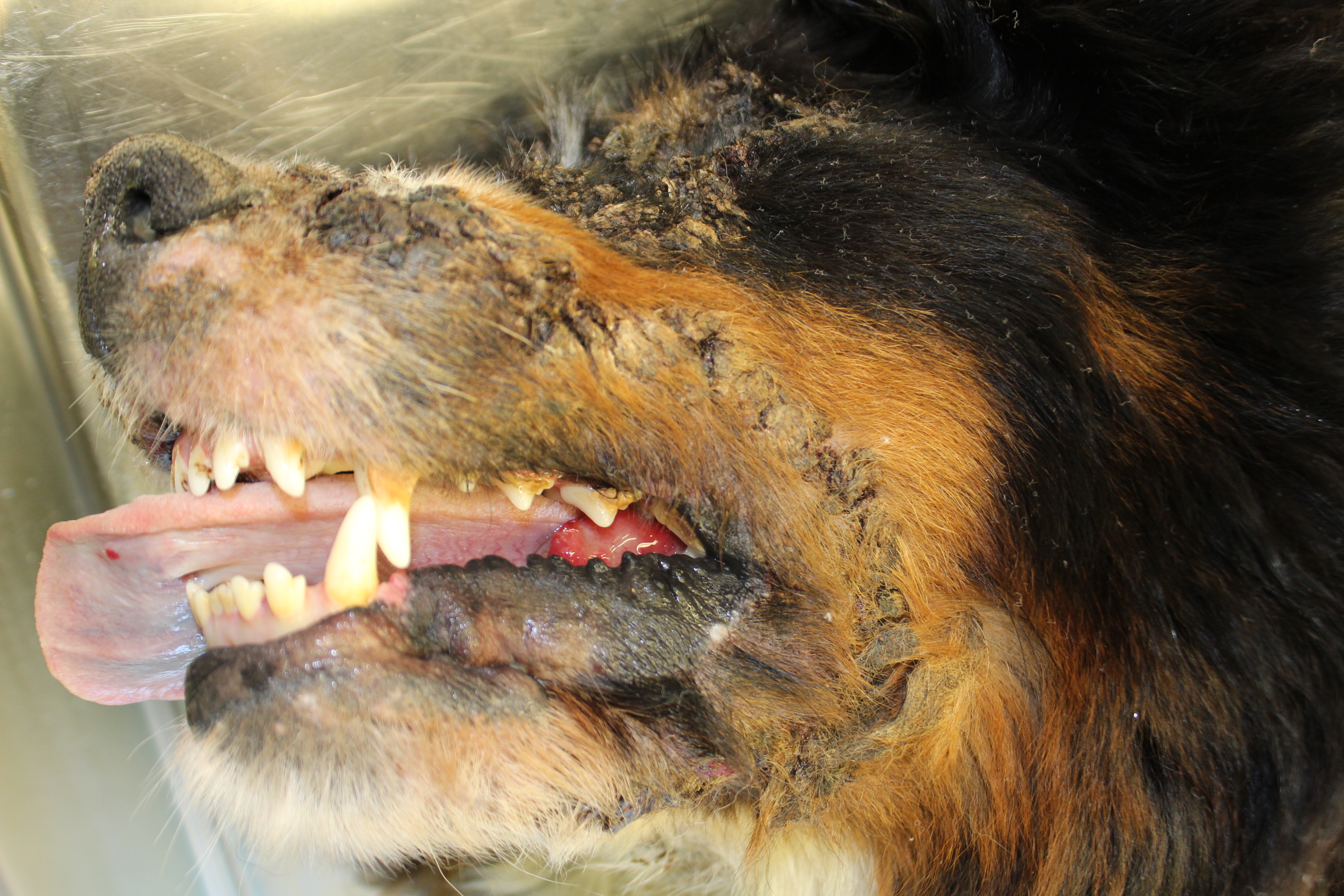
<p class=MsoNormal style='text-align:justify;line-height:115%'><b>Signalment:</b>
4-year-old spayed female Bernese mountain dog (<i>Canis familiaris</i>).</p>
<p class=MsoNormal> </p>
<p class=MsoNormal style='text-align:justify;line-height:115%'><b>History:</b> The
animal had a one year history of skin changes including multifocal alopecia,
crust formation and discolorations, whose underlying cause could not be
identified. The animal had been treated with high doses of dexadreson and
cyclosporine, resulting in a mild and slow improvement of the skin lesions. Two
weeks before death the animal started limping and a rupture of the cruciate
ligament was suspected. The animal was euthanized because of the questionable
prognosis of the skin lesions in combination with the cruciate rupture and
recurrent episodes of fever of unknown origin (temperature >39.8°C).</p>
<p class=MsoNormal style='text-align:justify;line-height:115%'> </p>
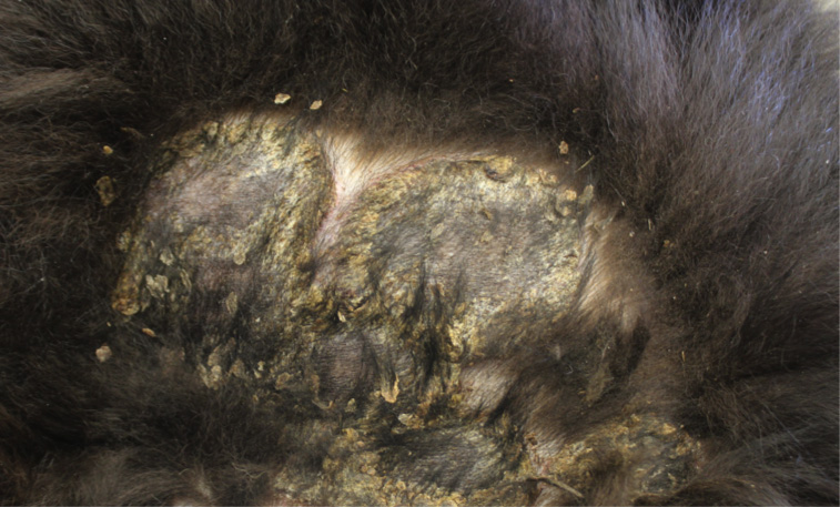
<p class=MsoNormal><b><span style='color:black'>Gross Pathology</span></b><span
style='color:black'>:<b> </b></span>On both sides of the trunk extending to the
axillas and groin, on the nasal bridge, on the ears, and around the eyes and
mouth, there were multiple, sharply demarcated, hairless or partially hairless
areas of skin with brown to grey discoloration and crust formation. Around
these hairless areas, the fur was clotted with crusty, brown material. Bilaterally
adjacent to the caudal aspect of the tongue, there were red, papillary masses
of soft tissue and approximately 1 x 2 x 0.5 cm observed (histologically
identified as chronic-active necrotizing and suppurative inflammation with
granulation tissue formation). On the left cheek, there was a pale yellow, soft
mass in the subcutis (lipoma). On the right knee joint the drawer test was
positive with increased mobility of the joint. The joint was filled with
turbid, slightly flocculent synovial fluid and both anterior and posterior
cruciate ligaments were ruptured (right sided complete cruciate ligament
rupture with secondary chronic multifocal gonitis on the right side with
follicle formation). The liver was markedly enlarged and displayed round edges
(diagnosed histologically as steroid induced hepatopathy). </p>
<p class=MsoNormal> </p>
<p class=MsoNormal>In the right cranial lobe of the lung, there was a firm,
poorly demarcated structure palpable in the parenchyma. On the serosal surface
of the left middle lobe, there were multiple plaque-like, sharply
circumscribed, brown depositions of material observed (histologically
identified as multifocal calcifications of pulmonary basal membranes with
reactive histiocytic inflammation). </p>
<p class=MsoNormal style='text-align:justify;line-height:115%'><b><span
style='color:black'> </span></b></p>
<p class=MsoNormal style='text-align:justify;line-height:115%'><b><span
style='color:black'>Laboratory results: </span></b></p>
<p class=MsoNormal>Ultrasound and radiography: no abnormalities detected.</p>
<p class=MsoNormal>Hematology/blood chemistry: no abnormalities detected</p>
<p class=MsoNormal> </p>
<p class=MsoNormal style='line-height:115%'><b>Microscopic Description:</b></p>
<p class=MsoNormal style='line-height:115%'><b> </b></p>
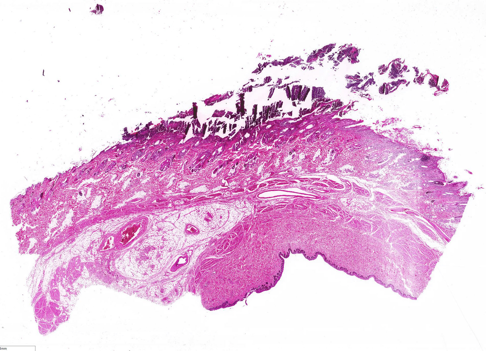
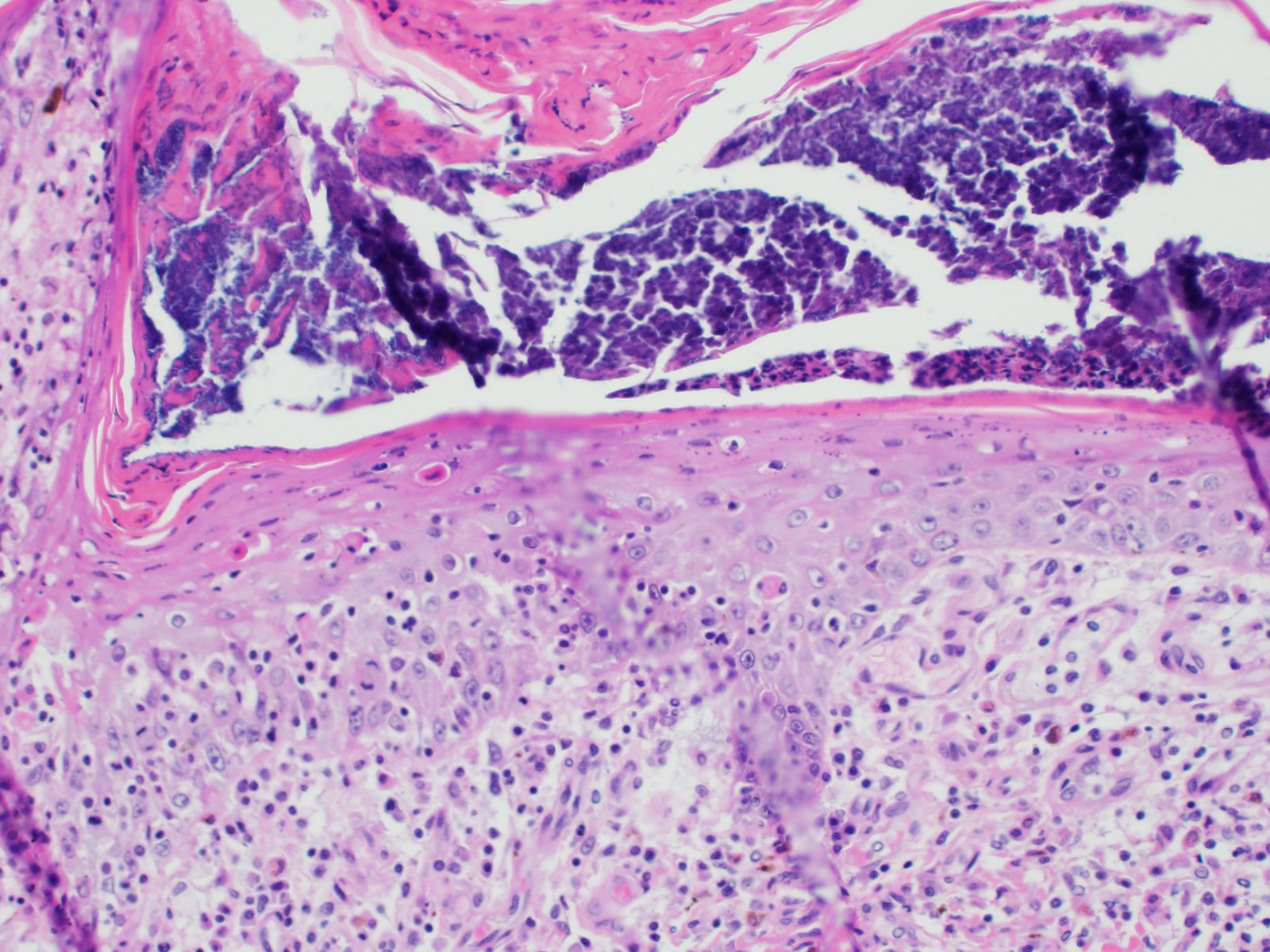
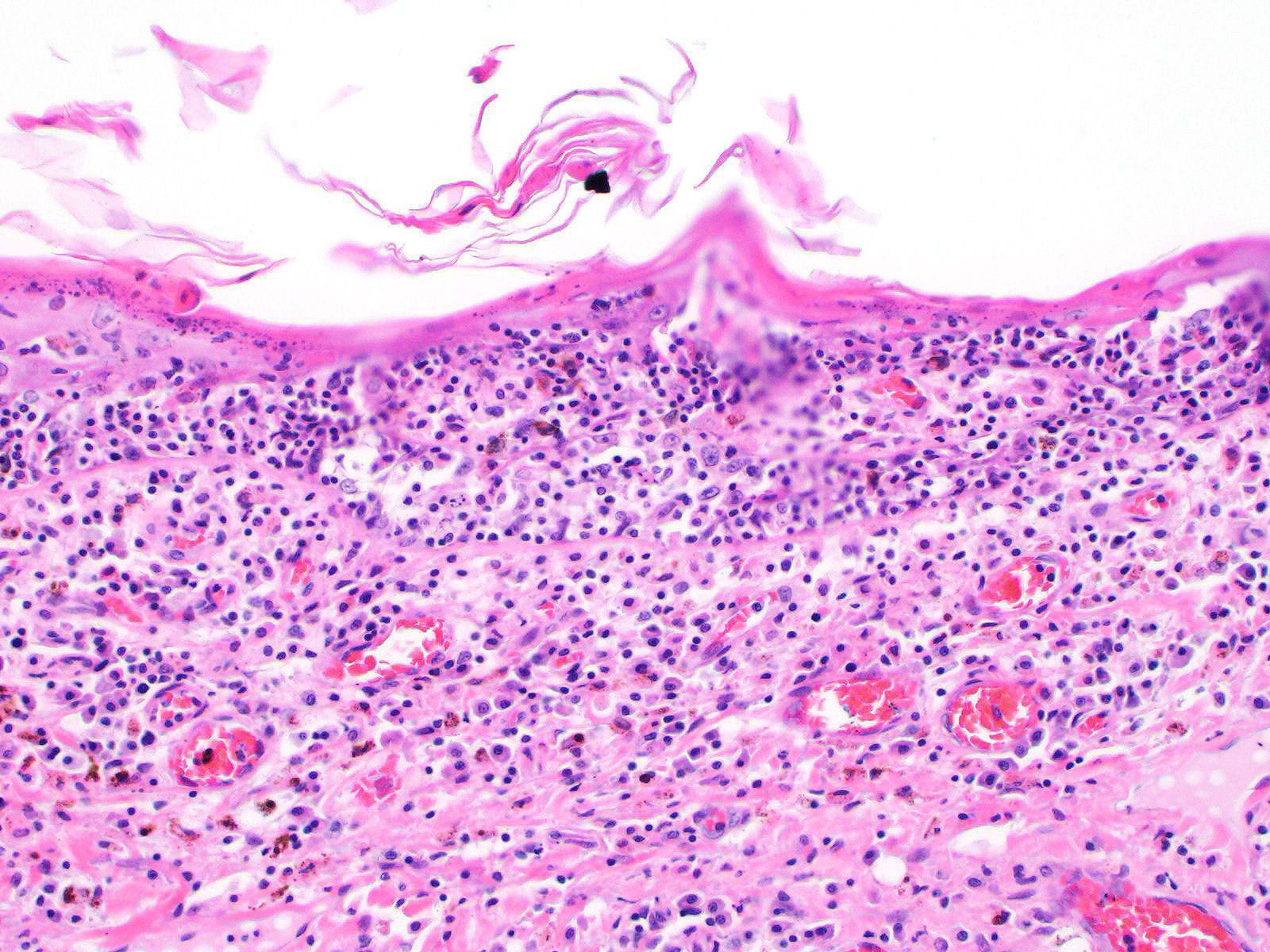
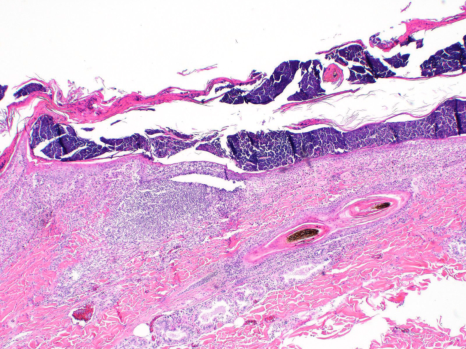
<p class=MsoNormal>All layers of the epidermis and also the follicular
infundibular epithelium contain rounded, hypereosinophilic keratinocytes with
pyknotic nuclei (apoptosis). Multifocally, apoptotic keratinocytes are
surrounded by lymphocytic infiltrations (satellitosis). Within the epidermis
there are multifocal accumulations of neutrophils, separating the superficial
from the deeper epidermal layers (interpreted as pustule formation). The
superficial dermis and the dermo-epidermal junction show ribbon-like, severe
infiltrations of lymphocytes, plasma cells and fewer macrophages, separating
the dermo-epidermal junction into areas which are variably clearly visible or
severely obscured. Lymphoid infiltrates are observed in the basal layer and
macrophages containing brown pigment (melanin) are visible in the dermis
(melanin incontinence). Multifocally the stratified layer is thickened and
multifocally nuclei can be observed also in the superficial keratinocytes
(parakeratotic and orthokeratotic hyperkeratosis). Furthermore, the epidermal
surface is covered by serocellular crusts and accumulations of (partially
degenerate) neutrophils. In the superficial areas, low numbers of thick-walled,
round structures with a clear center and approximately 5- 10 <span
style='color:black'>μm</span> in diameter (interpreted as <i>Candida </i>spores)
are observed. The neutrophilic infiltrates extend multifocally deep into the
dermis or into the hair follicles (secondary suppurative pyoderma and
folliculitis). </p>
<p class=MsoNormal style='line-height:115%'><b> </b></p>
<p class=MsoNormal style='line-height:115%'><b><span style='color:black'>Contributor
Morphologic Diagnosis: </span></b></p>
<p class=MsoNormal><span lang=EN-GB>Skin, chronic multifocal to coalescing
interface dermatitis, with apoptotic keratinocytes, lymphocytic satellitosis
and with secondary crust formation, parakeratotic hyperkeratosis, suppurative
pyoderma and folliculitis (consistent with erythema multiforme)</span></p>
<p class=MsoNormal><b> </b></p>
<p class=MsoNoSpacing style='line-height:115%'><b><span style='font-size:12.0pt;
line-height:115%;font-family:"Times New Roman",serif;color:black'> </span></b></p>
<p class=MsoNormal><b><span style='color:black'>Contributor Comment: </span></b>Erythema
multiforme is a rare skin disease in dogs and cats and has also been described in
horses, cattle, swine, ferrets and anecdotally in goats.<sup>3,6,7</sup> The
nomenclature and definitions of erythema multiforme (EM), Stevens-Johnson
syndrome (SJS) and toxic epidermal necrosis (TEN) in veterinary literature are
conflicting and therefore confusing.<sup>7,8</sup> In human medicine, EM, SJS
and TEN were considered to be different severities of the same disease.
Nowadays it is accepted that EM and SJS/TEN represent separate conditions.<sup>8</sup>
</p>
<p class=MsoNormal> </p>
<p class=MsoNormal>The pathogenesis of erythema multiforme in animals is still
poorly understood.<sup>3,6,7,8</sup> Possible etiologies include adverse drug
reactions (CADR), e.g. against sulphonamides, other antibiotics, and levamisole.
Neoplasia, food (including commercial dog food and beef/soy diet),
nutraceutical products and infections are reported triggers for erythema
multiforme in canine patients.<sup>3,4,6,7,8</sup> However, proven causalities
by re-challenge are rare and a multicentric study revealed that only 19% of the
canine EM cases were drug related.<sup>5</sup> The histological characteristics
and pattern indicate a misdirected immune response against keratinocytes, which
is lymphocyte-mediated with direct cytotoxicity of target cells.<sup>6,8</sup> </p>
<p class=MsoNormal> </p>
<p class=MsoNormal>Clinical lesions </p>
<p class=MsoNormal>In dogs, lesions are normally bilateral and involve the
trunk, groin and axilla and also the inner pinna, footpads and mucocutaneous
junctions.<sup>3,8</sup> Canine and feline lesions consist of erythematous
macules, papules and plaques. Lesion borders are indurated and lesion centers
are clear with discolorations to cyanotic or purpuric. Central crusting of
lesions is common, but in canine patients this may extend to heavily crusted
and/or scaly plaques.<sup>7,8</sup> </p>
<p class=MsoNormal> </p>
<p class=MsoNormal>Histology â typical lesions</p>
<p class=MsoNormal>Microscopic lesions in canine erythema multiforme are
similar to those in human patients.<sup>3,8</sup> Classic lesions include
interface dermatitis, with cell death occurring in all epidermal (suprabasilar
and basal) layers, accompanied by satellitosis (lymphoid infiltrates around
apoptotic keratinocytes).<sup>3,6,7,8</sup> Intraepidermal mononuclear cells
are mainly lymphoid, but Langerhans cells have also been identified.<sup>3,8.</sup>
In canine patients the follicular infundibular epithelium is regularly affected
and hyperkeratosis and parakeratosis are common, which is not the case in human
patients.<sup>3,7,8</sup> Yager et al. further suggest that hydropic
degeneration of the basal layer may not be such a prominent feature in canine
patients compared to humans.<sup>8</sup> </p>
<p class=MsoNormal> </p>
<p class=MsoNormal>Diagnosis and differential diagnoses </p>
<p class=MsoNormal>Erythema multiforme in the dog includes a wide range of
clinical lesions, leading to a long list of differential diagnoses such as
urticaria, demodicosis, dermatophytosis, bacterial folliculitis, superficial
spreading pyoderma and bullous autoimmune skin diseases.<sup>3,7,8</sup> The
presence of scaling-crusting lesions additionally includes superficial
necrolytic dermatitis, zinc-responsive dermatoses or other cornification
disorders as possible differential diagnoses.<sup>3,8</sup> Diagnosis of canine
erythema multiforme therefore is often based on a combination of anamnesis,
gross, and histological findings.<sup>3,6</sup> Yager et al. emphasize the
importance of taking the anamnesis and gross lesions into consideration when
giving a diagnosis of erythema multiforme.<sup>8</sup> </p>
<p class=MsoNormal style='line-height:115%'> </p>
<p class=MsoNormal style='line-height:115%'><b><span style='color:black'>Contributing
Institution:</span></b></p>
<p class=MsoNormal>Institute of Veterinary Pathology</p>
<p class=MsoNormal>Vetsuisse Faculty (University of Zurich)</p>
<p class=MsoNormal><span lang=EN-GB>Winterthurerstrasse 258</span><span
lang=DE-CH>, CH-8057 Zurich</span></p>
<p class=MsoNormal><span lang=DE-CH>Fax number +41 44 635 89 34</span></p>
<p class=MsoNormal>http://www.vetpathology.uzh.ch</p>
<p class=MsoNormal><b><br>
JPC</b> <b>Diagnosis<span style='color:#00B050'>: </span></b>1. <span
style='color:black'>Haired skin: Apoptosis, transepidermal, epidermal and
follicular, diffuse, severe, with neutrophilic and lymphohistiocytic interface
dermati</span>tis.</p>
<p class=MsoNormal><b> </b></p>
<p class=MsoNormal>2. Haired skin: Dermatitis, suppurative, multifocal to
coalescing, severe,<span style='color:black'> with diffuse moderate ortho-and
parakeratotic hyperkeratosis and bacterial cocci.</span></p>
<p class=MsoNormal style='text-align:justify'><b> </b></p>
<p class=MsoNormal style='line-height:115%'> </p>
<p class=MsoNormal style='line-height:115%'> </p>
<p class=EndNoteBibliography style='line-height:115%'><b><span
style='font-size:12.0pt;line-height:115%;font-family:"Times New Roman",serif'>JPC
Comment</span></b><span style='font-size:12.0pt;line-height:115%;font-family:
"Times New Roman",serif'>:</span><sup><span style='font-size:12.0pt;line-height:
115%;font-family:"Times New Roman",serif;color:#333333'> </span></sup><span
style='font-size:12.0pt;line-height:115%;font-family:"Times New Roman",serif'> The
contributor has compiled an excellent overview of the three entities of
erythema multiforme, Stevens-Johnson syndrome (SJS) and toxic epidermal
necrosis (TEN), syndromes about which there remains to this day a lot of
disagreement in the veterinary literature. Comparison with the human disease
has advanced knowledge in these chronic and occasionally fatal syndromes and
provided an excellent starting point, but the differences in the human disease
and its veterinary counterpart are profound as well. Moreover, the literature
lacks a critical number of well-documented cases of these diseases, and many of
the cases in the older literature may, upon further review in light of more
recent advances in veterinary dermatology, may have been incorrectly
diagnosed. </span></p>
<p class=EndNoteBibliography style='line-height:115%'><span style='font-size:
12.0pt;line-height:115%;font-family:"Times New Roman",serif'>In spite of
significant disagreement in the veterinary literature from the last decade on
these uncommon diseases, there are considerable points of agreement (many
already mentioned by the contributor, but worthy of mentioning again):<br>
<br>
1. Erythema multiforme (EM) and STS/TEN represent independent and different diseases,
rather than opposite poles of a spectrum of immune-mediated disease.<br>
2. Both EM and STS/TEN are mediated at least in part (STS/TEN) or in toto by
cytotoxic lymphocytes directed against altered keratocyte antigens.<br>
3. There is considerable overlap in the histologic diagnosis of these
diseases, and these findings must be closely correlated with clinical findings
and history for a definitive diagnosis.<br>
4. The histologic diagnosis of erythema multiforme is not a straightforward
diagnosis and bears a number of differential diagnoses that should be
considered before this diagnosis is rendered.<br>
<br>
</span></p>
<p class=EndNoteBibliography style='line-height:115%'><span style='font-size:
12.0pt;line-height:115%;font-family:"Times New Roman",serif'>The reliable
diagnosis of EM via STS/TEN has been confusing in both human and veterinary
medicine for years. Early attempts at classification of these diseases<sup>5</sup>
were based on human schema and proposed classification on five categories: type
of skin lesions, distribution, mucosal involvement, systemic signs, and
precipitating factors. Areas of significant variation include types of lesions
(in which epidermal detachment is useful for identifying STS/TEN) and mucosal
involvement (in which an absence of mucosal involvement is seen only with EM.
It may, however be seen with EM, so its presence is not of diagnostic utility).
On a purely academic note, one of the few differentiating factors between
Stevens-Johnson syndrome (STS) and toxic epidermal necrolysis (TEN) (and likely
the reason that they are so often lumped together) is that STS should have
<10% epidermal detachment) and TEN >30%. Regarding mucosal involvement,
several classifications have tried to characterize mucosal involvement, either
due to severity or the number of mucosal sites affected, but this criteria is
still under evaluation.<sup>1,8</sup> Significant overlap occurs in the
remaining categories. </span></p>
<p class=EndNoteBibliography style='line-height:115%'><span style='font-size:
12.0pt;line-height:115%;font-family:"Times New Roman",serif'>There is also
significant disagreement and uncertainty yet remaining in the exact
pathogenesis of the lesions in EM and STS/TEN. EM is characterized by lymphocytic
targeting of individual keratinocytes, which SJS/TEN lesions are lymphocyte
poor, with extensive areas of epidermal necrosis and lifting. Early SJS/TEN
lesions resemble the pattern seen in EM, while later lesions with large
confluent areas of necrosis suggest a progression to either waves of apoptosis,<sup>8</sup>
soluble mediators of inflammation such as reactive oxygen species, granulysin, and
soluble Fas ligand<sup>, </sup>or programmed cell death (necroptosis) A recent
publication established the death of keratinocytes in TEN to be an apoptotic
event, as seen in its human counterpart<sup>2</sup>, but this does not explain
the complete pathogenesis of this disease.</span></p>
<p class=EndNoteBibliography style='line-height:115%'><span style='font-size:
12.0pt;line-height:115%;font-family:"Times New Roman",serif'>As complex as the
diagnosis of STS/TEN may be, the correct diagnosis of erythema multiforme may
be even more complex due to the variable histologic presentation and potential
differential diagnosis. Lymphocytic-driven keratinocyte apoptosis at all
levels of the epidermis and indeed, full-thickness epidermal necrosis may also
be seen in EM lesions. The potential for hyperkeratosis in EM cases (also
known as âhyperkeratoticâ or âold dogâ EM) also brings cornification or
clinical scaling disorders into the differential diagnosis.<sup>8</sup> Luckily,
true cases of this condition, according to the moderator, appear to be
extremely rare in the veterinary literature. Moreover, cases complicated by
other secondary bacterial diseases or opportunistic infectious agents (as
illustrated by this particular case) pose an additional diagnostic challenge.</span></p>
<p class=MsoNormal style='line-height:115%'><span lang=EN>The moderator
stressed the importance of a good history as well as an optimal sample (often
from the center of the lesion in which devitalization and lesion development in
the differentiation of EM/SJS/TEM on surgical biopsy. As these three lesions
may also resemble each other on a single biopsy sample, in the absence of a
good history and clinical distribution of lesions, the prudent surgical
pathology may withdraw to a conclusion that the biopsy likely is within the
EM/SJS/TEM spectrum, but refrain from the desire to place it in one of the
three categories. In the cat, exfoliative dermatitis associated with thymoma
(and even a few without) may also present as a cytotoxic dermatitis which
resembles EM. </span></p>
<p class=MsoNormal style='line-height:115%'><span lang=EN> </span></p>
<p class=MsoNormal style='line-height:115%'><span lang=EN> </span></p>
<p class=MsoNormal style='line-height:115%'> </p>
<p class=MsoNormal style='line-height:115%'><b>References:</b> </p>
<p class=MsoNormal style='text-align:justify;line-height:115%'> </p>
<p class=MsoListParagraphCxSpFirst style='text-indent:-.25in'><span
style='font-size:12.0pt;line-height:115%;font-family:"Times New Roman",serif'>1)<span
style='font:7.0pt "Times New Roman"'> </span></span><span
style='font-size:12.0pt;line-height:115%;font-family:"Times New Roman",serif'>Banovic
F, Olivry T, Bazzle L., Tobias JR, Atlee, B, Zabel S, Hensel N, Linder KE.
Clinical and microscopic characteristics of canine toxic epidermal necrolysis.
<i>Vet Pathol</i> 2015; 53(2):321-330.</span></p>
<p class=MsoListParagraphCxSpMiddle style='text-indent:-.25in'><span
style='font-size:12.0pt;line-height:115%;font-family:"Times New Roman",serif'>2)<span
style='font:7.0pt "Times New Roman"'> </span></span><span
style='font-size:12.0pt;line-height:115%;font-family:"Times New Roman",serif'>Banovic
F, Dunston S, Linder KE, Rakich P, Olivry, T. Apoptosis as a mechanism for keratinocyte
death in canine toxic epidermal necrolysis. <i>Vet Pathol</i> 2017; 54(2):
249-253.</span></p>
<p class=MsoListParagraphCxSpMiddle style='text-indent:-.25in'><span
style='font-size:12.0pt;line-height:115%;font-family:"Times New Roman",serif'>3)<span
style='font:7.0pt "Times New Roman"'> </span></span><span
style='font-size:12.0pt;line-height:115%;font-family:"Times New Roman",serif'>Boehm
TMSA, Klinger, CJ, Udraite L, Mueller RS. Targeting the skin â erythema
multiforme in dogs and cats. <i>Tierärztl Prax Kleintiere.</i> 2017;
45:352-356.</span></p>
<p class=MsoListParagraphCxSpMiddle style='text-indent:-.25in'><span
style='font-size:12.0pt;line-height:115%;font-family:"Times New Roman",serif'>4)<span
style='font:7.0pt "Times New Roman"'> </span></span><span
style='font-size:12.0pt;line-height:115%;font-family:"Times New Roman",serif'>Favrot
C, Olivry T, Dunston SM, Degorce-Rubiales F, Guy JS. Parvovirus Infection of
Keratinocytes as a Cause of Canine Erythema multiforme. <i>Vet Pathol.</i>
2000; 37:647-649</span></p>
<p class=MsoListParagraphCxSpMiddle style='text-indent:-.25in'><span
style='font-size:12.0pt;line-height:115%;font-family:"Times New Roman",serif'>5)<span
style='font:7.0pt "Times New Roman"'> </span></span><span
style='font-size:12.0pt;line-height:115%;font-family:"Times New Roman",serif'>Hinn
AC, Olivry T, Luther PB et al. Erythema multiforme, Stevens-Johnson syndrome
and toxic epidermal necrolysis in the dog: clinical classification, drug
exposure, and histopathologic correlations. <i>J Vet Allergy Clin Immunol</i>
1998:6:13-20</span></p>
<p class=MsoListParagraphCxSpMiddle style='text-indent:-.25in'><span
style='font-size:12.0pt;line-height:115%;font-family:"Times New Roman",serif'>6)<span
style='font:7.0pt "Times New Roman"'> </span></span><span
lang=FR-CH style='font-size:12.0pt;line-height:115%;font-family:"Times New Roman",serif'>Itoh
T, Nibe K, Kojimoto A, et al. </span><span style='font-size:12.0pt;line-height:
115%;font-family:"Times New Roman",serif'>Erythema Multiforme Possibly
Triggered by Food Substances in a Dog. <i>J. Vet. Med. Sci.</i> 2006;
68(8):869-871</span></p>
<p class=MsoListParagraphCxSpMiddle style='text-indent:-.25in'><span
style='font-size:12.0pt;line-height:115%;font-family:"Times New Roman",serif'>7)<span
style='font:7.0pt "Times New Roman"'> </span></span><span
style='font-size:12.0pt;line-height:115%;font-family:"Times New Roman",serif'>Jubb,
Kennedy, Palmer. <i>Pathology of Domestic Animals</i>. 6<sup>th</sup> ed. St.
Louis, Elsevier Saunders; 2016. </span></p>
<p class=MsoListParagraphCxSpLast style='text-indent:-.25in'><span
style='font-size:12.0pt;line-height:115%;font-family:"Times New Roman",serif'>8)<span
style='font:7.0pt "Times New Roman"'> </span></span><span
style='font-size:12.0pt;line-height:115%;font-family:"Times New Roman",serif'> Yager
J.A, Erythema multiforme, Stevens-Johnson syndrome and toxic epidermal
necrolysis: a comparative review. Vet dermatol 2014; 25: 406-e64. </span></p>
<p class=EndNoteBibliography><b> </b></p>
</div>
</body>
</html>