WSC 23-24 CONF 10, CASE I:
Signalment:
Adult female betta fish, piscine (Betta splendens)
History:
This adult, female betta fish exhibited erratic and abnormal swimming behavior for 17.5 days. The owner described the fish as happy-go-lucky since being purchased at the local fish store. The fish even swam up to be caressed on the head at times. One day, after 2 years, the owner noted the fish had difficulty swimming. The owner also noticed that the fish had a greatly distended coelomic cavity. The fish was subsequently euthanized.
Gross Pathology:
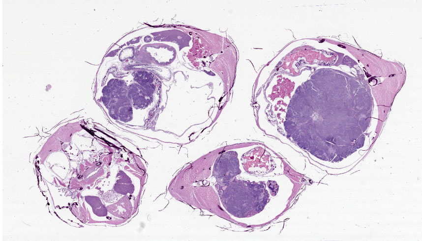
There was an approximately 1 cm diameter distension of the coelomic cavity. On cut section, the mass was tan and solid.
Microscopic Description:
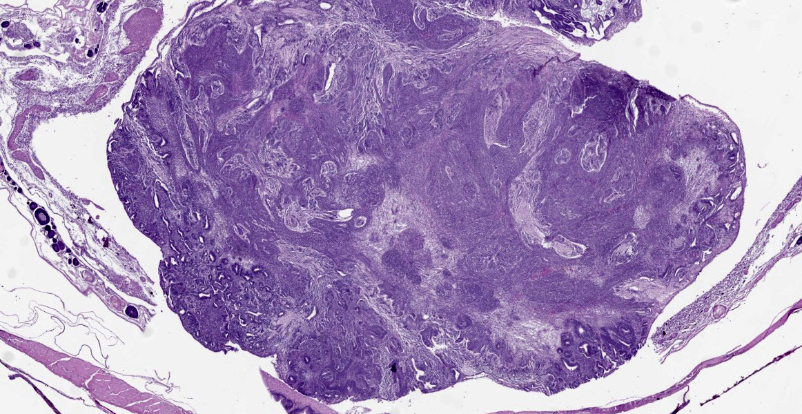
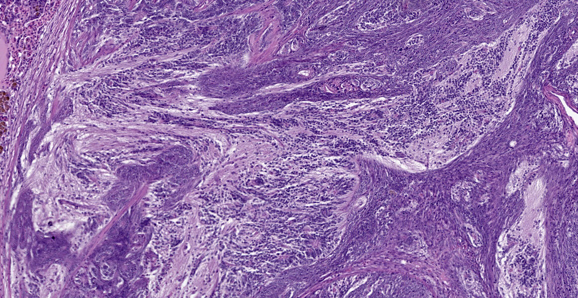
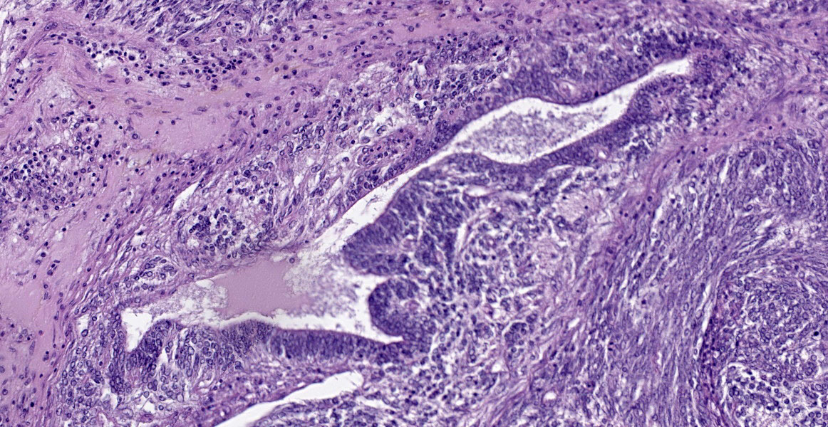
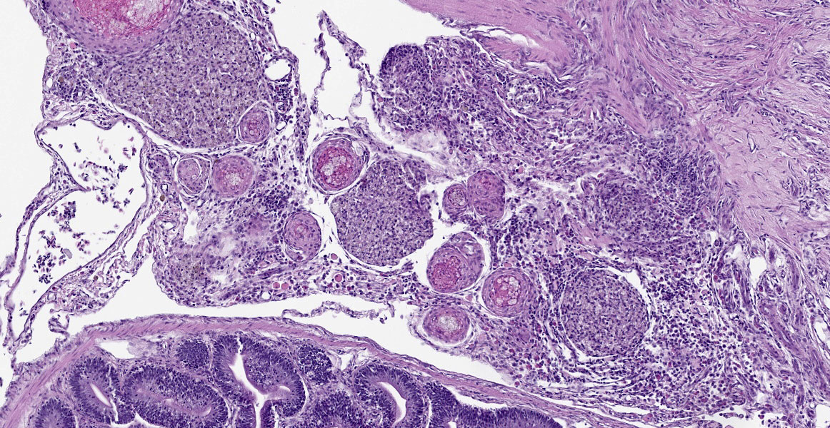
Kidney: Expanding and occupying greater than 90% of the coelomic cavity, compressing and displacing adjacent organs, and arising from the kidney, is a well-demarcated, unencapsulated, densely cellular, 0.75 cm diameter neoplasm composed of three distinct cell populations supported on a variably fine fibrovascular stroma. The first cell population is composed of polygonal cells (blastemal cells) which irregularly merge into two other populations. An epithelial cell population is arranged in palisading, branching, and tubule-like formations that often resemble lumens.
The third population resembles small, compact tufts of cells (glomeruloid structures). Spindle to stellate cells are scattered throughout the neoplasm (embryonal mesenchyme). Polygonal blastemal cells have indistinct cell borders, a high nuclear to cytoplasmic ratio, a scant amount of eosinophilic cytoplasm, round to oval nuclei, and dense chromatin with indistinct nucleoli. Epithelial cells are cuboidal to columnar with variably distinct cell borders, a moderate amount of eosinophilic cytoplasm, round to oval nuclei with stippled chromatin and variably distinct nucleoli. Embryonal mesenchymal cells are spindle to stellate and loosely arranged, with indistinct cell borders, scant eosinophilic cytoplasm with elongate to oval nuclei and indistinct nucleoli. Mitotic figures are not present in the neoplasm. Peripherally, ova are multifocally degenerate and are atrophied with variably sized, irregularly shaped oocyte borders.
Transmurally and multifocally expanding the stomach wall are variably dense aggregates of macrophages interspersed with areas of spindle cells and fibrous connective tissue. Multifocally throughout the stomach wall and extending into the coelomic cavity are 40-100um diameter granulomas characterized by a core of brightly eosinophilic necrotic debris, foamy macrophages, degenerate inflammatory cells, and mineral which are surrounded by variably dense concentric lamellations of spindle cells and macrophages. Multifocally, there are less developed granulomas that contain foamy macrophages with a light yellow-brown hemosiderin-like pigment. Scattered throughout the ovaries are multiple granulomas as previously described.
Contributor’s Morphologic Diagnoses:
1. Betta fish, kidney: Nephroblastoma.
2. Betta fish, ovaries, stomach and coelomic cavity: Granulomas, multiple, most likely Mycobacteria spp.
Contributor’s Comment:
Nephroblastoma is considered a common primary tumor of pigs, chicken, and fish with varying reports in dogs, cats, and cattle.7 In this case, spontaneous nephroblastoma in Betta splendens (“Siamese fighting fish”) has been reported with other fish species, to include Japanese eels (Anguilla japonica), koi (Cyprinus carpio), and striped bass (Morone saxatilis).2,4,6,12 Nephroblastomas arise from neoplastic transformation of nephrogenic stem cells, which give rise to the characteristic blend of three cell populations that attempt to parallel the histo-anatomic components of the kidney through epithelial, blastemal, and mesenchymal cells. The cause of these tumors in fish is considered spontaneous, but has also been attributed to carcinogens.5 Other than coelomic enlargement, the clinical presentation of nephroblastoma in fish varies.
Mycobacteriosis affects wild and cultured freshwater, marine, and brackish fish worldwide, and frequently manifests as a chronic, progressive, and systemic disease.10 Transmission routes vary but may include cannibalism of infected fish, consumption of contaminated feed and/or detritus, or shedding from other aquatic vertebrates.
Although many Mycobacteria species have been isolated from fish, the most frequent and significant fish mycobacterioses include M. chelonae, M. fortuitum, and M. marinum.1 Granuloma formation due to Mycobacteria infection in fish can be identified grossly (grayish-white, miliary nodules) and histologically in a variety of organs to include, but not limited to, the spleen, kidney, liver, and coelomic cavity. The histologic appearance of granulomas in most fish species is characterized by an outer wall of concentrically layered epithelioid macrophages, necrotic centers, and variable numbers of acid-fast bacilli within the core. The epithelial cell characteristics of these epithelioid macrophages is demonstrated by positive immunoreactivity for cytokeratin.8,9
The cause of death in this fish is most likely multi-factorial and attributed to pathophysiologic changes associated with tumor compression of vital organs and granuloma formation in multiple organs.
JPC Diagnosis:
1. Ovaries, posterior kidney, and coelom: Teratoma.
2. Ovaries: Follicular degeneration and necrosis, multifocal, chronic, moderate (follicular stasis/egg binding).
3. Stomach and coelom: Granulomatous and fibrosing gastritis, transmural, regionally extensive, severe, with regional coelomitis and intralesional birefringent debris (foreign body).
JPC Comment:
We agree with the contributor that there are three populations of cells within the examined section; however, we interpret these populations to originate from the three primordial germ layers, and thus prefer a diagnosis of teratoma. The examined section contains multiple large areas of neural differentiation (ectoderm), sheets of primitive mesenchymal tissue (mesoderm), and numerous tubular structures (endoderm). The teratoma is adhered to and infiltrates both the posterior kidney and the ovary. Intracoelomic teratomas usually originate from the ovary in female fish, making ovarian origin most likely for this tumor.11 As in this case, teratomas in fish often present as large coelomic masses causing coelomic distention.2,11
While most cases of granulomatous inflammation in fish should be stained with acid fast stains to rule out mycobacteriosis, the distribution of granulomas in this case would be an unusual presentation. Mycobacteriosis in fish typically causes discrete granulomas or sheets of macrophages in the liver, spleen, kidney, skin, and coelom. In this case, infection and fibrosis track through the gastric wall in a manner most consistent with trauma (foreign body). The H&E slide was examined under polarized light and birefringent material was noted in the granulomas in the coelomic cavity adjacent to the gastric lesion, further supporting foreign body trauma. Acid fast stains were applied to the lesions and were negative.
We note the contributor’s reference to granulomas in the ovaries; however, in our examined sections, these structures consist mostly of shrunken and necrotic ovarian follicles being engulfed by macrophages. This is a common lesion of follicular degeneration/egg binding and was likely caused by the mass effect of the teratoma preventing normal follicular release.
This week’s conference was moderated by Dr. Elise LaDouceur, Chief of Extramural Projects and Research at the Joint Pathology Center. Conference participants were of two schools, with all participants diagnosing either nephroblastoma or teratoma. All participants were eventually convinced of teratoma by the large areas of neural differentiation and ciliated epithelium.
Discussion also centered on whether the tumor was arising from or invading into the posterior kidney and ovary. While this was impossible to determine from the examined sections, in all species, teratomas most commonly arise in the gonads, making ovarian origin mostly likely.
Participants discussed egg binding, also known as follicular stasis. This condition in teleost fish has many causes, including mass effect, inflammation, or lack of access to nesting areas.
References:
1. Decostere A, Hermans K, Haesebrouck F. Piscine mycobacteriosis: a literature review covering the agent and the disease it causes in fish and humans. Vet Microbiol. 2004;99(3-4):159-166.
2. Groff JM. Neoplasia in fishes. Vet Clin North Am Exot Anim Pract. 2004;7(3): 705-706.
3. Helmboldt CF, Wyand DC. Nephroblastoma in a striped bass. J Wildl Dis. 1971; 7:162–165.
4. Lombardini ED, Law M, Lewis BS. Nephroblastoma in two Siamese fighting fish (Betta splendens). Fish Pathol. 2010;45:137–139.
5. Lombardini ED, Hard GC, Harshbarger JC. Neoplasms of the urinary tract in fish. Vet Pathol. 2014;51(5):1000-1012.
6. Masahito P, Ishikawa T, Okamoto N, et al. Nephroblastomas in the Japanese eel, Anguilla japonica Temminck et Schlegel. Cancer Res. 1992;52:2575–2579.
7. Meuten DJ, Everitt J, Inskeep W, et al. Histological classification of tumors of the urinary system of domestic animals. Armed Forces Institute of Pathology; 2004.
8. Noga EJ, Dykstra MJ, Wright JF. Chronic inflammatory cells with epithelial cell characteristics in teleost fishes. Vet Pathol. 1989;26(5):429-437.
9. Polinas M, Padrós F, Merella P, et al. Stages of granulomatous response against histozoic metazoan parasites in mullets (Osteichthyes: Mugilidae). Animals (Basel). 2021;11(6):1501.
10. Puk K, Guz L. Occurrence of Mycobacterium spp. in ornamental fish. Ann Agric Environ Med. 2020;27(4):535-539.
11. Romanucci M, Arbuatti A, Massimini M, Defourny SP, Della Salda L. Ovarian teratoma in an adult female Zoogoneticus tequila (Webb & Miller 1998): histological and immunohistochemical features. J Fish Dis. 2017;40(6):859-862.
12. Stegeman N, Heatley JJ, Rodrigues A, et al. Nephroblastoma in a koi (Cyprinus carpio). J Exotic Pet Med. 2010;19:29803.