Signalment:  

10-month gestation placenta, Thoroughbred, Equus caballus, equineThe mare was due to foal on January 20, 2003, but the mare foaled on December 28, 2002.


Gross Description:  

A complete placenta is submitted for examination. Along the chorionic surface of the body and non-pregnant horn are multifocal regions of denuded villi covered with a thick mucoid exudate. Adjacent villi are thickened and edematous. No other significant gross lesions are observed.


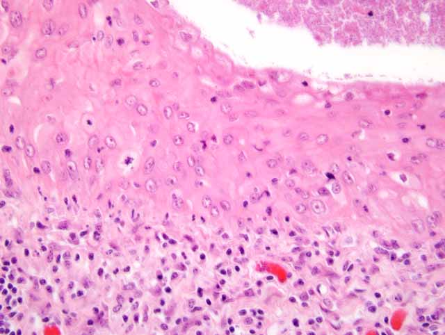
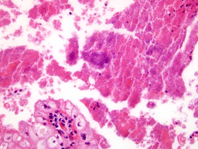
Histopathologic Description:

Allantochorion: Amorphous eosinophilic material with scattered aggregates of bacteria, intact and degenerate neutrophils, and cellular debris multifocally overlies the chorionic surface. The chorionic villi, lined by hypertrophic trophoblasts with mild to moderate cytoplasmic vacuoles, are multifocally blunted. Aggregates of moderate to large numbers of lymphocytes, plasma cells, and neutrophils occupy the superficial stroma adjacent to the villi and villous projections. The supporting stroma is characterized by diffuse, moderate edema, an infiltrate of low to moderate numbers of lymphocytes, neo-vascularization and moderate fibroplasia.


Morphologic Diagnosis:  

Severe multifocal necrotizing exudative placentitis; Equus caballus


Lab Results:  


Bacteriology
-Placenta: Cellulosimicrobium cellulans formerly known as Oerskovia xanthineolytica (numerous)
-FA testing for the detection of Leptospira interrogans on the placenta was negative. 


Condition:  

Cellulosimicrobium cellulans


Contributor Comment:  

Cellulosimicrobium cellulans is a gram positive, branching, motile, oxidase negative, catalase positive, non-acid fast-bacillus. When grown on agar, the colonies have a characteristic yellow color.(3) The bacteria are found in soil and have been associated with middle and late term abortions, as well as premature births in horses.(1) Lesions are typically but not always seen in the both the fetus and placenta. Fetal lesions include firm, expanded lungs, and mildly enlarged friable livers with occasional pale foci along the capsular surface that extend into the parenchyma.(1) Lesions observed in the allantochorion include a brown mucoid exudate overlying well demarcated areas of denuded villi along the chorion. In previous reports, portions of the chorion affected include areas adjacent to the cervical star, along the body, or encompassing the horns. Microscopically, the chorionic surface is covered by an eosinophilic exudate and had blunted villi and marked inflammation of both the villi and supporting stroma. Often seen with the placentitis is a pyogranulomatous pneumonia characterized by multinucleated giant cells, macrophages, and neutrophils occupying the lumens of alveoli, bronchi, and bronchioles.(1)

Given the presence of an exudative placentitis, the distribution of the placental lesions, and the characteristics of Cellulosimicrobium cellulans, there are similarities between the bacteria described here and placentitis resultant of a Crossiella equi infection. Pyogranulomatous pneumonia, as seen with Cellulosimicrobium cellulans, does not accompany nocardioform placentitis.

Common bacterial agents resulting in placentitis and abortion in the horse include Leptospira spp, nocardioform, Escherichia coli, Streptococcus zooepidemicus, Pseudomonas aeruginosa, Streptococcus equisimilis, Enterobacter agglomerans, Klebsiella pneumoniae, and alpha- hemolytic Streptococcus.(2)


JPC Diagnosis:  

Placenta, allantochorion: Placentitis, necrotizing, subacute, diffuse, moderate, with squamous metaplasia(Fig. 3-1), fibrin, edema, and large colonies of coccobacilli(Fig. 3-2)


Conference Comment:  

Cellulosimircobium cellulans, formerly named Oerskovia xanthineolytica, is rarely found in humans and is generally an opportunistic pathogen in immunocompromised hosts.(1) This organism is being recognized more frequently as a cause of equine abortion. 

The bulk of the conference discussion centered on the outbreak of abortions in Kentucky in the spring of 2001 and 2002 dubbed Mare Reproductive Loss Syndrome (MRLS). MRLS caused large numbers of abortions reaching epidemic proportions in Kentucky.(4) The economic loss alone has been estimated at almost $500 million. 

Mares with MRLS abort late in gestation or at term and the fetuses are still enclosed within the placenta and in good condition. Mares show no overt clinical illness prior to abortion. In the fetus, gross lesions include hemorrhages in the chorion, amnion, and amniotic segment of the umbilical cord, pleura, and heart, hypema, and uninflated lungs. Additionally, the amniotic cord is often dull gray and thickened. Histologically, macrophages and neutrophils are seen in ulcerated areas on the surface of the amniotic cord in association with bacteria. The allantochorion also has similar lesions. Funisitis, amnionitis, pneumonia, fetal bacteremia, and chorionitis are also evident microscopically. 

The most commonly implicated cause for MRLS is the eastern tent caterpillar (ETC). Feeding of the eastern tent caterpillar to pigs has resulted in abortions in one study. Similar studies in horses have demonstrated that the feeding of the exoskeleton of the eastern tent caterpillar causes abortion in mares. One of the most current hypotheses is that a portion of the caterpillar cuticle is responsible for its abortifacient effects.(4)


References:

1. Bolin DC, Donahue JM, Vickers ML, Giles RC, Harrison L, Jackson C, Poonacha KB, Roberts JF, Sebastian MM, Sells SF, Tramontin R, Williams NM: Equine abortion and premature birth associated with Cellulosimicrobium cellulans infection. J Vet Diagn Invest 16:333-336, 2004
2. Hong CB, Donahue JB, Giles RC, Petrites-Murphy MB, Poonacha KB, Roberts AW, Smith BJ, Tramontin RR, Tuttle PA, Swerczek TW: Etiology and pathology of equine placentitis. J Vet Diagn Invest 5:56-63, 1993
3. Rihs JD, McNeil MM, Brown JM, Yu VL: Oerskovia xanthineolytica implicated in peritonitis associated with peritoneal dialysis: case report and review of Oerskovia infections in humans. J Clin Microbiol 28:1934-1937, 1990
4. Schlafer DH, Miller RB: Female genital system. In: Jubb, Kennedy, and Palmer's Pathology of Domestic Animals, ed. Maxie MG, vol 3, pp. 506-507. Elsevier Limited, Philadelphia, PA, 2007


Click the slide to view.


3-1. Chorioallantois, horse.


3-2. Chorioallantois, horse.


Back | VP Home | Contact Us |