Joint Pathology Center
Veterinary Pathology Services
Wednesday Slide Conference
2017-2018
Conference 4
September 20th, 2017
CASE III: CASE 2 (JPC 4101313).
Signalment: 8-month-old, heifer, Nelore, Bos taurus indicus, Bovine.
History: Three sick cows were submitted to the Veterinary Hospital of the Universidade Federal de Minas Gerais (UFMG), as well as samples of the spinal cord of an additional cow from the same farm, collected during a field necropsy. These 4 cows were from a farm in Minas Gerais state, with a herd of 3,000 cattle, where in the past 3 years (2013-2016), 35 cows died after presenting clinical signs characterized by ataxia, paresis and paralysis of the pelvic limbs, emaciation, and sternal recumbency. Two of these cattle were euthanatized due to the severe ataxia, inability to stand, and emaciation. The herd was vaccinated against foot-and-mouth disease twice a year.
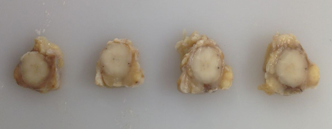
Gross Pathology: The cow was in poor body condition. Locally extensive areas of the skeletal muscle of the thoracic region (longissimus dorsi muscle) were replaced by numerous 0.3-0.8 mm in diameter, yellow and firm coalescent nodules (pyogranulomas) surrounded by moderate amounts of white and firm tissue (fibrous connective tissue). On the cut surface, some nodules contained yellowish and viscous fluid (purulent exudate) or whitish and viscous fluid (similar to the oily adjuvant of the foot-and-mouth disease vaccine). In the medullary canal of the subjacent vertebrae, extending from the intervertebral foramen to the epidural space and dura mater, there were pyogranulomas identical to those described in the skeletal muscle. The remaining dura mater was thickened and firm (fibrosis).
Laboratory results: Laboratory results are pending.
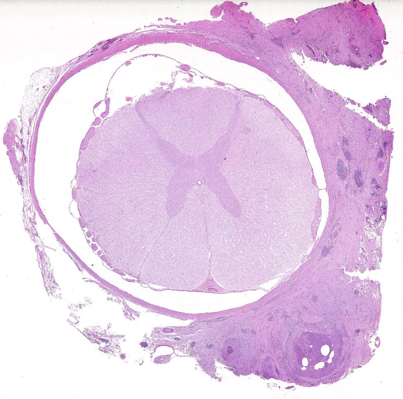
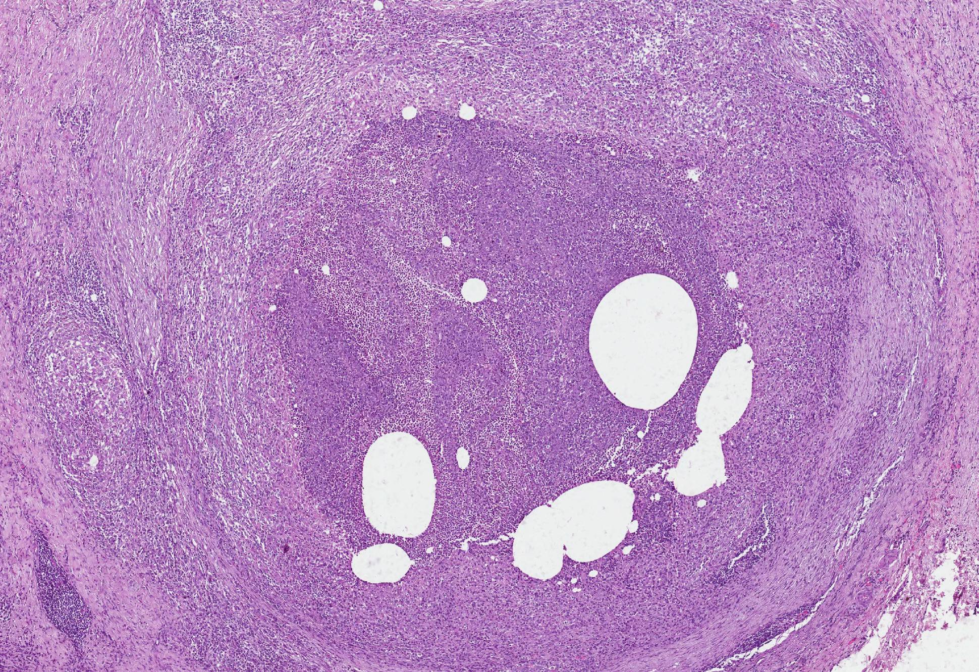
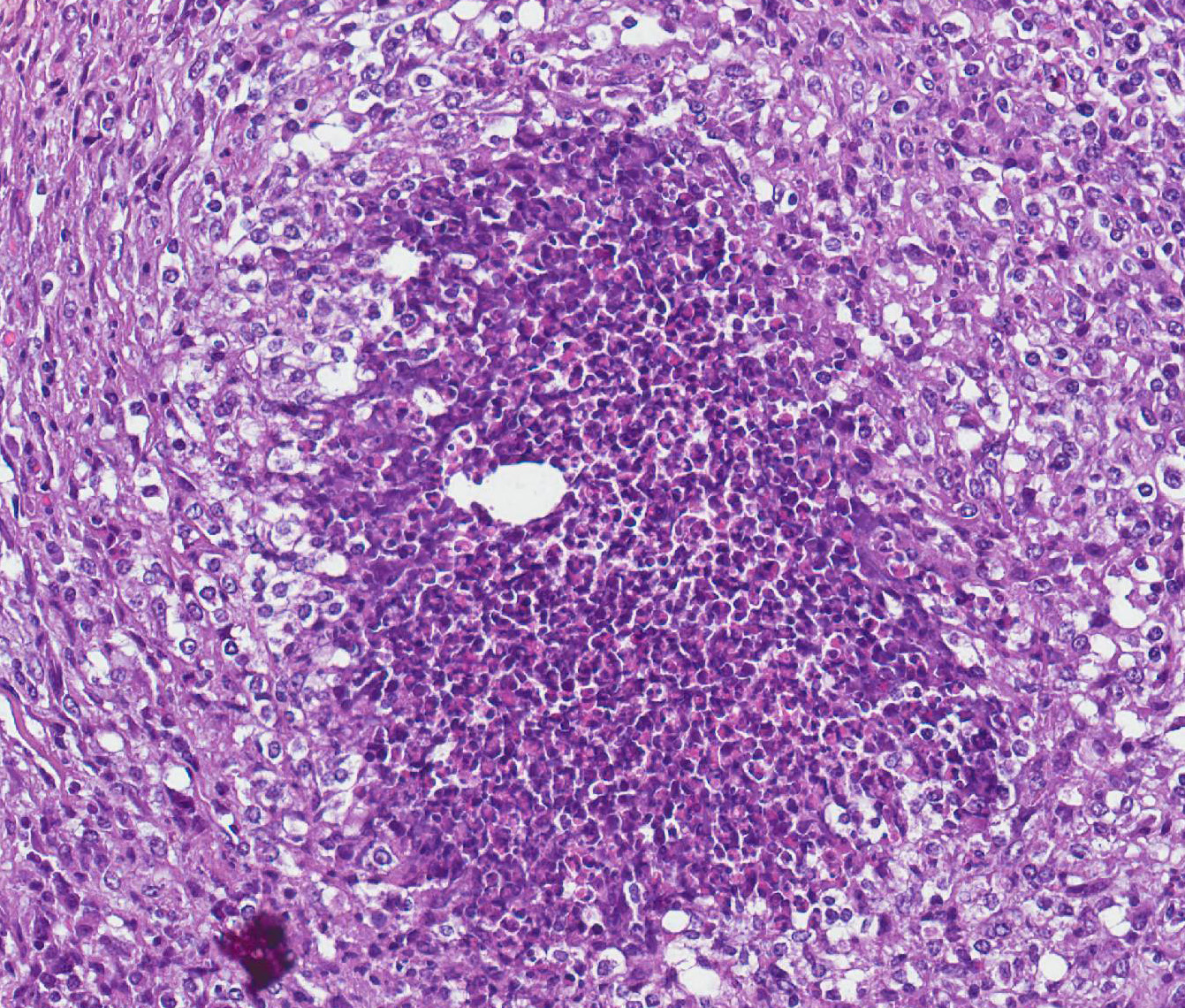
Microscopic Description: Meninges: The dura mater is expanded, and partially or completely effaced by extensive areas of pyogranulomatous inflammation with proliferation of fibrous connective tissue. The pyogranulomas are composed by a central clear vacuole of variable sizes (ranging from 30 to 300 µm) (consistent with the space left by the oil adjuvant droplets), surrounded by variable numbers of degenerated and viable neutrophils, with aggregates of necrotic material and mineralization, and, more externally, by large numbers of epithelioid macrophages and fewer multinucleated giant cells, lymphocytes and plasma cells. These structures are further surrounded by a thick layer of dense fibrous connective tissue. Extensive areas of the dura mater are thickened by fibrous connective tissue infiltrated by low to moderate numbers of lymphocytes, plasma cells, and macrophages. Pyogranulomas and fibrous tissuesinvade or compress the adjacent nerve fibers. In the white matter of the affected sections of the spinal cord, there are numerous well-defined, large and clear vacuoles (dilated periaxonal spaces) containing either swollen axons (spheroids) or foamy macrophages (digestion chambers).
Contributors Morphologic Diagnoses: 1. Meninges (dura mater): Pachymeningitis, pyogranulomatous and fibrosing, multifocal to coalescent, marked, with intralesional vacuoles (consistent with oil adjuvant droplets).
2. Spinal cord: Wallerian degeneration, multifocal, moderate, with spheroids and digestion chambers.
Contributors Comment: Clinical signs and gross and histopathological findings, in these four cows, were compatible with compressive myelopathy due to pyogranulomatous reaction to the oily adjuvant of a vaccine. The history of previous application of the foot-and-mouth disease vaccine in the thoracic region (site of the muscular pyogranulomas) indicated its involvement with these lesions.
Compressive myelopathies in ruminants have been associated with several causes of space occupying lesions within the medullary canal, including abscesses, granulomas, physical traumas, malformations, and neoplasms1, 2, 3. While traumas and abscesses are apparently more common in feedlot cattle, calves, and small ruminants; neoplasms, mainly lymphomas, occur more frequently in adult dairy cows3. Compressive myelopathies due to postvaccinal granulomas are uncommon in cattle and occur mainly in association with foot-and-mouth disease (FMD) vaccine adjuvant2, 3, 4. Cases of post-vaccinal granulomas have also been related to water-in-oil adjuvant of a vaccine against Escherichia coli and Campylobacter fetus spp. veneralis5 and of a vaccine against E. coli and Clostridium perfringens type C1.
Clinical signs of compressive myelopathy related to postvaccinal granulomas include ataxia, paresis and paralysis of pelvic limbs, permanent recumbency, and progressive loss of the muscular tone.2, 3, 4, 5 The beginning of the clinical signs occurs up to 60 days after the vaccination.3 In the reports of the condition in Brazil, the mortality rate ranged from 0.83% to 6.0%.2, 3, 4 Due to the similarity of the clinical signs, this condition must be included as a differential diagnosis of other two important neurological diseases in Brazilian cattle herds, rabies and botulism.4
An important factor for the development of the medullary lesions was the inappropriate administration of the vaccine in the muscle of a paravertebral area in the thoracic and lumbar regions. According to the orientation from the manufacturers of the vaccine and from the guidelines of the National Program for the Eradication of foot-and-mouth disease6, this vaccine must be applied subcutaneously or intramuscularly, in the lateral cervical region. Even when applied in the recommended location, subcutaneous and muscular lesions are frequently observed in the sites of application. These lesions are either granulomas or abscesses and are an important source of economic losses due to the cost to trimming the lesion in slaughterhouses.1 According to the owner of these cows, the application of the vaccine in the thoracic region was performed to avoid evident subcutaneous and muscular lesions in the cervical area and to facilitate the procedure when it was performed in a basic cattle handling system with straight race.
The presence of typical intralesional vacuoles (interpreted as the space left by the oily adjuvant of the vaccine, removed during the processing for the histopathological analysis) and the absence of infectious organisms in special stains (Grocott methenamine silver, Giemsa, or Ziehl-Neelsen acid-fast stains) corroborate the association of the lesions to the adjuvant. Adjuvants are important components of the vaccines and act nonspecifically, increasing the immune response against injected antigens. The adjuvant used in the FMD vaccine, that was responsible for the lesions observed in the cases, is reported as a water-in-oil emulsion.3 The water-in-oil adjuvant used in a Clostridium perfringens type CE. coli bacterin-toxoid vaccine and in a Rotavirus and Coronavirus vaccine, was also able to induce muscular lesions, such pyogranulomas, fibrosis, mineralization and necrosis.5 Occasionally, adjuvants can cause other adverse effects, including, anaphylaxis, lymphoplasmacytic inflammation and neoplasms.1
Despite some studies hypothesizing an association between needle insertion into the intervertebral foramen with lesions in the medullary canal, histologic findings indicate a progression of the adjuvant due to constant rupture of the granulomas. This hypothesis is corroborated by the observation of ruptured granulomas, presence of degenerated neutrophils within the granulomas, and occasional free vacuoles among the granulomas. Migration through the tissues is a well-known property of water-in-oil adjuvants.1, 3
Vaccination against FMD is one of most important policies for animal health in the beef cattle industry in Brazil. FMD is a highly contagious viral disease affecting cloven-hoofed animals. It has great potential for causing severe economic loss, due its importance for commercial trade, and the requirement for total elimination of the affected herds. Brazil has no outbreaks of FMD since 2005, when outbreaks in two states led to the sacrifice of 39,845 cattle. Currently, the country has 4 zones (corresponding for 76.1% of the national territory) certified as free of the disease with use of vaccination and 1 zone (corresponding for 1.1% of the national territory) as free of the disease without using vaccination.6
JPC Diagnoses: 1. Spinal cord, epidural space: Pyogranulomas, multiple, with clear vacuoles, Nelore, bovine.
2. Spinal cord: Wallerian degeneration, multifocal, mild with dilated myelin sheaths and swollen axons.
Conference Comment: The contributor provided an excellent review of the gross and microscopic lesions associated with tissue migration of water-in-oil adjuvant pyogranulomas.
As mentioned above, adjuvant and associated inflammation can spread into the intervertebral foramina by direct extension through progressive rupture of the pyogranulomas and reformation of the fibrous capsule.
The risk of injection site granulomas appears to be higher in vaccines with bacterial components in them. This is theorized to be due to soft tissue damage from the bacterial endotoxin that abets extension of the inflammation through tissue planes.5
Conference attendees noted Wallerian degeneration (dilated myelin sheaths, swollen axons, Gitter cells in digestion chambers phagocytizing myelin) in the white matter of the spinal cord with grey matter that was relatively unaffected. These changes are characteristic of chronic compression, whereas acute compression predominately affects the grey matter.5
Veterinary School
Universidade Federal de Minas Gerais
References:
1. MAPA 2017. Ministério da Agricultura , Pecuária e Abastecimento Programa Nacional de Erradicação da Febre Aftosa. Available online: www.agricultura.gov.br/assuntos/sanidade-animal-e-vegetal/programas-de-saude-animal/programa-nacional-de-erradicação-da-febre-aftosa-pnefa
2. Marques ALA, Simões SVD, Maia LA. Compressão medular em bovinos associada a vacinação contra febre aftosa. Ciência Rural. 2012;42(10):1851-1854
3. McAllister MM, O´Toole D, Griggs K. Myositis, lameness and paraparesis associated with use of an oil-adjucant bacterin in beef cattle. J Am Vet Med Assoc. 1995;207(7):936-938.
4. OToole D, Steadman L, Raisbeck et al. Myositis, lameness and recumbency after use of water-in-oil adjuvanted vaccines in near term beef cattle. J. Vet Diagn Invest. 2005;17:23-31.
5. OToole D, McAllister MM, Griggs K. Iatrogenic compressive lumbar myelopathy and radiculopathy in adult cattle following injection of an adjuvanted bacterin into loin muscle: histopathology and ultrastructure. J. Vet Diagn Invest. 1995;7:237-244.
6. Panziera W, Rissi DR, Galiza G. et al. Pathology in practice. J Am Vet Med Assoc. 2016;249(5):483-485.
7. Ubiali DG, Cruz RAS, Lana MVC et al. Spinal cord compression in cattle after the use of an oily vaccine. Pesq Vet Bras. 2011;31(11):997-999.