Signalment:
5-year-old, female, American Staffordshire terrier dog (
Canis familiaris)The dog presented with imbalance, a stiff gait, urinary incontinence, anorexia, inappetence and lethargy. Electromyography showed lesions at the promixal muscles of the forelimbs and the masticatory muscles. MRI revealed no abnormalities. On radiographic examination, masses were detected in the lung, multiple ribs and the dorsal processes of T2 and T5. Osteophytes were seen radiographically on the forelimb and scapulae.
Gross Description:
Numerous flat, subperiostal, bony proliferations were detected on the diaphysis and metaphysis of humeri, scapulae, femora and tibiae. These dense proliferations varied in size from 3 mm to 2 cm in length. Multiple irregular, osseus proliferations were present on several ribs, processus dorsalis of T2 and T5 and corpus spinalis of L2. The anteroventral lung lobes showed bilaterally poorly demarcated, infiltrative growing, firm, white-tan masses.
Histopathologic Description:
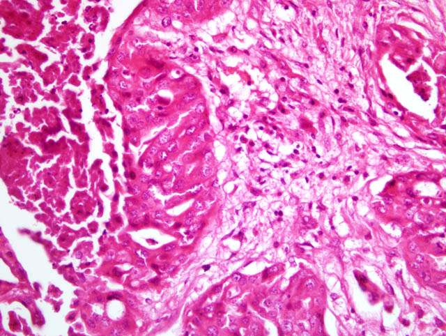
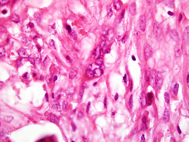
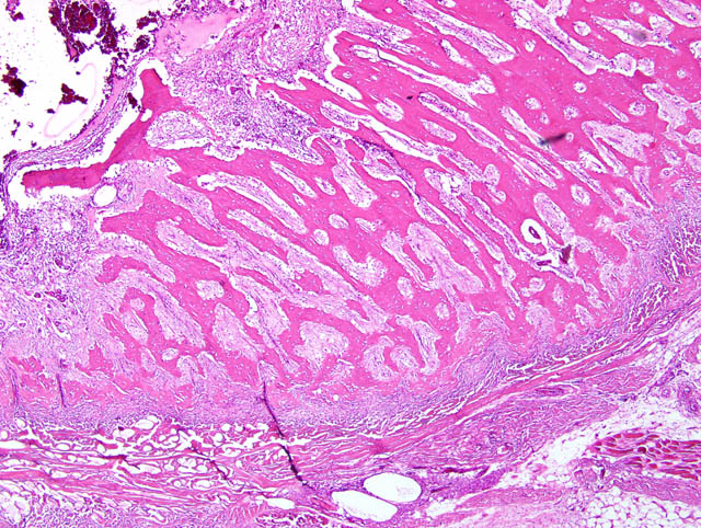
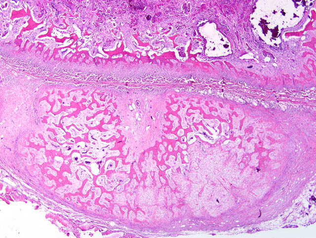
Rib. Two main lesions are present. The first consist of circumferential proliferation of trabecular woven bone perpendicular to the cortex (periosteal new bone formation). The cortical bone is decreased in thickness and shows a high porosity (osteopenia). Within the medullary cavity, a neoplastic mass is present (second lesion). It consists of neoplastic cells showing papillary growth in the existing bone marrow. There is an increase in fibrous tissue. Neoplastic cells are cuboidal to polygonal, 20 μm in diameter with a moderate amount of eosinophilic, poorly demarcated cytoplasm. The nucleus is round with a single prominent nucleolus and finely stippled chromatin. There is marked anisokaryosis and anisocytosis. The mitotic index is 1 per HPF. Multifocally, there are numerous islands of tumor cells with central necrosis and infiltration of neutrophils.
Morphologic Diagnosis:
Reactive periosteal new bone formation. Bone metastasis of papillary adenocarcinoma. Lesions consistent with pulmonary hypertrophic osteopathy.
Condition:
Metastatic papillary adenocarcinoma, hypertrophic osteopathy
Contributor Comment:
Hypertrophic osteopathy is also known as hypertrophic pulmonary disease and is characterized by new bone formation along the distal parts of the limbs. It occurs in humans and several animal species. Unlike in humans, where the condition is also called hypertrophic osteoarthropathy, articular surfaces are not affected in animals. It is a paraneoplastic condition generally associated with a lesion located in the thoracic cavity. In dogs it has been described in association with renal pelvis carcinoma(1,3), congenital megaesophagus(6), parasitic infection(4) and endocarditis.(2) Hypertrophic osteopathy is considered to be a reversible condition if the underlying lesion is corrected. The pathogenesis of the condition is still unclear.(2)
JPC Diagnosis:
Bone, rib: Carcinoma, metastatic.
Conference Comment:
In concurrence with the contributor, conference participants identified a metastatic carcinoma, and many, including the conference moderator, favored the contributors more specific diagnosis of metastatic adenocarcinoma based on the formation of vague tubules within the neoplasm. In the absence of a clinical history, other participants suspected a metastatic transitional cell carcinoma based on the presence of signet ring cells among the neoplastic population; given the history of urinary incontinence and presence of metastasis to the thoracic vertebrae, this remains a reasonable possibility. While very common in humans, metastatic bone disease is considered rare in domestic species, with carcinomas of the mammary gland, thyroid gland, prostate gland, ovary, and lung being the most common sites of origin.(5) Ultimately, we favored the less specific diagnosis of carcinoma based on the histologic features in the preponderance of slides available for evaluation, including the absence of distinct tubules in most sections. Additionally, some slides contain multinucleate neoplastic cells.
Conference attendees carefully considered the contributors diagnosis of hypertrophic osteopathy (HO); however, most participants interpreted the florid periosteal new bone as a reactive lesion secondary to invasion by metastatic carcinoma cells, rather than the paraneoplastic condition of hypertrophic osteopathy, for the following histologic and clinical reasons:(5)
- In the submitted case, the vast majority of the pre-existing cortical bone is lost and replaced by woven bone, and there are moderate numbers of osteoclasts within Howships lacunae, indicating active bone resorption. By contrast, in HO, pre-existing lamellar cortical bone generally remains intact, with the formation of periosteal trabeculae of new woven bone oriented perpendicular to the cortex.
- In many of the submitted slides, there is both endosteal and periosteal new bone growth, which is best appreciated at low magnification; endosteal new bone growth is not typical of HO.
- The rib is not a predilection site for the development of HO lesions. Periosteal new bone formation attributed to the condition of HO is typically confined to the limbs, and preferentially affects the metacarpals, metatarsals, radius, ulna, and tibia, with sparing of the articular surfaces, upper limbs, and phalanges.
While we cannot exclude the possibility of HO at other anatomic sites in this dog, the contributors gross description of bony proliferation exclusively affecting the bones of the upper limbs would be highly unusual, because HO classically spares the upper limbs, or affects them late in the disease process following initial and more severe lesions on the distal limbs. We thank the contributor for submitting this very interesting and edifying case.
References:
1. Chiang YC, Liu CH, Ho Sy, Lin CT, Yeh LS: Hypertrophic osteopathy associated with disseminated metastases of renal cell carcinoma in the dog: a case report. J Vet Med Sci
69(2):209-212, 2007
2. Dunn ME, Blond L, Letard D, DiFruscia R: Hypertrophic osteopathy associated with infective endocarditis in an adult boxer dog. J Small Anim Pract
48:99-103, 2007
3. Grillo TP, Brandao CVs, Mamprim MJ, de Jesus CMN, Santos TC, Minto BW: Hypertrophic osteopathy associated with renal pelvis transitional cell carcinoma in a dog. Can Vet J
48:745-747, 2007
4. Mylonakis ME, Rallis T, Koutinas AF: Canine spirocercosis. Compend Contin Educ Vet
30(2):111-116, 2008
5. Thompson K: Bones and joints.
In: Jubb, Kennedy, and Palmers Pathology of Domestic Animals, ed. Maxie MG, 5th ed., vol. 1, pp. 107-108, 127-129. Elsevier Saunders, Philadelphia, PA, 2007
6. Watrous Bj, Blumenfeld B: Congenital megaesophagus with hypertrophic osteopathy in a 6-year-old dog. Vet Radiol Ultrasound
43(6):545-549, 2002