WSC 23-24, CONFERENCE 10, CASE III:
Signalment:
Juvenile, male Harbour porpoise, cetacean (Phocoena phocoena)
History:
This animal was found dead on the beach.
Gross Pathology:
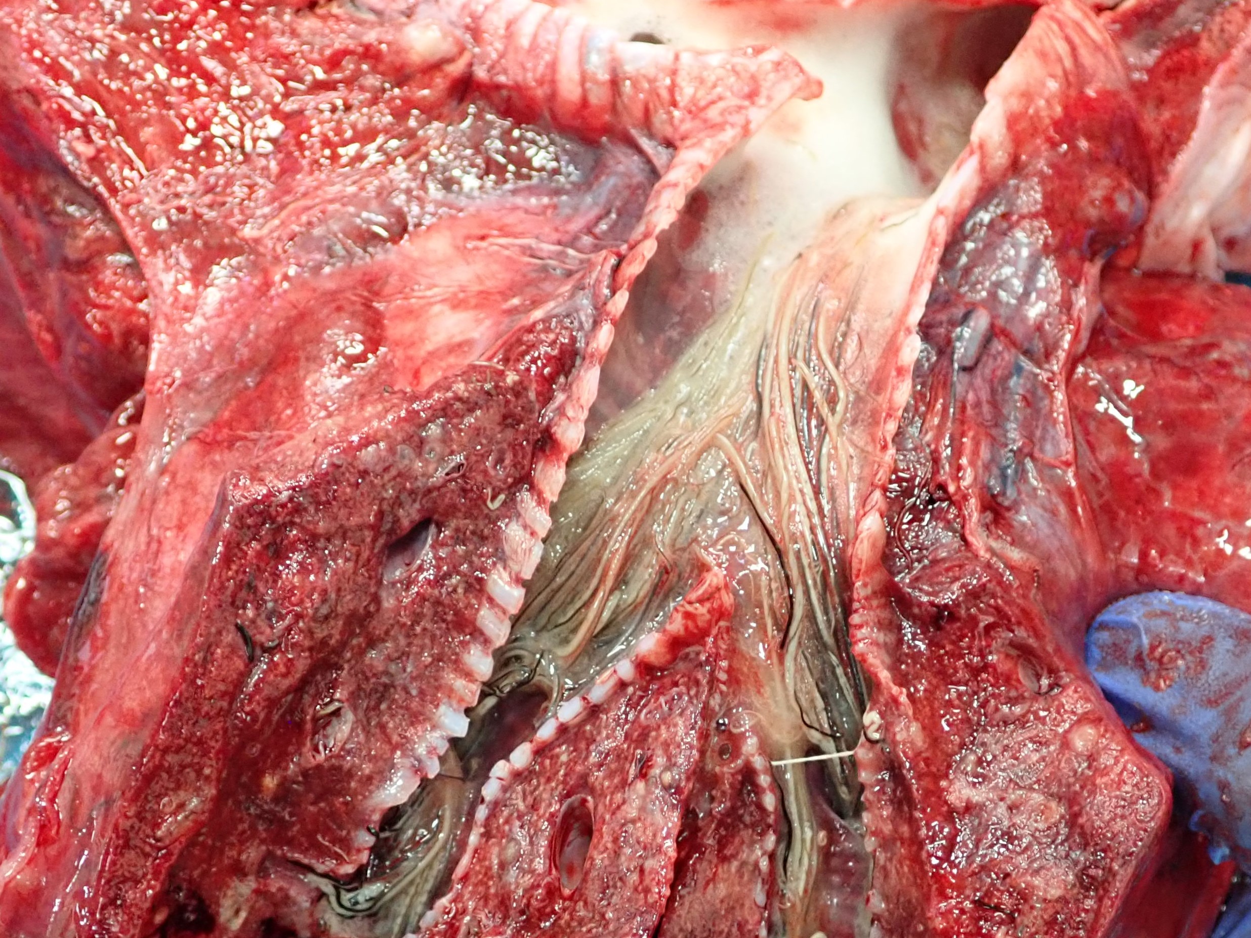
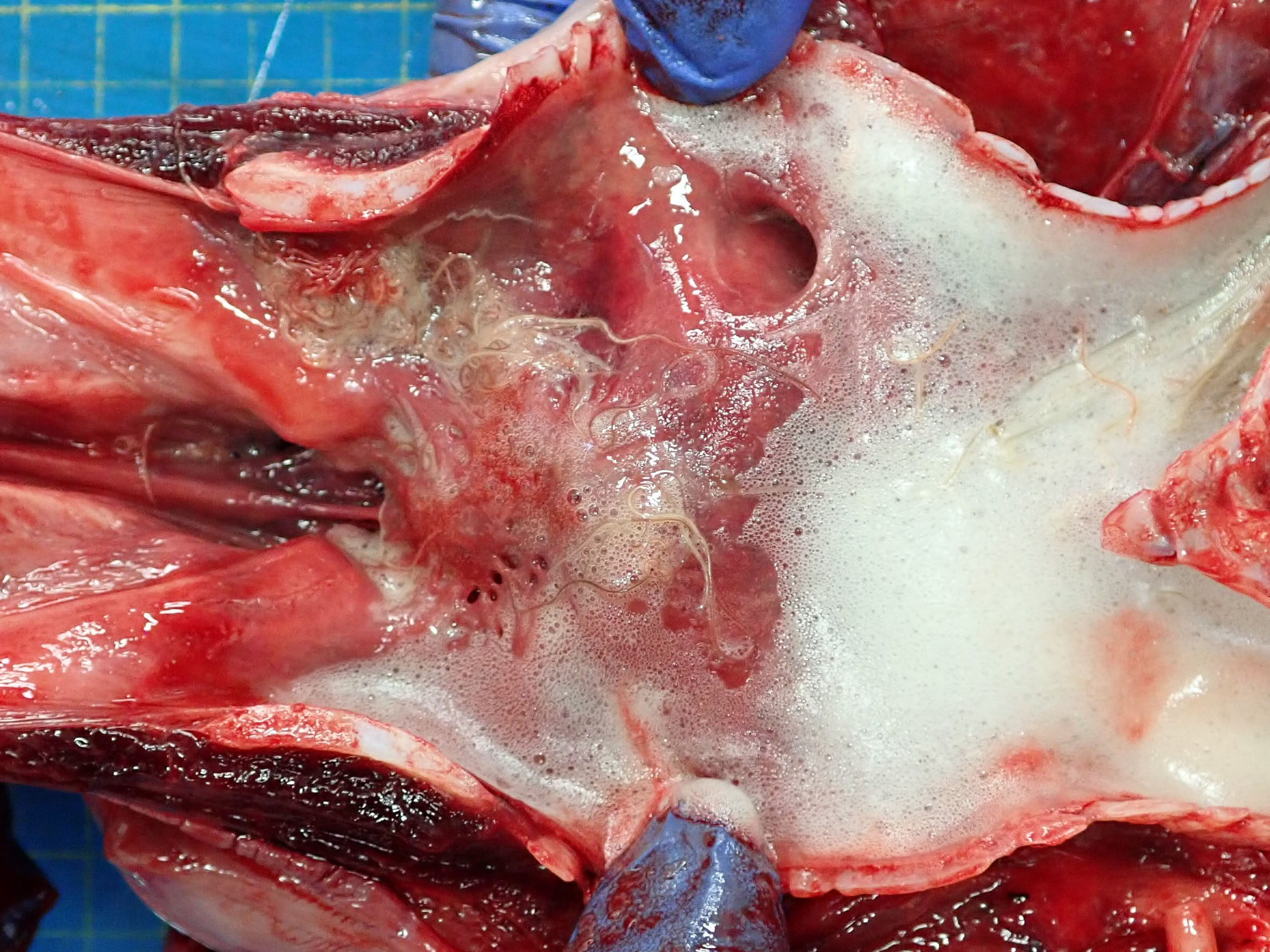
There was white frothy material in the trachea which extended to just proximal to the tracheal bifurcation (mild to moderate pulmonary oedema). There was a moderate amount of white frothy material in the primary bronchi (pulmonary oedema). There were myriad, large (approx. 6 cm long and 1 mm diameter) nematodes within the primary, secondary and tertiary bronchi.In the smaller bronchi the lumens were frequently filled with nematodes. The right lung was diffusely slightly darker red than the left (hypostasis). The left lung exhibited several parallel dark and light red stripes (rib imprints). Throughout the lung parenchyma, bilaterally, there were multiple firm foci ranging from 5 to 15 mm diameter, which were red to cream on cut surface, and sometimes contained nematodes (severe verminous pneumonia). Other lesions included: gastritis, multifocal, subacute, moderate, with intralesional nematodes and mucosal ulceration; inner ear, nematodiasis, bilateral, subacute, moderate to severe; abdominal cavity, haemoperitoneum, acute, diffuse, mild to moderate; mesenteric lymph nodes, lymphadenomegaly, diffuse, subacute, moderate; skin, abrasion, multifocal, acute, mild to moderate; liver, scar, multifocal, chronic, minimal.
Laboratory Results:
Lung: Light growth of Salmonella spp. obtained from cultures.
Microscopic Description:
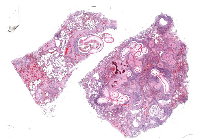
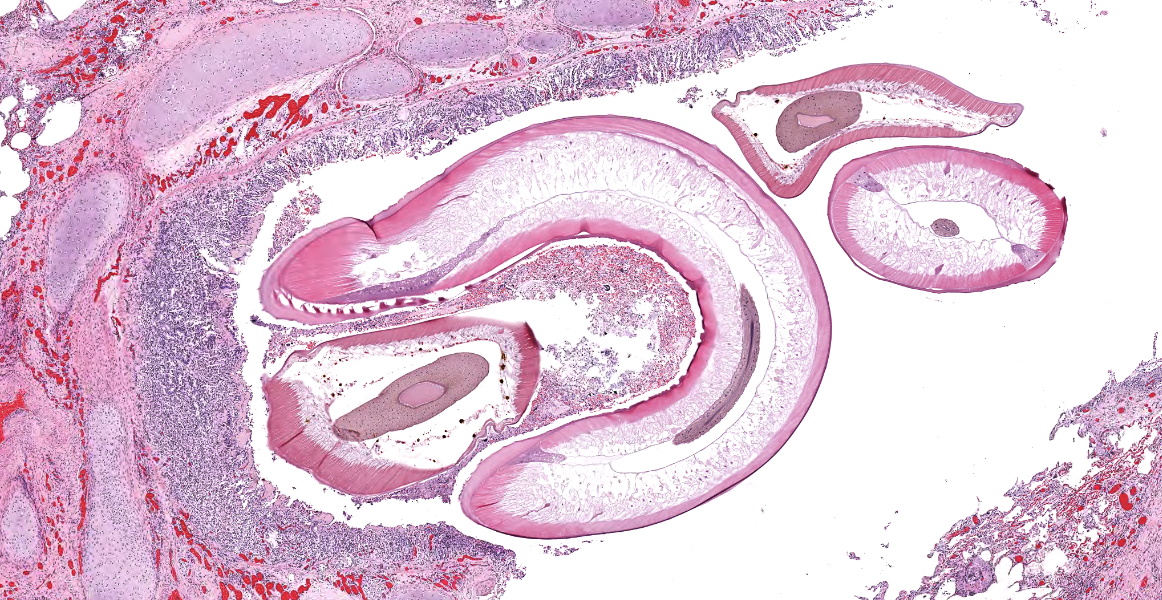
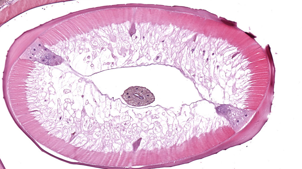
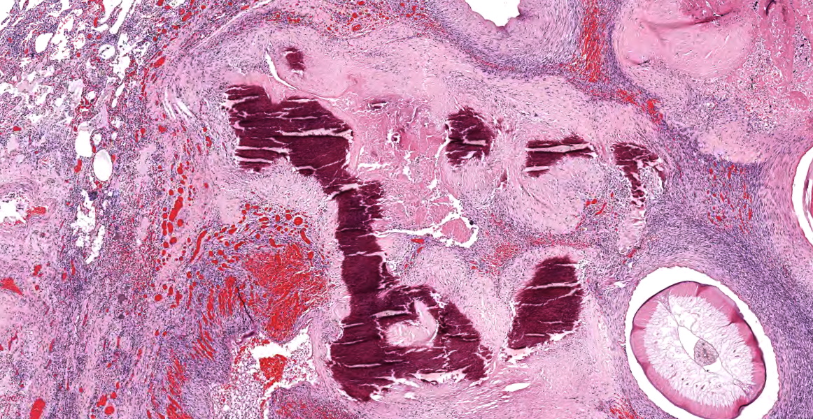
Two sections of lung: Multiple secondary and tertiary bronchi contain multiple intraluminal 1 mm diameter nematodes characterised by the presence of a 15 μm thick cuticle, coelomyarian musculature, and lateral cords. Bronchi also contain immature individuals and embryonated eggs, as well as degenerated sloughed epithelial cells and extravasated erythrocytes (haemorrhage). There is moderate diffuse infiltration of eosinophils and fewer lymphocytes, plasma cells and macrophages in the epithelium and lamina propria of the affected bronchi.
Approximately 50% of the section examined is effaced by multifocal to coalescing accumulations of a dense light eosinophilic material surrounded by elongated cells (fibroblasts), foreign body type multinucleated giant cells (up to 20 nuclei) and epithelioid macrophages (granulomas). These areas occasionally contain a moderate to large amount of eosinophilic to basophilic angular to crystalline material (mineralisation).
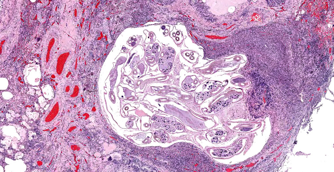
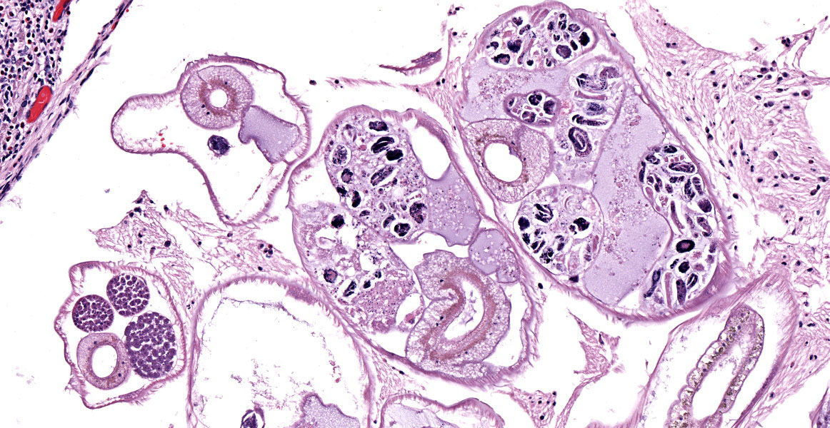
Multifocally, bronchioles and alveoli are filled with a moderate to large amount of necrotic cellular debris, a moderate number of eosinophils, macrophages with foamy cytoplasm, lymphocytes, light eosinophilic amorphous material (oedema), light eosinophilic fibrillary material (fibrin) and, occasionally, 0.5 um maximum length extracellular basophilic coccobacilli. These areas are often associated with the presence of thin-walled cysts (up to 1.5 mm diameter) within the parenchyma, which contain multiple small (200 μm diameter) nematodes in both transverse and longitudinal section. These nematodes have a 5-10 μm cuticle with coelomyarian musculature, and lateral cords. The coelom contains an intestinal tract and reproductive tract with larvae. There is also a mixed inflammatory infiltration on the alveolar wall, composed of eosinophils, lymphocytes and plasma cells and multifocal areas of dark basophilic angular to crystalline material (mineralisation).
Pulmonary arteries show marked smooth muscle hyperplasia with reduction of the arterial lumen. Alveolar capillaries show proliferation and congestion. There are also areas of atelectasis and alveolar distension with rupture of alveolar wall (emphysema).
Contributor’s Morphologic Diagnosis:
Lung: Bronchopneumonia, granulomatous and eosinophilic, multifocal, chronic, severe, with intralesional nematodes, fibrosis and mineralisation.
Contributor’s Comment:
The histological features represent a case of granulomatous and eosinophilic bronchopneumonia associated with lungworms in a harbor porpoise. In the present case, Salmonella enterica Group B has also been isolated from the pulmonary lesions and is likely to be a clinically significant finding.
Lungworms are a common finding in wild cetaceans, with species of nematodes reported in up to 69% of stranded Harbour porpoises (Phocoena phocoena), all from the family Pseudaliidae.6,7Pseudalius inflexus and Torynurus convolutus are found in the bronchi, bronchioles and blood vessels, whereas Halocercus invaginatus inhabits the pulmonary parenchyma.2,9In this case, it appears most likely that the nematodes in the upper airways and the bronchioles were P. inflexus and T. convolutus and those within the parenchyma were H. invaginatus.
Verminous pneumonia is a common finding although is not frequently thought to be the primary cause of death. However, there can be severe changes, resulting in extensive fibrosis and mineralisation, as seen in this case. There is evidence that increased parasitic burden is associated with high levels of pollutants (polychlorinated biphenyls) which are associated with immunosuppression.3 In addition, some studies suggest that lungworms may act as a potential means of transfer of bacterial infections in harbour porpoise, with Salmonella enterica Group B, as cultured in the current case, identified as potentially spread in this way.4-6 The case presented here reinforces the theory that parasites should be considered as potential vectors of zoonotic bacterial infections in marine mammals.
Contributing Institution:
The University of Liverpool
https://www.liverpool.ac.uk/veterinary-science/
JPC Diagnosis:
- Lung: Bronchopneumonia, eosinophilic, multifocal, with intrabronchial metastrongyle nematodes.
- Lung: Pneumonia, interstitial, eosinophilic, multifocal, chronic, severe, with intraparenchymal adult metastrongyle nematodes.
- Lung: Pneumonia, interstitial, eosinophilic, multifocal severe, with intraparenchymal nematodes.
JPC Comment:
This case provides an excellent example of lungworm parasitism in a harbour porpoise, a marine species found in the North Atlantic Ocean that typically bears a heavy pulmonary parasite burden.3 Studies of the harbour porpoise have found that parasites are most common in the respiratory system, and lungworms have been found in virtually all harbour porpoises older than 1 year, most often in the bronchial tree and the pulmonary blood vessels.9 As in this case, parasites within the pulmonary parenchyma typically incite an eosinophilic and granulomatous bronchointerstitial pneumonia. When found in the pulmonary blood vessels, nematodes can cause chronic thrombosis and vasculitis, often with calcification of thrombotic material.9
In general, far less is known about the biology of pulmonary parasites of marine mammals compared to their terrestrial counterparts; however, two of the nematodes examined here belong to the metastrongyle family which, collectively, are far less mysterious. Metastrongyles, or “lungworms,” have coelomyarian musculature, an external cuticle that is typically smooth, an intestine lined by few multinucleated cells, and accessory hypodermal cords. Mature females can produce either eggs or fully developed embryos; species that produce eggs deposit them into host tissues where they embryonate.10 Other notable metastrongyles of veterinary importance include Angiostrongylus cantonensis (rat), Dictyocaulus viviparus (ruminants), Muellerius capillaris (small ruminants), Metastrongylus apri (pigs), Aelurostrongylus abstrusus (cats), and Filaroides hirthi (dogs).
This case incited robust discussion among conference participants. Participants identified three different nematode species in section: the large nematodes within the airways and two, much smaller nematode species within the parenchyma. Some participants remarked that many areas identified initially as granulomas could represent foci of severe vasculitis, possibly secondary to intravascular lungworms. Of the three nematodes identified in section, participants felt that the smallest of the three, which were approximately 60um in diameter, had features consistent with filarid parasites, which could account for the possible vasculitis. Post-conference consultation with Dr. Christopher Gardiner confirmed that these nematodes are consistent with filarid nematodes, though we were unable to speciate them with the minimal features present in section.
The two largest nematodes in section are metastronglyes and contain very nice examples of the classic metastrongyle morphologic features described above. The largest nematodes are over 1mm in diameter and are morphologically consistent with Pseudalius inflexus; however, the lack of gonads within the examined section did not allow for definitive speciation. Interestingly, there are reports of Pseudalius inflexus causing similar lesions in Burmeister’s porpoises; those cases were accompanied by vasculitis and thrombosed arteries with a morphologic appearance similar to many of the presumptive occlusive vascular changes in this section.1
Finally, the mid-sized nematode population, with a diameter of approximately 225um, contain classic metastrongyle features and female gonads contain developing larvae (see Figure 3-8). Similar to the previously discussed nematodes, we were unable to speciate these metastrongyles, though the species discussed by the contributor appear to be the most common causes of marine mammal pulmonary nematodiasis.
Conference participants debated combining the morphologic diagnoses or creating separate diagnoses based on etiology. As the three different nematodes each had different pathologic effects, participants preferred separate morphologic diagnoses. And while participants felt strongly that a significant vasculitis was evident in the examined section, vasculitis was not included in the diagnosis due to the inability to confirm vasculitis with immunohistochemical stains on this entirely digital submission.
References:
- Alvarado-Rybak M, Toro F, Abarca P, Paredes E, Espanol-Jimenez S, Seguel M. Pathological findings in cetaceans sporadically stranded along the Chilean coast. Front Mar Sci. 2020;7.
- Balbuena JA, Aspholm PE, Andersen KI, Bjorge A. Lung-worms (Nematoda: Pseudaliidae) of harbour porpoises (Phocoena phocoena) in Norwegian waters: patterns of colonization. Parasitology. 1994;108:343-349.
- Bull JC, Jepson PD, Sauna RK, et al. The relationship between polychlorinated biphenyls in blubber and levels of nematode infestations in harbour porpoises, Phocoena phocoena. Parasitology. 2006; 132:565-573.
- Davison N, Barnett J, Rule B, Chappell S, Wise G. Group B Salmonella in lung-worms from a harbour porpoise (Phocoena phocoena). Vet Rec. 2010; 167:351-352.
- Davison NJ, Simpson VR, Chappell S, et al. Prevalence of a host-adapted group B Salmonella enterica in harbour porpoises (Phocoena phocoena) from the south-west coast of England. Vet Rec. 2010; 167: 173-176.
- Foster G, Patterson IA, Munro DS. Monophasic group B Salmonella species infecting harbour porpoises (Phocoena phocoena) inhabiting Scottish coastal waters. Vet Microbiol. 1999;65: 227-231.
- Jepson PD, Baker JR, Kuiken T, Simpson VR, Kennedy S, Bennett PM. Pulmonary pathology of harbour porpoises (Phocoena phocoena) stranded in England and Wales between 1990 and 1996. Vet Rec. 2000;146:721-728.
- Reckendorf A, Everaarts E, Bunskoek P, et al. Lungworm infections in harbour porpoises (Phocoena phocoena) in the German Wadden Sea between 2006 and 2018, and serodiagnostic tests. Int J Parasitol Parasites Wildl. 2021;14: 53-61.
- Siebert U, Wunschmann A, Weiss R, Frank H, Benke H, Frese K. Post-mortem findings in harbour porpoises (Phocoena phocoena) from the German North and Baltic Seas. J Comp Pathol. 2001;124: 102-114.
- Gardiner CH, Poynton SL. An Atlas of Metozoan Parasites in Animal Tissues. Armed Forces Institute of Patholoy;2006.