Joint Pathology Center
Veterinary Pathology Services
Wednesday Slide Conference
2018-2019
Conference 3
Sept 12, 2018
CASE III: 12L-15812 (JPC 4067411-00).
Signalment: 12-year-old, female, Ragdoll cat, (Felis catus)
History: Three days period of nausea and vomiting, followed by unconsciousness, convulsions and finally death.
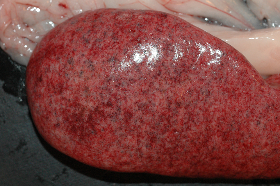
Gross Pathology: Spleen: Splenomegaly, with pale pink / white marbled parenchyme. The liver showed disseminated, 2-5 mm sized, partly confluent pale white subcapsular and parenchymal areas. Within the gastrointestinal tract, a focal acute ulcerative gastritis, and a multifocal chronic ulcerative duodenitis was present. The duodenal lymph nodes showed mild enlargement and a homogeneous pale cream cut surface.
Laboratory results: None.
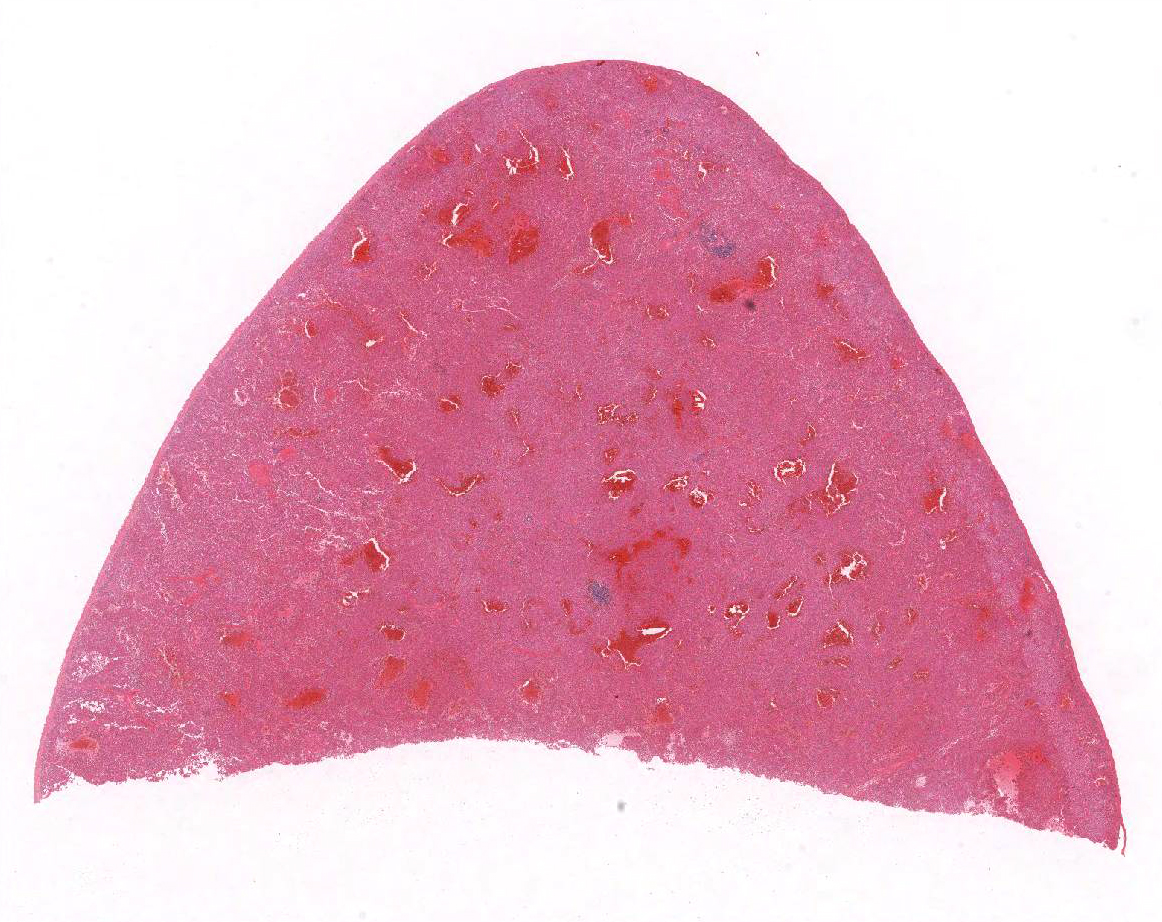
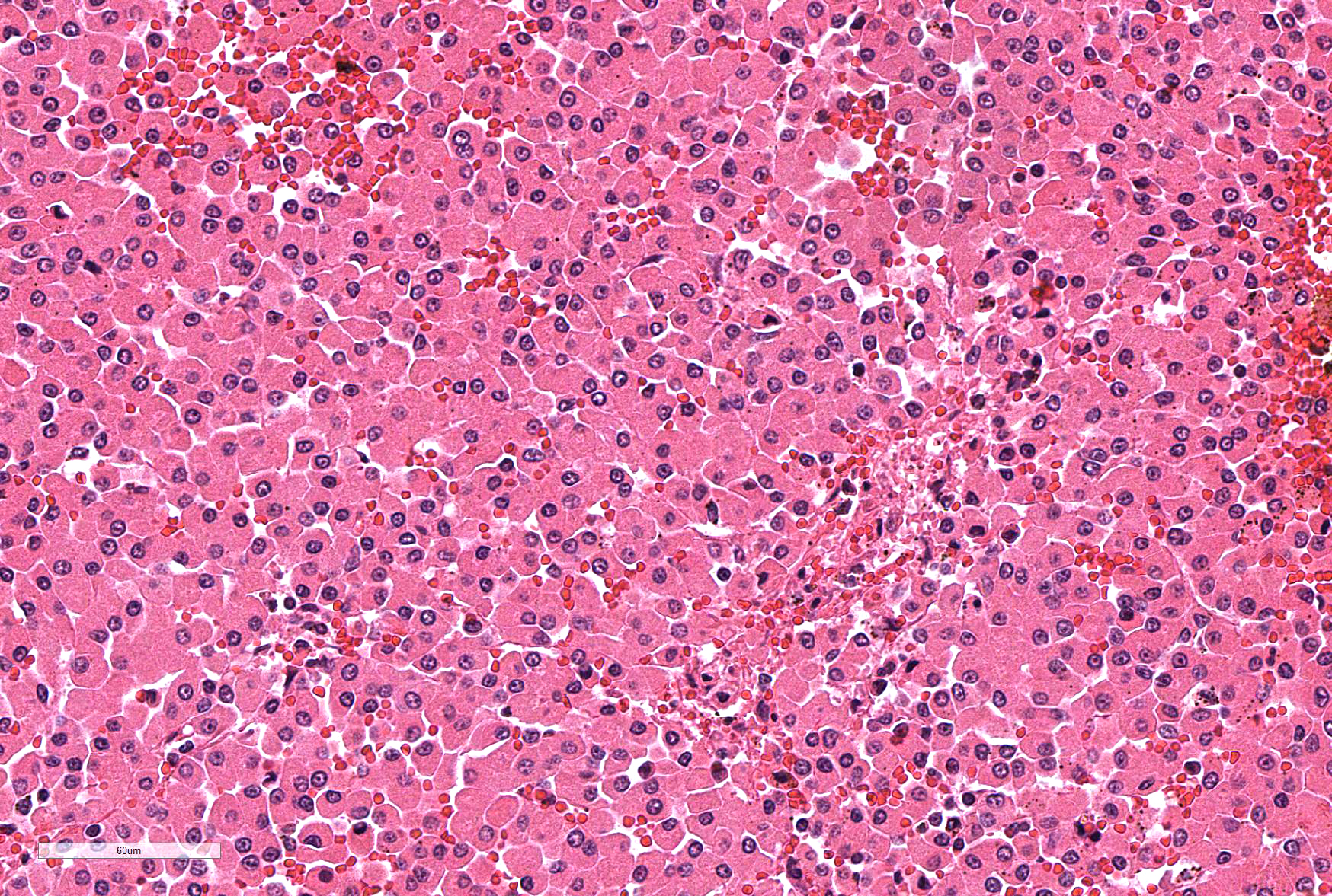
Microscopic Description: Section of spleen with mild mesothelial hyperplasia and mild capsular fibrosis. The splenic architecture (red and white pulp) is widely effaced by infiltration of a population of monomorphic large, approximately 20-40µm sized, plump, round cells with fine granular eosinophilic cytoplasm and round, small, hypochromatic, eccentric nuclei with one nucleolus. Mitotic figures are rare, focal apoptotic cells, mild diffuse hemosiderosis and hyperaemia is present.
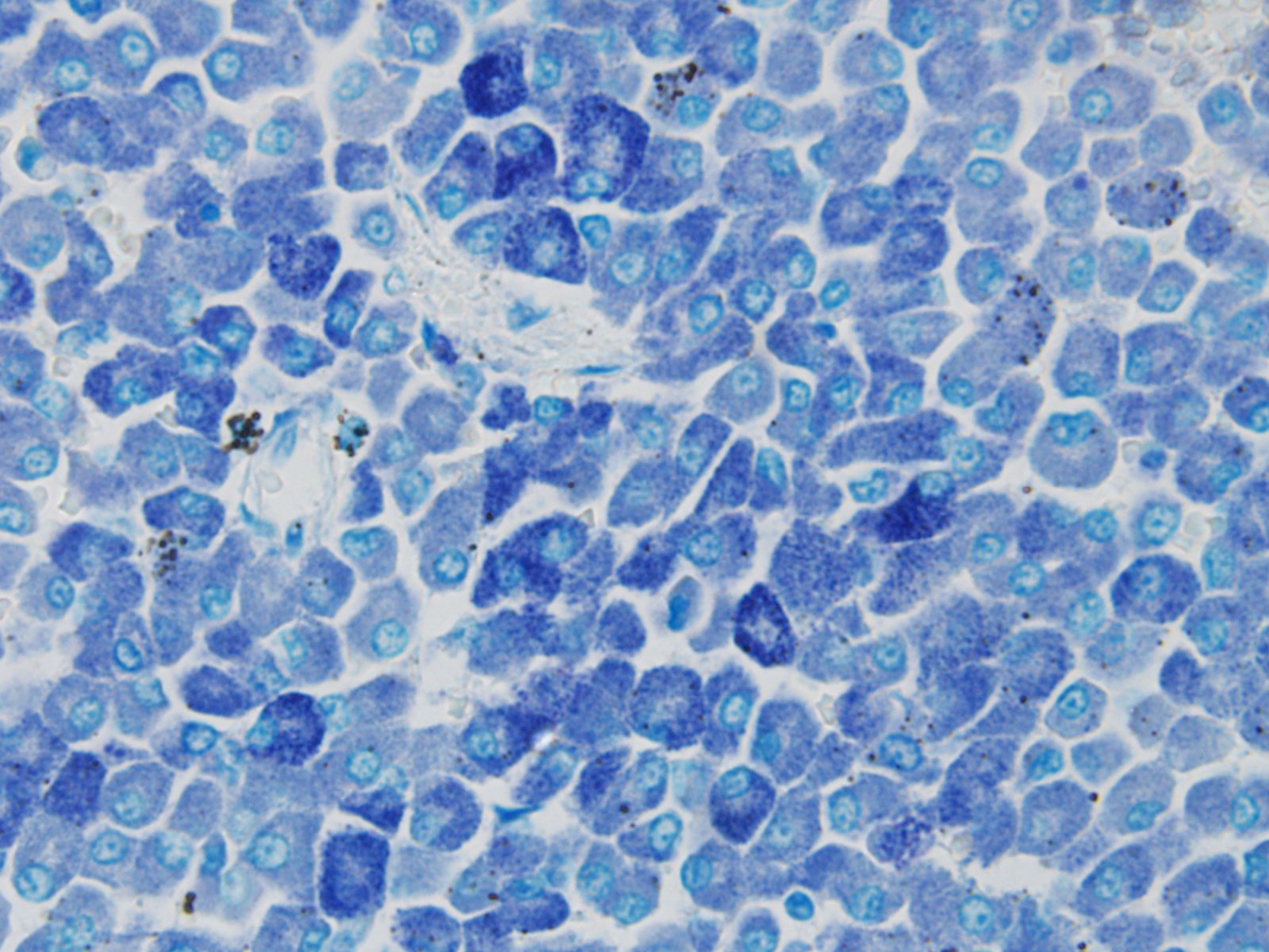
Special stains: Toluidine blue stain: Neoplastic cells reveal variable amounts of metachromatic fine cytoplasmic granula.
Immunohistochemistry:
- Anti-Mast Cell Tryptase: Neoplastic cells exhibit variable degrees of weak positive reaction
- CD3: Neoplastic cells: negative reaction
- CD45R: Neoplastic cells: negative reaction
Contributor’s Morphologic Diagnoses: Diffuse visceral (splenic) mast cell tumor with mild mesothelial hyperplasia and capsular fibrosis, Ragdoll, Felis catus.
Contributor’s Comment: According to the information sheet of Veterinary Society of Surgical Oncology (VSSO, http://www.vsso.org/Splenic_MCT.html), about 50% of feline visceral mast cell tumours are located in the spleen. Other sites of visceral mast cell tumours are the mediastinum, lymph nodes, and intestines. Visceral mast cell tumours present in three forms, smooth, diffuse, and nodular. These neoplasms often metastasize to liver (90%), visceral lymph nodes (73%), bone marrow (23%-40%), lung (20%), and intestine (17%), often accompanied by pleural and peritoneal effusions. The cutaneous involvement of mast cell tumours in cats with primary visceral MCT is less frequent (18%).
In the present case, metastases in mesenteric lymph nodes and bone marrow was observed. Within sections of stomach and duodenal ulcerations, no neoplastic mast cells could be demonstrated. large numbers of neoplastic mast cells as observed in the spleen, were observed within hepatic sinuses.
JPC Diagnosis: Spleen: Mast cell tumor.
Conference Comment: Review of the recent literature reveals a number of terms which are interchangeably used to describe non-cutaneous mast cell tumors in the cat. Visceral mast cell tumor, systemic mastocytosis, and mast cell leukemia are all in common usage; the use of the term mast cell leukemia implies mastocytemia. Mastocytemia is not uncommon in cats with visceral mast cell tumors; rates of up to 68% have been reported.4
Splenic involvement of mast cell tumors ranges from 15-50% in various reviews.1,5 The liver and intestine are also common sites, and multiorgan disease is also seen. Although cats with visceral tumors may have concomitant cutaneous tumors as well, visceral mast cell tumors are considered a separate disease from cutaneous mast cell tumor; evidence that visceral tumors arise from a cutaneous primary is lacking in the veterinary literature.
Grossly, visceral mast cell tumors often cause diffuse organ enlargement, but may sometimes appear nodular.1 Histologically, neoplastic mast cells have a finely granulated poorly stained cytoplasm and granules may not be recognizable on hematoxylin and eosin stained sections. Moreover, cytoplasmic granules may not stain with Toluidine blue or Giemsa. This is thought to be the case when neoplastic mast cells arise from the intestinal mucosa; special fixation in media other than formalin may be required for their cytoplasmic granules to be metachromatic.1
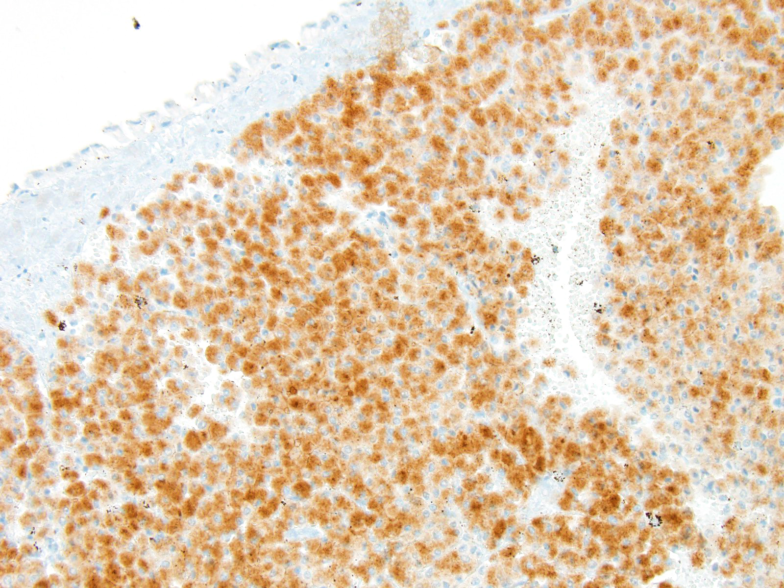
Immunohistochemical of evaluation of feline mast cell tumors may be frustrating. Reported rates of cytoplasmic expression of c-Kit ranges from 353 to 85%5. In the submitted case, Toluidine blue and Giemsa stains run at the JPC were strongly positive, and the tumors showed diffuse strong cytoplasmic expression of c-kit (toluidine blue and c-Kit illustrated in images 3-4 and 3-5, respectively.) One study demonstrated 14 missense c-kit mutations in 13 of 20 tumors; with 11 mutations located in exon 8, and 3 in exon 9.5 No correlation was observed between c-kit mutations and tumor differentiation, mitotic activity, or survival time.5
Contributing Institution:
University of Liverpool
http://www.liv.ac.uk/vetpathology/index.htm
References:
- Kiupel, M. Mast Cell Tumors. In: Meuten DJ, ed. Tumors in Domestic Animals, 5th Ames IA, Wiley and Sons 2017. pp 195-199.
- Lamm CG, Stern AW, Smith AJ, Cooper EJ, Ullom SW, Campbell GA. Disseminated cutaneous mast cell tumors with epitheliotropism and systemic mastocytosis in a domestic cat. J Vet Diagn Invest 2009; 21:710-715.
- Mallett CL, Northrup NC, Saba CF, Rodriguez CO, Rassnick KM, Gieger TL, Childress MO, Howerth EW. Immunohistochemical characterization of feline mast cell tumors. Vet Pathol 2012; 50(1): 106-109.
- Piviani M, Walton RM. Significance of mastocytemia in cats. Vet Clin Pathol 2013; 42:4-10.
- Sabattini S, Barzon G, Giantin M, Lopparelli RM, Dacasto M, Prata D, Bettini G. Kit receptor tyrosine kinase dysregulations in feline splenic mast cell tumors. Vet Comp Oncol 2017; 15(3):1051-1056.
- Veterinary Society of Surgical Oncology. http://www.vsso.org/Splenic_MCT.html