CASE 4: B20-121 (JPC 4152719-00)
Signalment:
12-year old, neutered, unknown sex, domestic shorthair, Felis catus, cat
History:
The cat had a tan mass in the lateroventral aspect of the left eye. Fibrin and hemorrhage were reported in the anterior chamber.
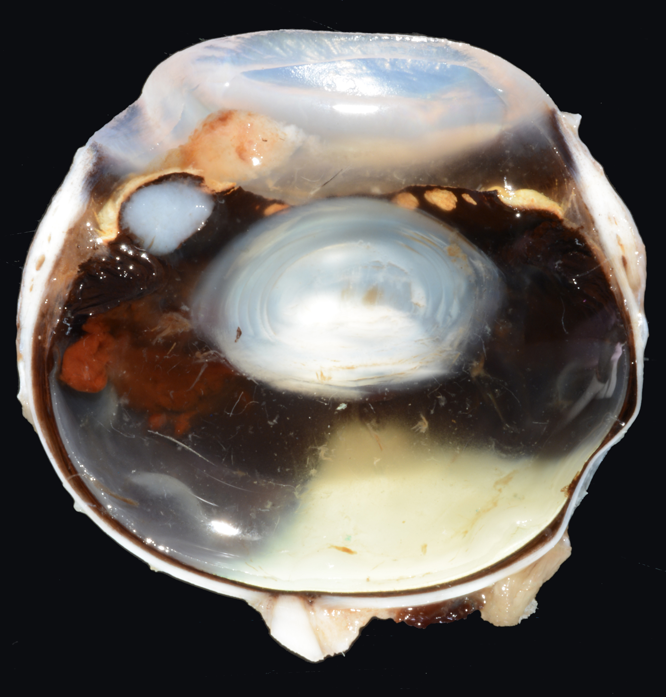
Gross Pathology:
There is a firm tan mass along the posterior surface of the iris. There is red gelatinous material (hemorrhage) in the adjacent vitreous. A small amount of tan material (fibrin) with red flecks (hemorrhage) is in the anterior chamber.
Laboratory results:
None.
Microscopic description:
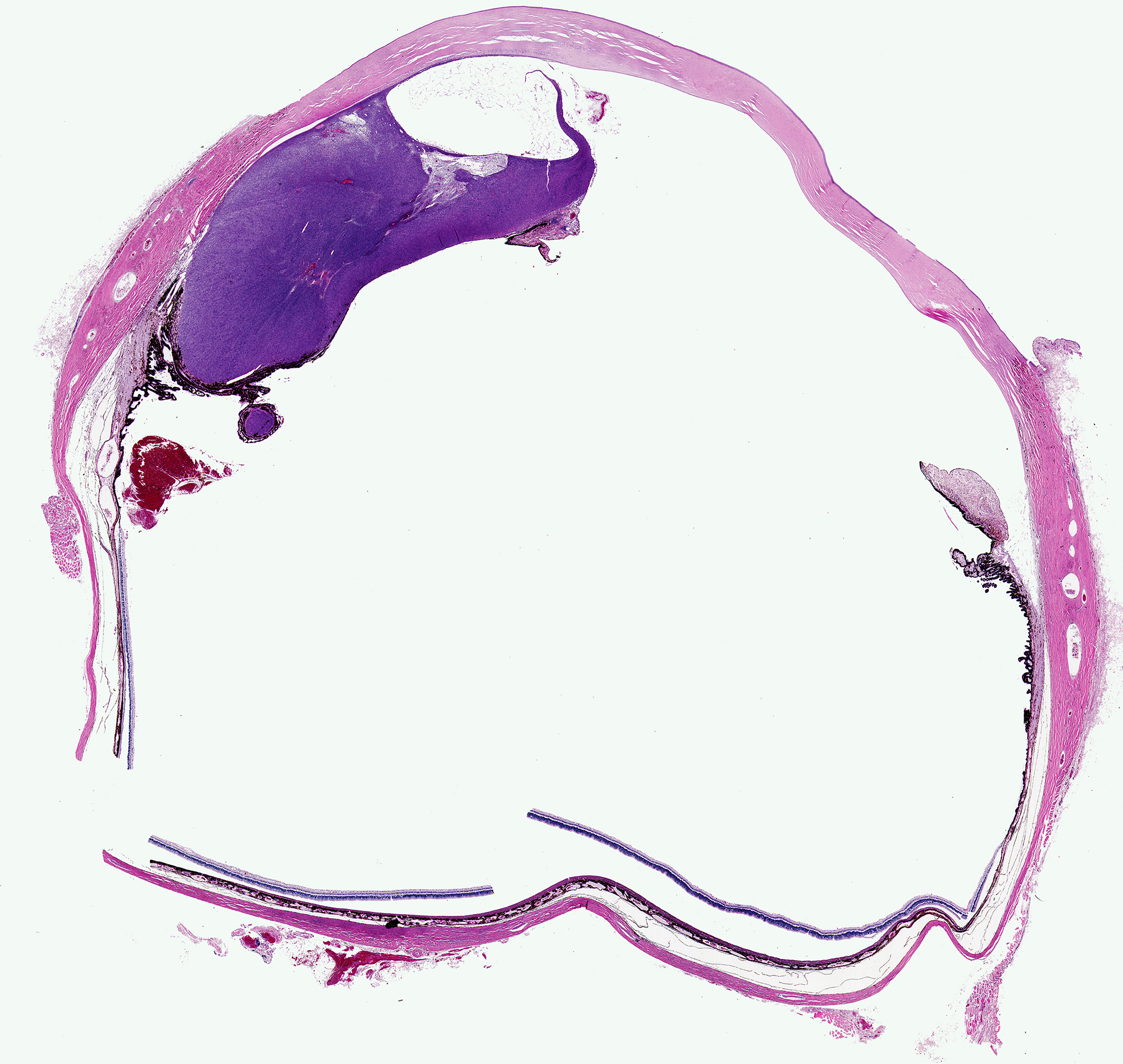
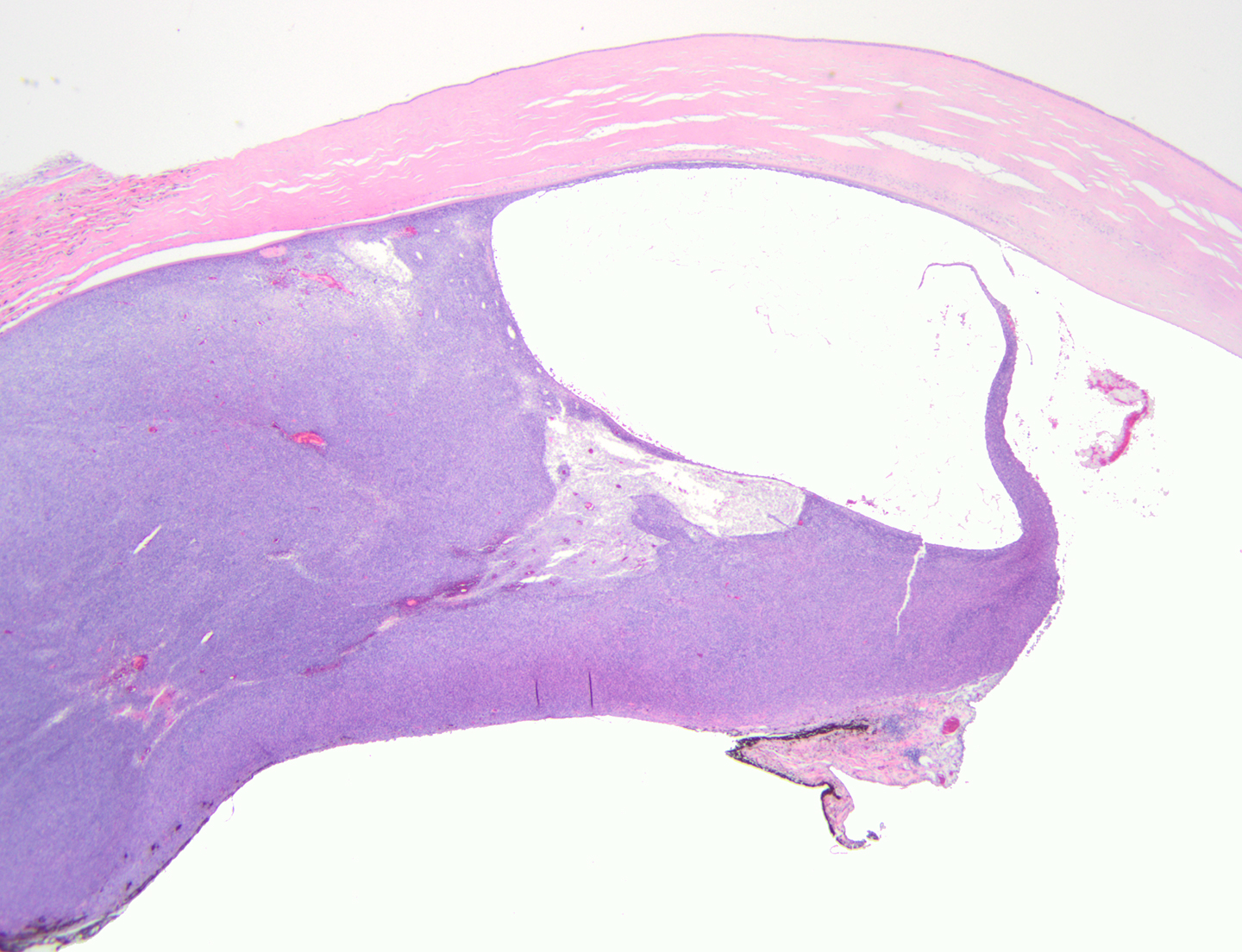
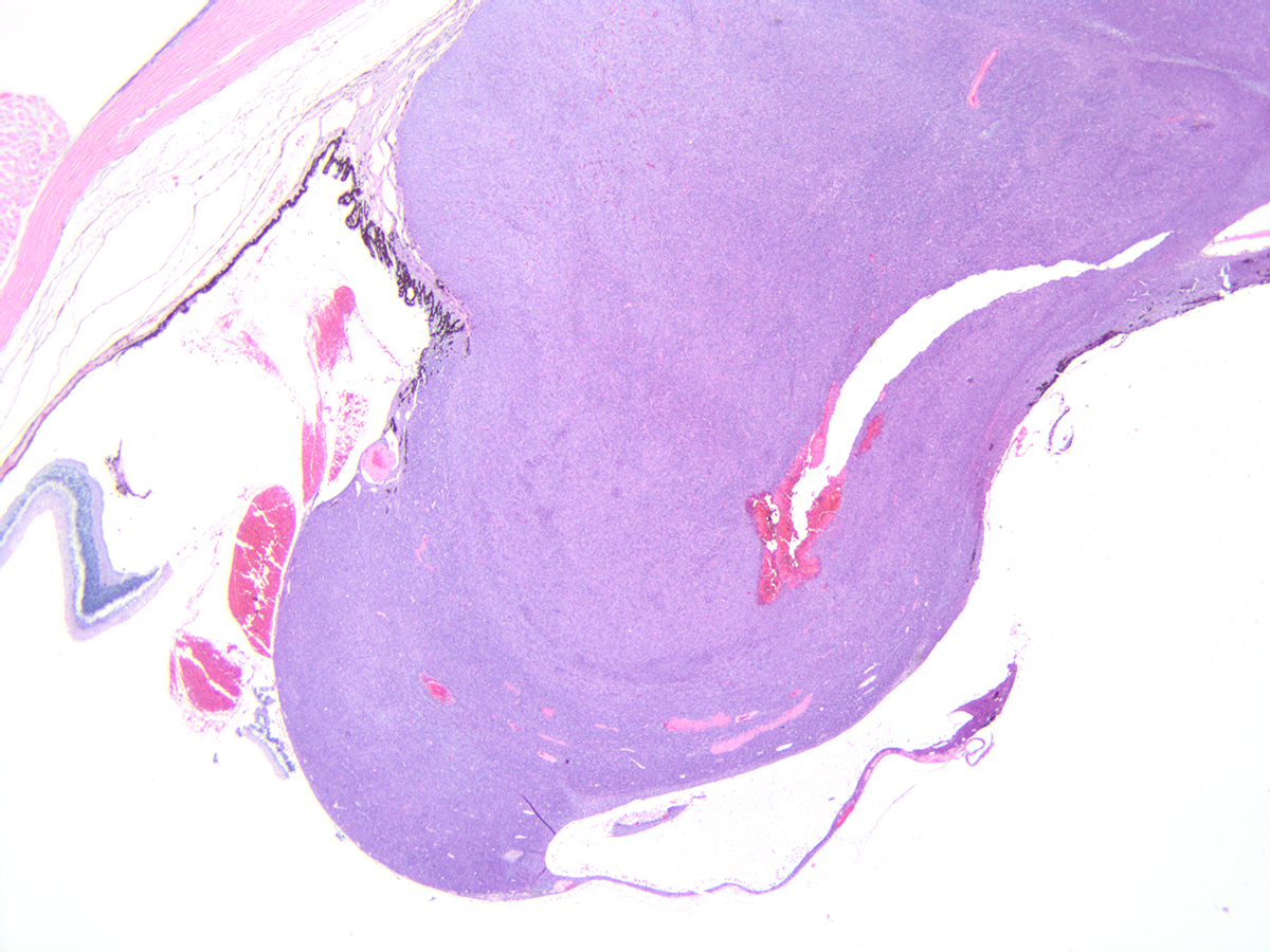
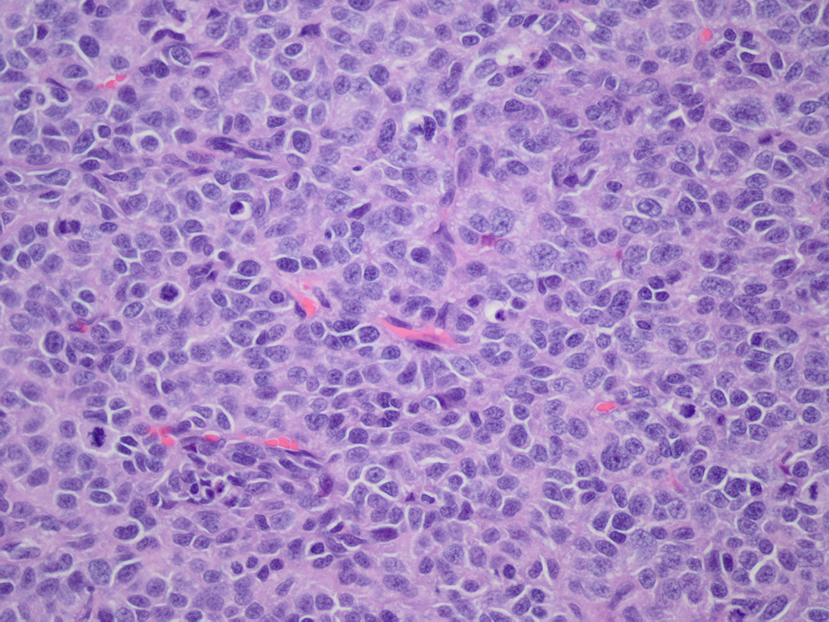
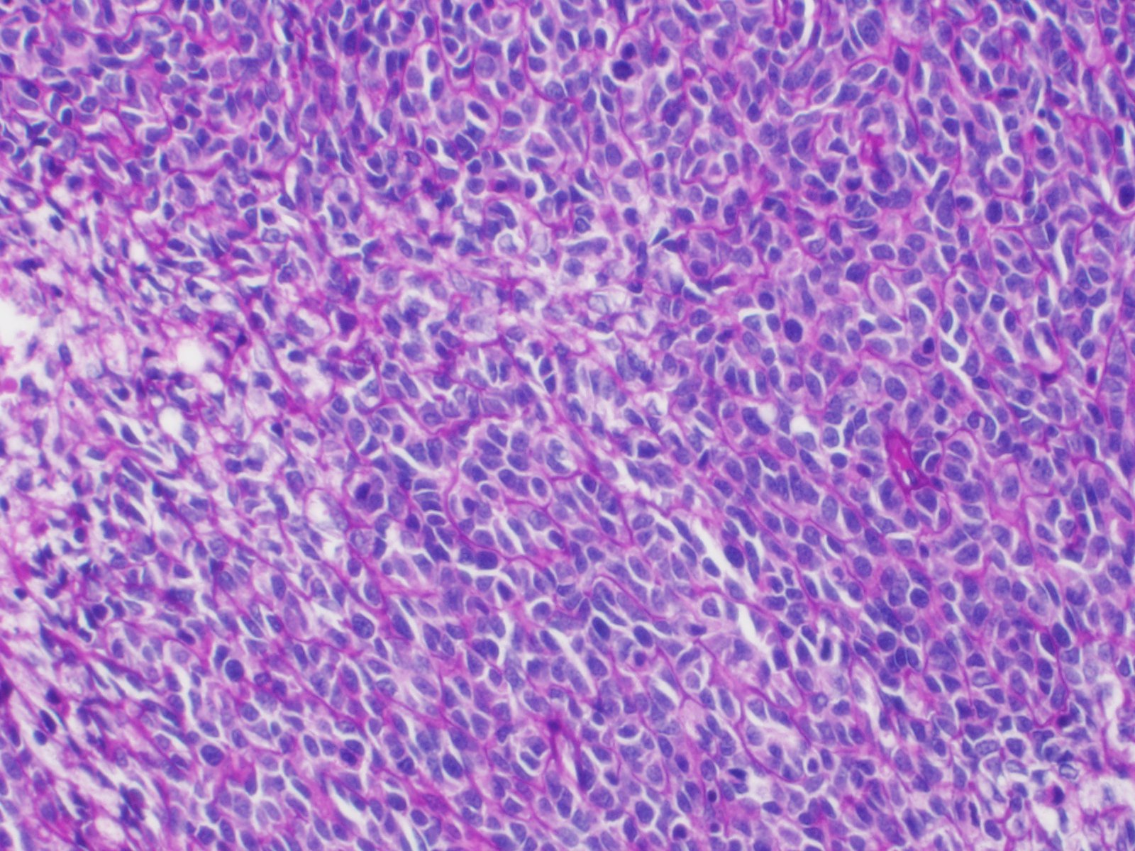
Expanding one side of the iris and ciliary body and compressing the iridociliary angle is a well-demarcated mass composed primarily of polygonal cells arranged in nests. The neoplastic cells have moderate amounts of eosinophilic cytoplasm. Nuclei have finely stippled chromatin and indistinct nucleoli. There are 45 mitotic figures in ten 400x fields. In some areas the cells are more spindleoid. In some sections, there are cystic areas within the mass. A periodic acid Schiff (PAS) stain highlights prominent basement membranes around the nest of neoplastic cells. The neoplastic cells are not immunoreactive to MelanA. In some areas the neoplastic cells are weakly immunoreactive to broad spectrum cytokeratin MNF116 or to vimentin. The associated iris leaflet is covered in a pre-iridal fibrovascular membrane. There are aggregates of lymphocytes in the iris stroma and ciliary body. There are varying amounts of fibrin and hemorrhage in the anterior chamber, posterior chamber and vitreous chamber. There is mild peripheral corneal vascularization. The lens is artifactually absent. The retina is multifocally artifactually detached.
Contributor's morphologic diagnosis:
Iridociliary adenoma
Contributor's comment:
Iridociliary tumors are uncommon in cats, and as in this case, occur primarily in older animals.3 In dogs and cats, iridociliary tumors are most typically benign,9 with adenocarcinomas being rarely reported.
Compared to canine iridociliary tumors, which usually have easily recognizable cords of polygonal epithelial cells, the neoplastic cells in feline iridociliary tumors tend to form solid sheets. In some tumors, cystic areas may be common.
The presence of basement membrane around the cells, which as in this case, can be highlighted with a PAS stain3,9 is helpful in refining the diagnosis and ruling out metastatic carcinomas.9
Iridociliary epithelium is derived from neuroectoderm and therefore is typically immunoreactive to the mesenchymal cell marker vimentin,3,9 and variably immunoreactive to the epithelial marker cytokeratin.3 Immunoreactivity of neoplastic epithelial cells to vimentin may be useful in differentiating iridociliary tumors from metastatic carcinomas, which otherwise would not be expected to express the vimentin. However, more aggressive iridociliary adenomas may express cytokeratin and or TERT (telomerase reverse transcriptase).3 Additionally, the neoplastic cells are usually immunoreactive to neuron specific enolase (NSE)3,9 and variably immunoreactive to S-100.3
Contributing Institution:
University of Tennessee, College of Veterinary Medicine, Department of Biomedical and Diagnostic Sciences http://www.vet.utk.edu/departments/path/index.php
JPC diagnosis:
Globe: Iridociliary adenoma.
JPC comment:
The moderator discussed a list of possible differentials with conference participants. Along with iridociliary adenoma/adenocarcinoma, melanoma, post-traumatic sarcoma, severe uveitis, and lymphoma should be considered. Ocular lymphomas were previously presumed to only manifest when an extension of systemic disease. However, more recently, an examination of cases of ocular lymphoma may support a subset of cases that are currently called "presumed solitary ocular lymphoma", or PSOL. These patients did not experience progression of lymphoma following enucleation, and the lymphoma phenotype was not associated with survival.4,8
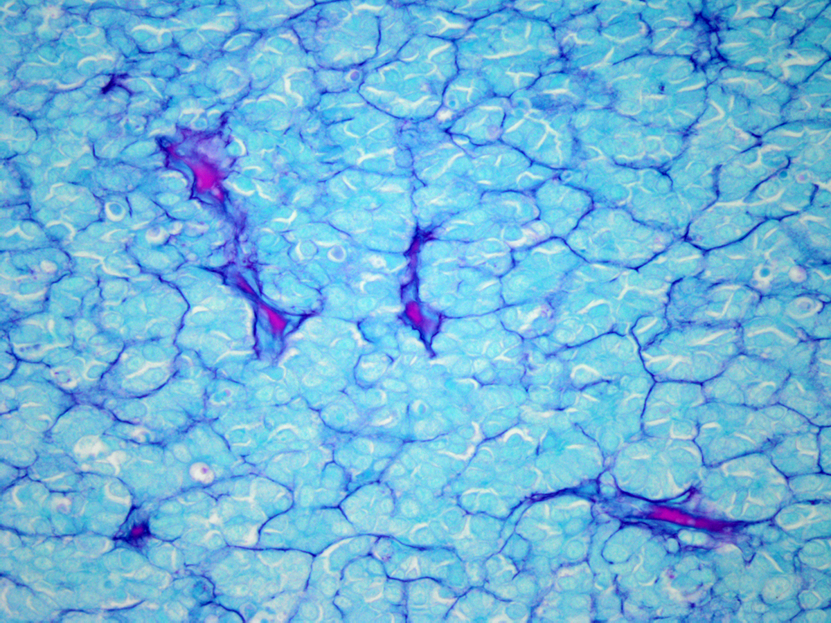
Iridociliary adenomas in the dog and cat, while uncommon, are the second most common primary intraocular tumor. Because these neoplasms are often associated with the formation of a pre-iridal fibrovascular membrane, aqueous outflow obstruction often results in glaucoma. In addition to PAS, vimentin, S100, NSE, and cytokeratin, alcian blue stain may provide additional evidence of epithelial differentiation. These neoplastic cells produce hyaluronic acid, which stains blue with alcian blue, and resists digestion with hyaluronidase.2
Analysis of a small sample of canine iridociliary epithelial tumors indicated that surgical excision is difficult, with about half of the patients having recurrence. There are currently no widely accepted treatment guidelines for anterior neoplasms in veterinary species. While human medicine currently favors conservative treatment and globe preservation, the most common treatment for animals is usually enucleation. Determining optimal treatments for veterinary species is limited by the rarity of these cases, limited access to skilled veterinary ophthalmologists, and lack of case follow-up.1
There have been a number of reports of iridociliary epithelial tumors in animals other than humans, dogs, and cats. A pleomorphic iridociliary adenocarcinoma was recently reported in a chinchilla (Chinchilla lanigera). This animal also had metastasis to its cervical lymph node.7 A series of four cases of iridociliary neoplasms in rabbits (Oryctolagus cuniculus) determined that, while uncommon, this species experiences both iridociliary adenomas and carcinomas.5 Iridociliary adenoma has also been documented in a Congo African Grey parrot (Psittacus erithacus).6
References:
- Beckwith-Cohen B, Bentley E, Dubielzig RR. Outcome of iridociliary epithelial tumour biopsies in dogs: a retrospective study. Veterinary Record. 2014;176(6):147.
- Dubielzig RR. Tumors of the Eye. In: Meuten DJ, ed. Tumors in Domestic Animals, 5th Ed. Ames, IA:John Wiley and Sons, Inc. 2017:908-910.
- Dubielzig RD, Ketring K, McLellan GJ and Albert DM. The Uvea. In: Veterinary Ocular Pathology. 1st ed. St. Louis, MO: Elsevier; 2010: 245.
- Ota-Kuroki, et al. Intraocular and periocular lymphoma in dogs and cats: a retrospective review of 21 cases (2001-2012). Vet Ophthalmol. 2013;1-8.
- Swisher SD, Klein H, Lennox AM, et al. Four cases of iridociliary tumors in domestic rabbits (Oryctolagus cuniculus). Veterinary Ophthalmology. 2018;21(6):646-651.
- Thielen LE, Sledge DG, Hess L. Ocular iridociliary adenoma in a Congo African Grey parrot (Psittacus erithacus). J of Avian Medicine and Surgery. 2019;33(3):278-284.
- Ueda K, Ueda A, Ozaki K. Pleomorphic iridociliary adenocarcinoma with metastasis to the cervical lymph node in a chinchilla (Chinchilla lanigera). J. Vet. Med. Sci. 2019;81(2):193-196.
- Wiggans, et al. Presumed solitary intraocular or conjunctival lymphoma in dogs and cats: 9 cases (1985-2013). JAVMA. 2014;244:460-470.
- Wilcock BP and Njaa BL. Special Senses. In: Jubb, Kennedy, and Palmer's Pathology of Domestic Animals. 6th ed. St. Louis, MO: Elsevier; 2016: vol 1:485.