Signalment:
Gross Description:
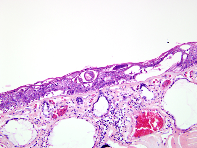
Histopathologic Description:
Morphologic Diagnosis:
Lab Results:
Condition:
Contributor Comment:
In general, the key histologic characteristics of aphasmid nematodes include a thin smooth external cuticle with hypodermal bacillary bands, unapparent musculature, an esophagus encased by a stichosome, and presence of a single uterus in females.(1) Additional specific microscopic features of Capillaria xenopodis/Pseudocapillaroides xenopi include sexual dimorphism (females are 4 times longer and 2 times wider than males), and the presence of unembryonated and embryonated eggs within the uterus (as opposed to presence of only unembryonated eggs in other aphasmid nematodes).(4)
Capillaria xenopodis/Pseudocapillaroides xenopi is the only reported nematode parasite of the epidermis of South African-clawed frogs, and is potentially a significant cause of morbidity and mortality in populations of South African-clawed frogs in the laboratory setting. Clinically, a small proportion of infected frogs in a laboratory animal population will display signs of Capillaria xenopodis/Pseudocapillaroides xenopi infection, which is often characterized as a nonspecific wasting syndrome (often lasting 3 to 4 months) that may include one or more of the following signs: lethargy, anorexia, skin color change, rough "flaky" skin, decreased egg production in females, and unexpected death. Often the first sign of this syndrome is the presence of large fragments/"flakes" of desquamated epithelium in the tank water.(3)
Histologically, Capillaria xenopodis/Pseudocapillaroides xenopi infection is characterized by profound epidermal hyperplasia (and variable associated epidermal/dermal inflammation) along with the presence of the nematodes and eggs in tunnels within the epidermis. This epidermal hyperplasia is thought to cause significant impairment of normal epidermal functions that result in overall debilitation and/or death of the animal. Specifically, the normal respiratory and metabolic functions (including gas exchange, waste removal, and osmotic balance) of the skin are compromised. Additionally, the normal physical barrier of the skin is compromised, often allowing for localized secondary skin infections and septicemia (including infection by Gram-negative bacteria, such as Aeromonas hydrophila, the cause of red leg).(3)
Diagnosis of infection can be made by direct examination of unstained skin sections (including the desquamated flakes in the tank water), and histopathology.Â
Capillaria xenopodis/Pseudocapillaroides xenopi has a direct lifecycle, with all stages of the lifecycle completed within the epidermis of the skin. Because of the direct lifecycle, autoinfection is also possible.(3) Levamisole has been described as an effective treatment for Capillaria xenopodis/Pseudocapillaroides xenopi infection in laboratory South African-clawed frogs.(2)
Other aphasmid parasites that primarily infect and cause hyperplasia of epidermal/epithelial surfaces include: Capillaria spp. (C. annulata, C. contorta, C. obsignata) in the crop and esophagus of birds; Capillaria philippinensis in the small intestines of humans; Capillaria bohmi in the nasal mucosa of canids; Trichosomoides crassicauda in the urinary bladder of rats; Anatrichosoma spp. of the skin and nasal mucosa of primates; and Gongylonema spp. in the nasal mucosa, oral mucosa, and esophagus of primates and cattle.Â
JPC Diagnosis:
Conference Comment:
Dr. Nichols discussed several aspects of amphibian skin during the conference, and it was noted that the skin of this animal was markedly hyperkeratotic and hyperplastic. Dr. Nichols opined that frog skin should not be more than a few cell layers thick with sparse to unnoticeable keratin. He also pointed out that frogs perform several physiologic processes with their skin to include respiration, thermoregulation, and electrolyte homeostasis. Due to this complex interaction with the external environment, it is easy to speculate that this type of skin lesion would be enough to cause mortality. Dr. Nichols also referenced the large serous glands containing eosinophilic secretory product in the dermis and the adjacent mucous glands containing clear to slightly amphophilic flocculent material. These glands combine to provide the slime cover of these aquatic amphibians.Â
Other aphasmids of note in domestic animals that the contributor did not mention include Trichuris sp., Trichinella sp., Dioctophyma sp., and Eustrongylides sp.(1)
References:
2. Iglauer F, et al: Anthelmintic treatment to eradicate cutaneous capillariasis in a colony of South African clawed frogs (Xenopus laevis). Lab Anim Sci 47:477-82, 1997
3. Reptiles & Amphibians, Fact Sheet, African Clawed Frog, National Zoo, http://nationalzoo.si.edu/Animals/ReptilesAmphibians/Facts/FactSheets/Africanclawedfrog.cfm Stephen LC, et al: Epidermal capillariasis in South African clawed frogs (Xenopus laevis). Lab Anim Sci 37:341-344, 1987