Signalment:
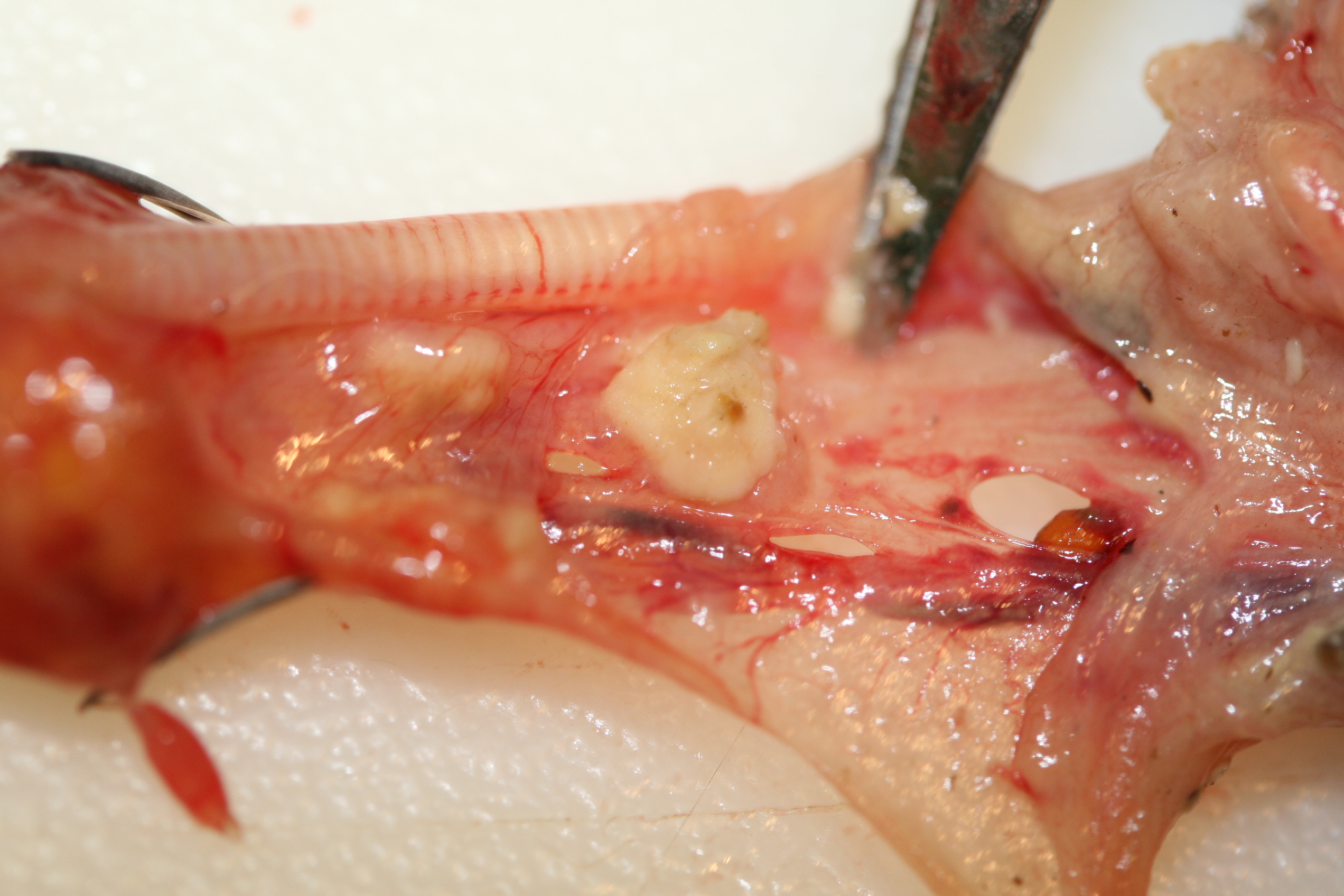
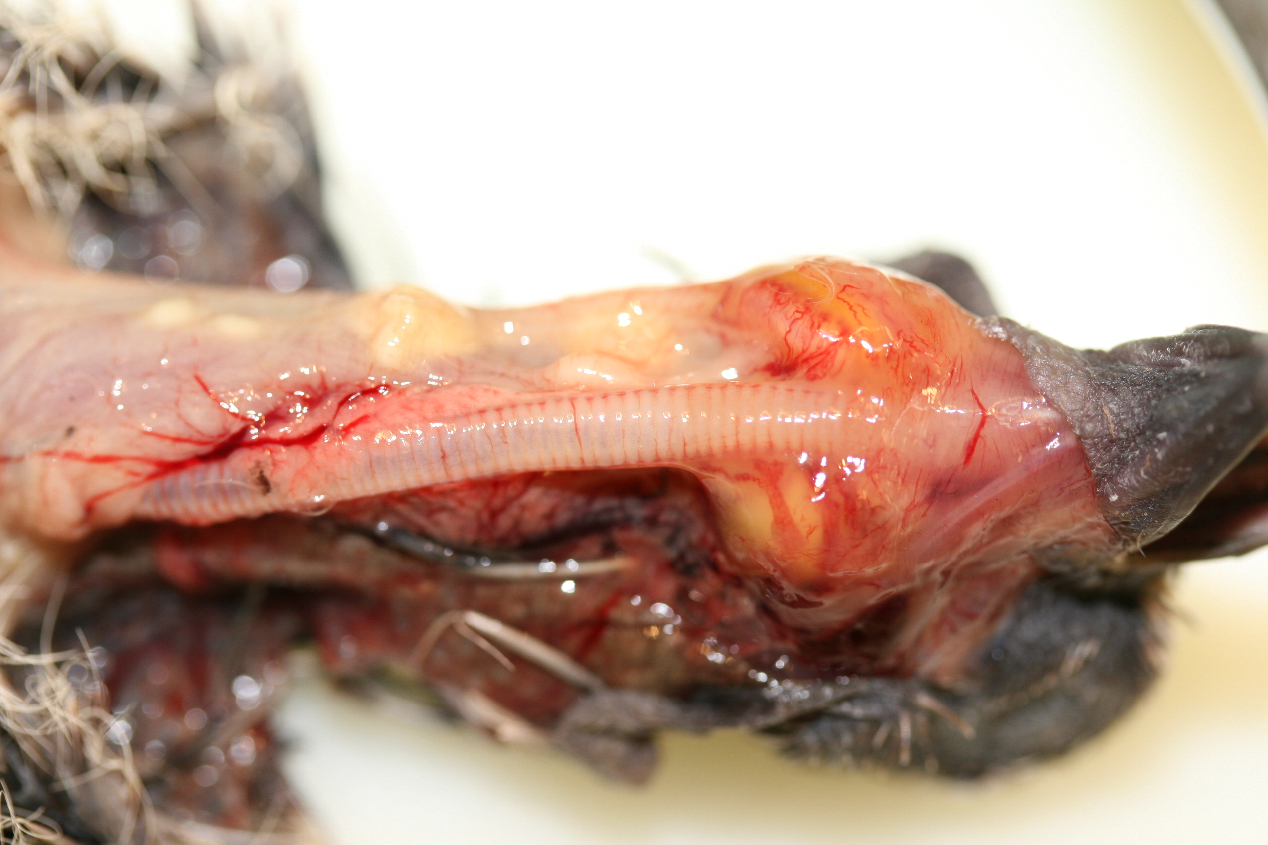
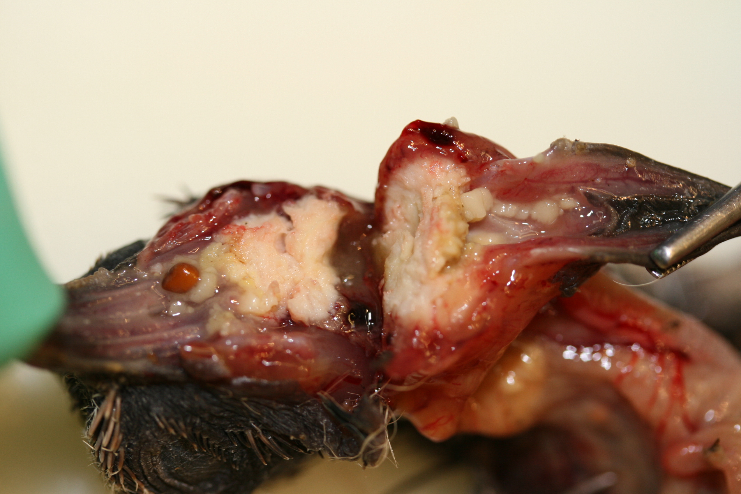
Gross Description:
Histopathologic Description:
ESOPHAGUS: In a focally extensive area, the esophageal epithelium is ulcerated and there is a transmural infiltration of inflammatory cells. Replacing the epithelium is a band of macrophages interspersed with 5 7 um diameter, ovoid to round protozoa, and a mat of bacteria at the luminal surface. Subjacent to this layer are many heterophils with few macrophages, necrotic debris and scattered protozoa. This inflammatory combination extends through the tunica muscularis to the serosa, which is expanded by heterophils and moderate amounts of mucinous material. The adjacent epithelium is moderately acanthotic with prominent intracellular edema. The serosa below the adjacent epithelium is expanded by a proliferation of capillaries with plump endothelial cells and diffusely the serosa is expanded by heterophils and mucinous material. Small clusters of macrophages, lymphocytes, and few plasma cells are present in the mucosal connective tissue of the remaining, intact esophageal mucosa.
Trachea: Essentially normal tissue for a juvenile bird with unmineralized tracheal rings.
Morphologic Diagnosis:
Condition:
Contributor Comment:
At necropsy, the flagellates may be seen on a wet mount from carcasses that are up to 24 hours old6. A cytologic flagellum stain or silver stains on histologic preps may also elucidate the organism.
JPC Diagnosis:
Conference Comment:
Ultrastructural features of Trichomonads include piriform to spherical shapes, four anterior flagella of unequal size, well-developed undulating membrane, an axostyle, and a pelta surrounding the periflagellar canal.8 The anterior flagellae emerge from the periflagellar canal which is reinforced by the pelta.8
Gross differentials for Trichomonas gallinae within the esophagus and crop include avian poxvirus, oral capillariasis, and candidiasis. Fibrinonecrotic lesions of wetpox, caused by avian poxvirus, are characterized by small white nodules to coalescing raised plaques with a diphtheritic membrane within the mouth, esophagus, trachea, pharynx, and larynx. Oral capillariasis produces almost identical gross lesions to that of T. gallinae and requires further testing to differentiate.2 Candida albicans causes gray-white pseudomembranous patches in the mouth, pharynx, esophagus, and, most frequently, the crop.7
References:
2. Heidenreich, M: Birds of Prey, Medicine and Management, pp. 131-132. Blackwell, Wissenschafts-Verlag GmbH, Berlin, 1995
3. Kocan RM, Herman CM: Trichomoniasis. In: Infectious and Parasitic Diseases of Wild Birds, eds. Davis JW, Anderson RC, Karstad L, Trainer DO, pp. 282-287. Iowa State University Press, Ames, IA, 1971
4. Lumeij JT: Gastroenterology, the esophagus and crop. In: Avian Medicine: Principles and Applications, eds. Ritchie BW, Harrison GJ, Harrison LR, pp. 491-497. Wingers Publishing Inc., Lake Worth, FL, 1994
5. Ostrand, WD, JA Bissonette, MR Conover: Trichomoniasis as a factor in mourning dove population decline in Fillmore, Utah. J Wildl Dis 31:87-89, 1995
6. Schulz, JH, AJ Bermudez, JJ Millspaugh: Monitoring presence and annual variation of trichomoniasis in mourning doves. Av Dis 49:387-389, 2005
7. Saif YM: Diseases of Poultry, 11th ed., pp. 896-898, 1006-1008, 1029-1030. Iowa State Press, Ames, Iowa, 2003
8. Tasca T, de Carli GA: Scanning electron microscopy study of Trichomonas gallinae. Vet Parasitol 118:37-42, 2003