Signalment:
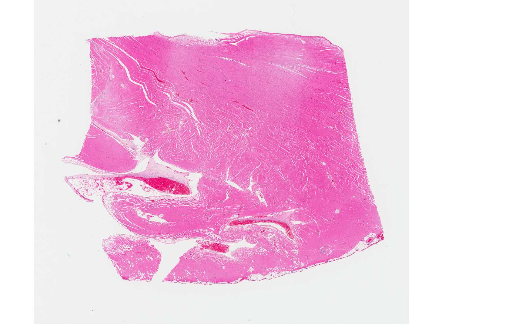
Gross Description:
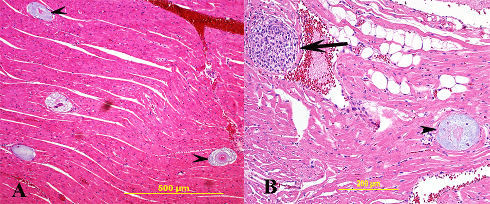
Histopathologic Description:
Morphologic Diagnosis:
Lab Results:
Condition:
Contributor Comment:
Hepatozoon americium causes American canine hepatozoonosis, which is a highly debilitating, tick-borne disease of dogs mainly in the south-central and south-eastern USA(4) although documented in other regions of the USA.(1) It is caused by Hepatozoon americium, a protozoan parasite, the definitive host of which is the tick Amblyomma maculatum.(3) In the United States, A. maculatum was traditionally endemic in states bordering the Gulf Coast and several states bordering the Atlantic coast including Georgia, Florida, and the southern portion of South Carolina. However, current data report establishment of the Gulf Coast tick in states farther inland including Oklahoma, Kansas, Arizona, Arkansas, Missouri, Indiana, Kentucky, and Tennessee and additional states along the Atlantic coast including Maryland, Virginia, and West Virginia.(1,4) Dogs get the disease by ingesting infected ticks.(3,4)
Clinically, infected dogs are often febrile, stiff, lethargic, and depressed. Wasting of body mass, marked in temporal muscles, and periosteal bone proliferation (hypertrophic osteopathy) are documented in dogs with chronic disease. Microscopically, trophozoite within macrophage-like cells in many tissues, mainly in striated muscles, apparently transforms the host cell into a mucopolysaccharide-producing entity that builds structures commonly called onion skin cysts. Parasite-containing cysts and lesions can be found in many tissues, but are consistently found in striated muscles.(4) Adipose and loose connective tissues are less commonly affected; rarely, other organs/ tissues such as lymph nodes, spleen, liver, and pancreas may be affected.(5,6) Mature meronts of a well-developed cyst of H. americium release merozoites, which incite local inflammation such as pyogranulomatous myositis, and are associated with a systemic reaction and overt illness.(4) Dogs may die due to secondary amyloidosis, glomerulonephritis(8) and associated other secondary lesions such as mineralization.
JPC Diagnosis:
Conference Comment:
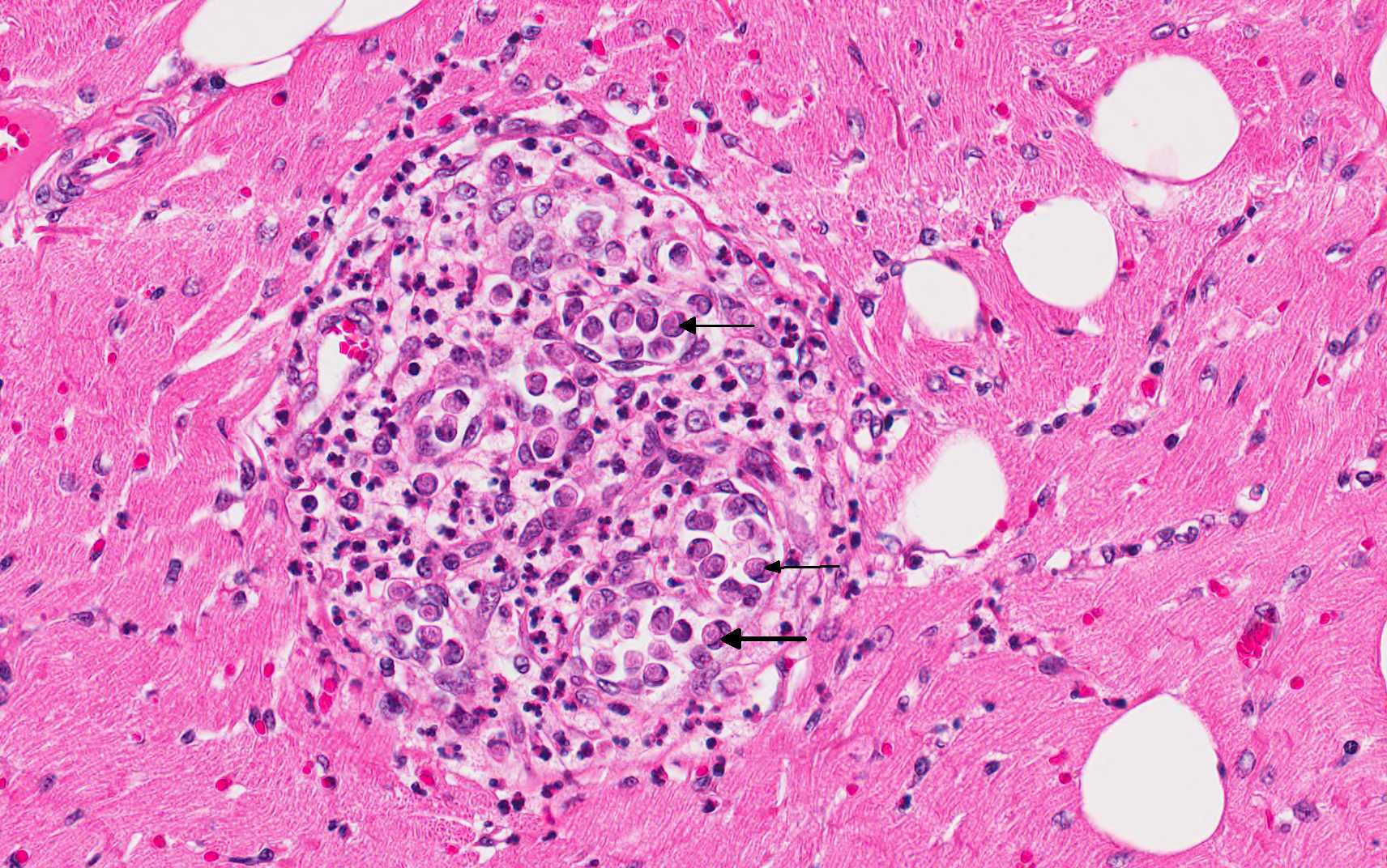
Merogony results in the production of merozoites that eventually give rise to gamonts that circulate in the blood where ticks can ingest them.(4) When merozoites are released from the cyst form, they incite a marked, localized, acute inflammatory response that eventually results in formation of a granuloma, but infected mononuclear cells are able to escape the granuloma.(3) The cysts are actually located between muscle fibers and contain a host cell, within which is the developing zoite stage. The host cell is surrounded by the lamellated structure composed of mucopolysaccharide in the onion skin cyst stage. Collagen fibers, fibroblasts and capillaries may be embedded within or closely associated with the lamellated cyst wall; but inflammatory cells are generally not associated with the large cysts. The developing parasites within the host cell, inside the cyst, bear ultrastructural features of developing apicomplexan trophozoites, although a parasitophorous vacuole is not observed. Both the merozoite asexual stage and the gamont sexual stage may be observed within different macrophages present in the granulomas which form after the tissue cysts rupture.(3)
As mentioned above, H. americium infection can result in periosteal bone proliferation. Most commonly, periosteal bone proliferation occurs in the diaphyseal regions of proximal limb bones but may also manifest in other locations such as the vertebrae. This finding is in contrast to hypertrophic osteopathy which occurs in the distal limb. Histologically, H. americanum-induced periosteal bone lesions are often symmetric, resemble hypertrophic osteopathy, and characterized by trabeculae of woven bone oriented perpendicular to the cortex. The lesions may be widespread and in locations unassociated with presence of the organism, suggesting a systemic effect of the infection as opposed to a local condition.(2) As compared above, H. americanum generally results in more severe disease than H. canis and is often fatal. Infection with H. americanum results in a profound neutrophilia and anemia, although the organisms may not be seen on a blood smear. In addition to muscle atrophy and bone pain, other clinico-pathologic findings in affected dogs include hyperglobulinemia, mucopurulent ocular discharge and uveitis. The disease may follow a waxing and waning course over time with periods of relapse correlating with release of merozoites and associated inflammation.(1,7)
Conference participants described the onion skin cysts as 200um in diameter, composed of lamellations of mucinous material, and separating cardiac myocytes. Small characteristic granulomas, present in some slides, were described as foci of macrophages containing tachyzoites which peripheralize the nucleus, with few neutrophils at the margin. A subset of slides also contain a small foci of fibrosis which is populated with low numbers of hemosiderin laden macrophages, lymphocytes and plasma cells. Multifocal areas of hemorrhage are also present. Rare foci of mineralization and myofiber degeneration are present in some sections.
References:
2. Craig LE, Dittmer KE, Thompson KG. In: Maxie MG, ed. Jubb, Kennedy, and Palmer's Pathology of Domestic Animals. 6th ed. Vol 1. St. Louis, MO: Elsevier; 2016:94.
3. Cummings CA, Panciera RJ, Kocan KM, Mathew JS, Ewing, SA. Characterization of Stages of Hepatozoon americanum and of Parasitized Canine Host Cells. Vet Pathol. 2005; 42:788-796.
4. Ewing SAE, Panciera RJ. American Canine Hepatozoonosis. Clin Microbiol Rev. 2003;16: 688697.
5. Panciera RJ, Ewing SA, Cummings CA, Kocan AA, Breshears MA, Fox JC. Observations on tissue stages of Hepatozoon americanum in 19 naturally infected dogs. Vet Parasitol. 1998; 78:265276.
6. Potter TH, Macintire, DK. Hepatozoon americium: an emerging disease in the south-central/southeastern United States. J Vet Emer Critical Care. 2010; 20: 70-76.
7. Valli VEO, Kiupel M, Bienzle D. Hematopoietic system. In: Maxie MG, ed. Jubb, Kennedy, and Palmer's Pathology of Domestic Animals. 6th ed. Vol 3. St. Louis, MO: Elsevier; 2016:109-111.
8. Van Vleet JF, Valentine BA. Muscle and tendons. Hepatozoonosis. In Maxie MG, ed. Jubb, Kennedy and Palmers pathology of domestic animals. 5th ed. Vol 1. Elsevier Saunders; 2007: 270-271.