Joint Pathology Center
Veterinary Pathology Services
Wednesday Slide Conference
2019-2020
Conference 14
15 January 2020
Dr.
Neel Aziz
Supervising Veterinary Pathologist
Smithsonian Conservation Biology Institute
National Zoological Park
Washington DC, 20008
CASE I: 18-4856 2 (JPC 4116557).
Signalment: Western Pond Turtle, female adult.
History: The turtle was found on a road in Waterloo campground in Lebanon, Oregon. Eyes were swollen bilaterally. Fed ensure and cat foot but ate very little. Treated with eye ointment and baytril.
Gross Pathology: A 468g male Western Pond Turtle (Actinemys marmorata) is necropsied on 10/6/2017 in fair body condition and mild post mortem autolysis. Eyelids are markedly swollen bilaterally with thick, light yellow, caseous material present under the lids. Globes are not appreciated grossly. Approximately 30% of the total lung parenchyma is red and firm and thick, light yellow, caseous material oozes out on cut surfaces.
Laboratory results: N/A.
Microscopic Description:
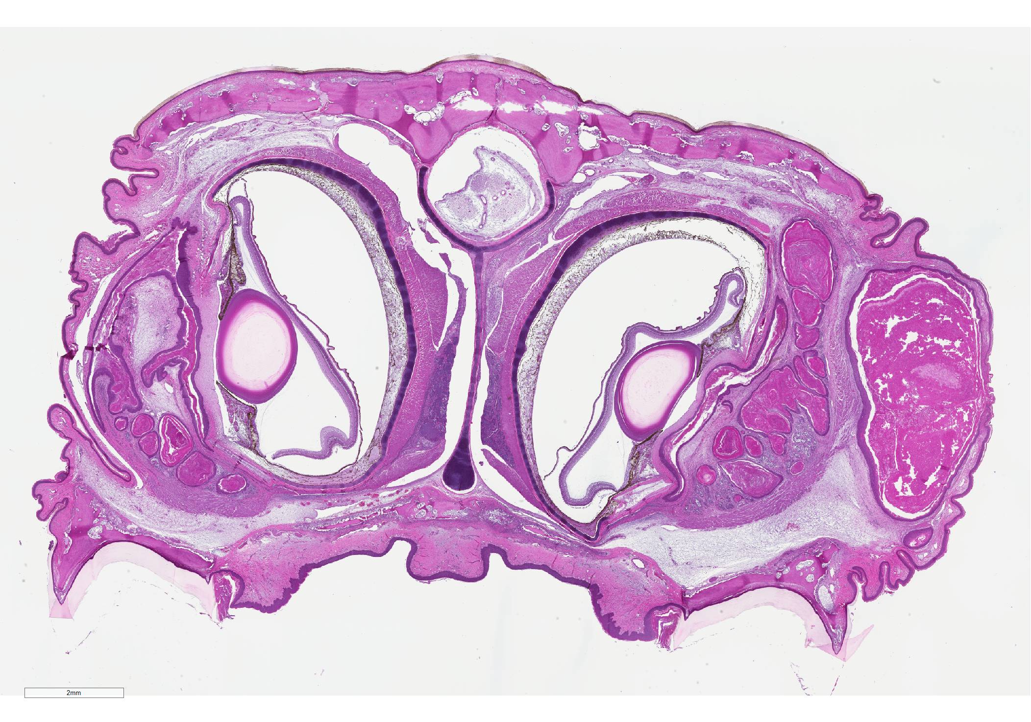
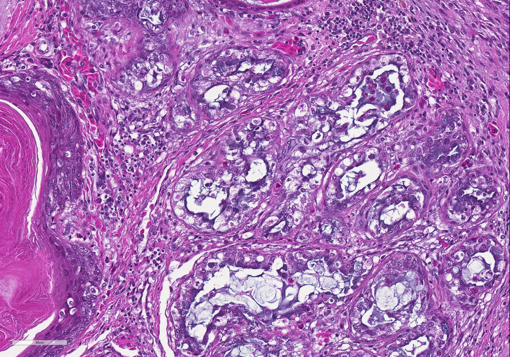
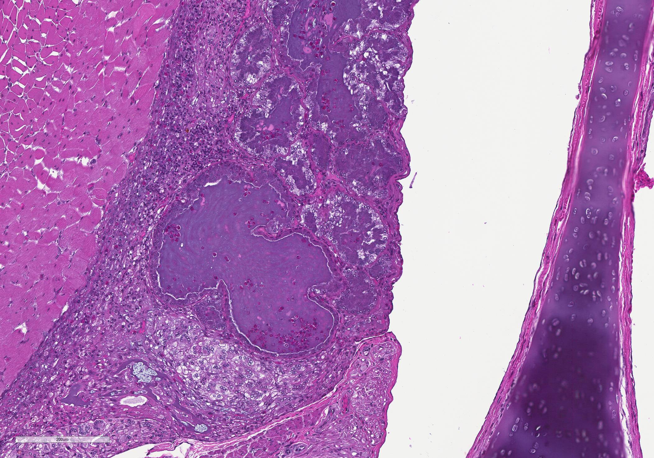
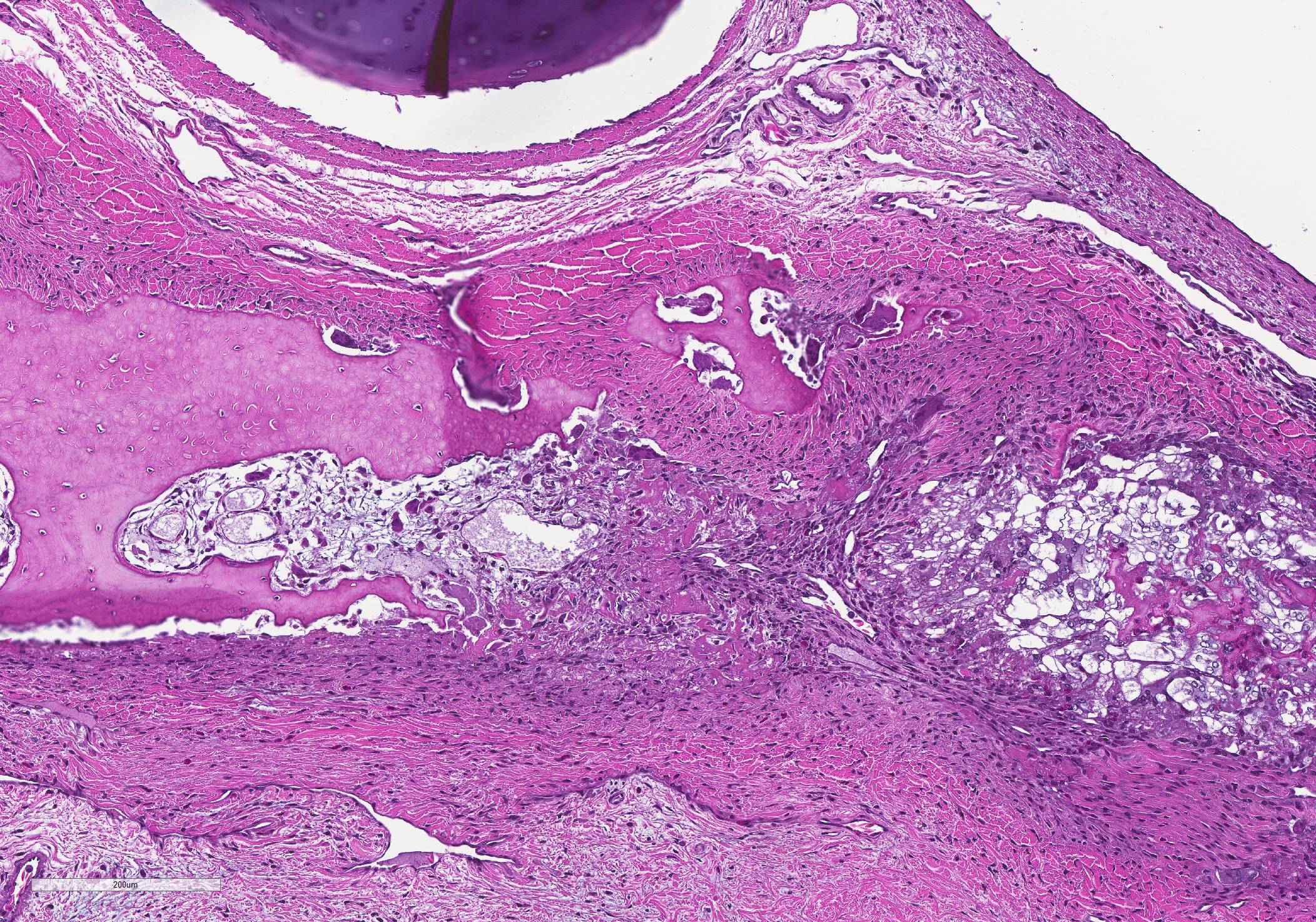
Cross sections of the skull, brain, and eyes. There is hyperplasia and hyperkeratosis of the eyelids and external ear canal. There is marked squamous metaplasia and layered keratin accumulation within the nasal vestibule, more prominent on one side, with dilation of some sinus mucous glands containing moderate numbers of heterophils. In one of these heterophil-rich foci there is rimming layer of epithelioid macrophages and multinucleate giant cells with small numbers of bacteria visible in the exudate. Other glands in the region (ophthalmic or sinus mucous glands) multifocally show moderate to severe squamous metaplasia and occasional keratin accumulation. There are multifocal, asymmetric areas of marked osteoclast activity.
Contributor Morphologic Diagnosis:
Whole body: Emaciation
Multiple organs: Squamous metaplasia consistent with hypovitaminosis A;
Sinuses: Severe diffuse heterophilic rhinitis with squamous metaplasia and keratin accumulation
Other lesions:
Lungs: severe chronic active heterophilic and proliferative bronchointerstitial pneumonia with bacteria
Eyelids/eye: Hyperplastic keratoconjunctivitis
Ears: Bilateral severe hyperplastic otitis media
Contributor Comment: Histopathology is consistent with hypovitaminosis A; squamous metaplasia with keratinization was observed in multiple tissues throughout the body. Other disease processes may be occurring simultaneously/secondarily, (opportunistic bacteria) but the most severe lesions were due to the vitamin deficiency.
The cause of the increased bone resorption/osteoclast activity is unknown. As the change is not generalized, a Vitamin D deficiency/hypocalcemia is unlikely. In addition, clinical reports of hypovitaminosis D in turtles mention paradoxical mineralization of soft tissues (including muscle, renal tubules, etc) and such changes are lacking here. In this case, osteoclasis often occurred near areas of squamous metaplasia, so perhaps local tissue swelling/pressure was the force for osseous remodeling.
Contributing Institution:
Oregon Veterinary Diagnostic Laboratory, Carlson College of Veterinary Medicine, Oregon State University, Corvallis, Oregon 97331. https://vetmed.oregonstate.edu/diagnostic
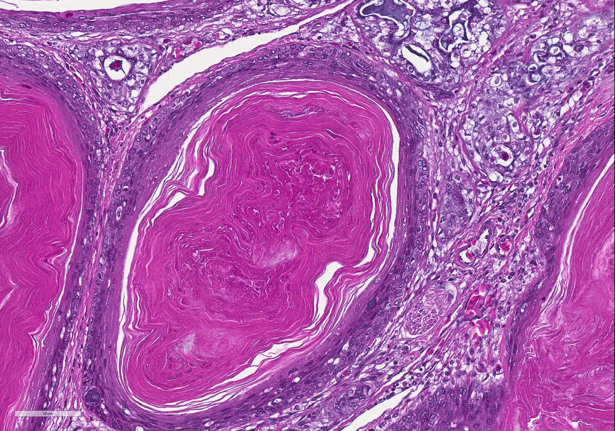
JPC Diagnosis: 1. Lacrimal glands and ducts: Squamous metaplasia, diffuse, severe, with marked hyperkeratosis, diffuse lacrimal gland degeneration, and conjunctival keratin aggregation.
2. Nasal vestibule: Adenitis, heterophilic, unilateral,
diffuse, moderate.
3. Skull, vomer and frontal bone: Osteopenia, diffuse, severe.
JPC
Comment: This
slide is an excellent example of squamous metaplasia seen in the lacrimal
glands in chelonians as a result of Viamin A deficiency.
The 1967 paper in Pathologia Veterinaria ( the forerunner of Veterinary
Pathology) by Elkan and Zwart is an outstanding reference on the systemic
effects of Vitamin A deficiency in the "young terrapin", as well as a tremendous
example of scientific prose, the like of which we do not see today in modern
publications.3 While a quotation of the entire first page is poor
form (and the reader can download it online), a short snippet is illustrative
of how far current medical writing has fallen - "...In vain do the textbooks on
herpetology mention the exacting dietary requirements of juvenile terrapins, in
vain their advice on how to meet these requirements...Neither the sellers nor the
buyers read these books, presuming that a plastic bowl and some 'ant's eggs'
suffice to keep a juvenile terrapin alive. The annual holocaust among the
imported terrapins proves the error of this assumption."3
As mentioned by the contributor, Vitamin A deficiency in chelonians is a systemic process, rather than just one confined to the structures of the orbit.3 Squamous metaplasia also occurs in the external ear, where it has been associated with aural "abscessation" in a number of chelonian species in association with a mixed flora of gram-negative bacilli, presumably ascending from the Eustachian tube.1,7 In addition, squamous metaplasia accompanied by excessive keratinzation occurs in other organs as well. Pancreatic ducts are thickened, hyperkeratotic, and ensheathed in heterophils. In the kidney, collecting ducts are distended and occluded by keratin debris, often resultin in severe nephritis. Blockage by keratin debris may also be seen in the ureters and urinary bladder. Degenerative and inflammatory changes were noted by Elkham in the thyroid and liver, but squamous metaplasia was not documented in these animals.3
Some participants
had difficulty in orienting themselves on the submitted section, and there was
considerable variation between sections (necessitated by the submission
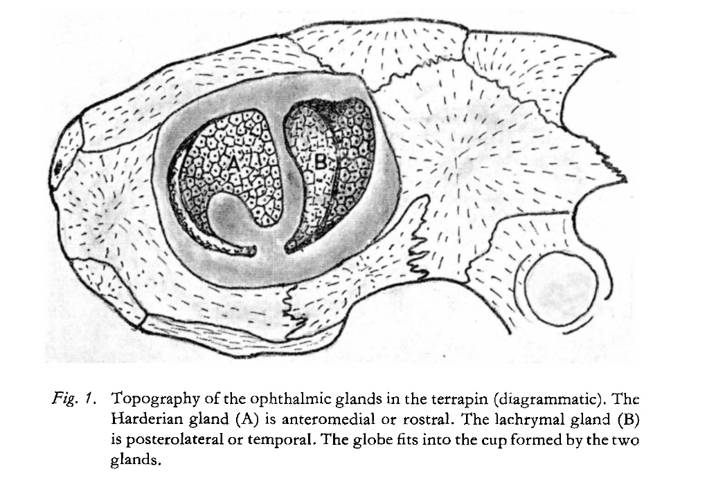
requirement of 165 sections.) As illustrated by Elkham and Zwart, the
chelonian globe fits into the cup formed by the two glands, the anteromedial
Harderian gland, and the posterolateral lacrimal gland.3 It is
preseumed that the lacrimal gland is present in the submitted sections due to
its position lateral to the globe. The ear canal is not present on these
sections. Moreover, at this stage of lesion development, the participants had
great difficulty determining which of the large keratin filled structures might
be derived originally from glands or ducts. Isolated mucus cells were present
within the walls of several dilated keratin-filled structures, which would not
be consistent from ductal derivation, but at this level of change, it is
difficult and likely unnecessary to be certain.
The effects of Vitamin A deficiency have been documented in a wide range of
poikilothermic and homothermic species. In crocodiles, squamous metaplasia
result in similar changes in the conjunctiva and kidenys, as well as nodular distentions
of the ostia of the lingual glands.2 In captive anurans, squamous
metaplasia may be seen in mucus glands of the skin, tongue, oral mucosa,
esophagus, cloaca, renal tubules, oviduct, and bladder.5 Periocular
gland squamous metaplasia has been seen in penguins.8 Squamous
metaplasia may be seen in other species of birds, including psittacines in a
wide range of tissues including glands of the oral cavity, esophagus, salivary
gland and respiratory tract. Protruding keratin masses from esophageal glands
are classsic lesions associated with Vitamin A deficiency in poultry. Squamous
metaplasia of Harderian glands, although not to the level seen in chelonians,
has also been seen in laboratory rodents as well.3 In suckling
calves, it has been incriminated in a sider range of ocular abnormalitis to
include cataract formation, lens luxation, micropthalmia, and reduction in the
size of the optic nerve.9
While it might be thought that in present days, Vitamin A deficiency and related opthalmic disease would be a rarity, the human species has found new ways to impair its ability to take in this important dietary ingredient. Starvation still rules in many parts of the world, but in developed nations, hypovitaminosis A is still seen, as a result of restrictive and monotonous diets (including vegan diets, ?cafeteria? and ?junk food? diets, and eating disorders.4 Malabsorptive syndromes, to include bariatric surgery also accounts for a number of cases each year. While signs of deficiency are system wide, well-documented ocular diseases associated with Vitamin A in humans include night blindness, xeropthalmia (?dry eye?) Bitot?s spot (a buildup of kerati8n located superficially in the conjunctiva associated with corneal drying), keratitis and keratomalacia.4
Several attending the conference suggested that bacterial infection or chronic irritation should also be considered in the potential differential diagnosis for this particular lesion in light of the lack of information on husbandry of this particular animal. Dr. Andrew Cartoceti of the National Zoo mentioned that samples for Vitamin A should be packaged in a brown container due to the susceptibility of the compound to sunlight. The bony resorption of the bones of the skull was attributed by the group to protein and caloric malnutrition.
References:
1. Brown JD, Richards JM, Robertson J, Holladay S, Sleeman JM. Pathology of aural abscesses in free-living Eastern box turtles. J Wildl Dis 2004; 40(4): 704-712.
2. Conley JK, Shilton CM. Crocodilia. In: Pathology of Wildlife and Zoo Animals Terio KA, McAloose D, St Leger J eds., 2019; London: Associated Press, p. 849
3. Elkan E, Zwart P. The ocular disease of young terrapins caused by Vitamin A deficiency. Pahol Vet 1967; 4:201-222.
4. Faustino JF, Rieiro-Silva A, Dalto RF, de Souza MM, Furtado JMF, de Melo Rocha G, Alves M, Rocha EM. Vitamin A and the eye: an old tale for modern times. Arch Brazil Opthamol 2016; 79(1):56-61.
5. Pessier, A. Amphibia. In: Pathology of Wildlife and Zoo Animals Terio KA, McAloose D, St Leger J eds., 2019; London: Associated Press, p. 921-922.
6. Reavill DR, Dorrestein G. Psittacines, Coliiformes, Musophagiformes, Cuculiformes. In: Pathology of Wildlife and Zoo Animals Terio KA, McAloose D, St Leger J eds., 2019; London: Associated Press, p. 770
7. Rodriguez CE, Duque AMH, Steinberg J, Woodburn DB. Chelonia. In: Pathology of Wildlife and Zoo Animals Terio KA, McAloose D, St Leger J eds., 2019; London: Associated Press, p. 831-832.
8. Stidworthy MF, Denk D. Sphisciformes, Gaviiformes, Podicipediformes, Procellariformes, and Pelecaniformes. In: Pathology of Wildlife and Zoo Animals Terio KA, McAloose D, St Leger J eds., 2019; London: Associated Press, p. 651.
9. No authors listed. Range of ocular deformities in calves due to hypovitaminosis A. Vet Record 2014; 174(10)244-247.