Conference #25
CASE II:
Signalment:
Juvenile female striped skunk (Mephitis mephitis)
History:
none
Gross Pathology:
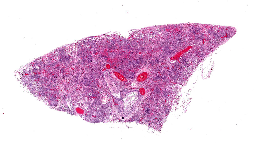
The skunk was in good body condition with normal fat stores, and in good post-mortem condition. There were small numbers of fleas in the fur. There was a small amount of cloudy pink nasal discharge bilaterally. The cranioventral lung lobes were bilaterally dark red, wet, heavy, and firm. The right cranial lung lobe was mildly enlarged. All lung lobes contained small numbers of slender white worms up to 2cm in length. There were 16 ascarids in the gastrointestinal tract, 10 of which were in the stomach.
Laboratory Results:
Aerobic culture of the lung yielded a heavy growth of a Pseudomonas bacteria. Another colony type grew as well, but could not be separated from the heavy Pseudomonas growth.
Microscopic Description:
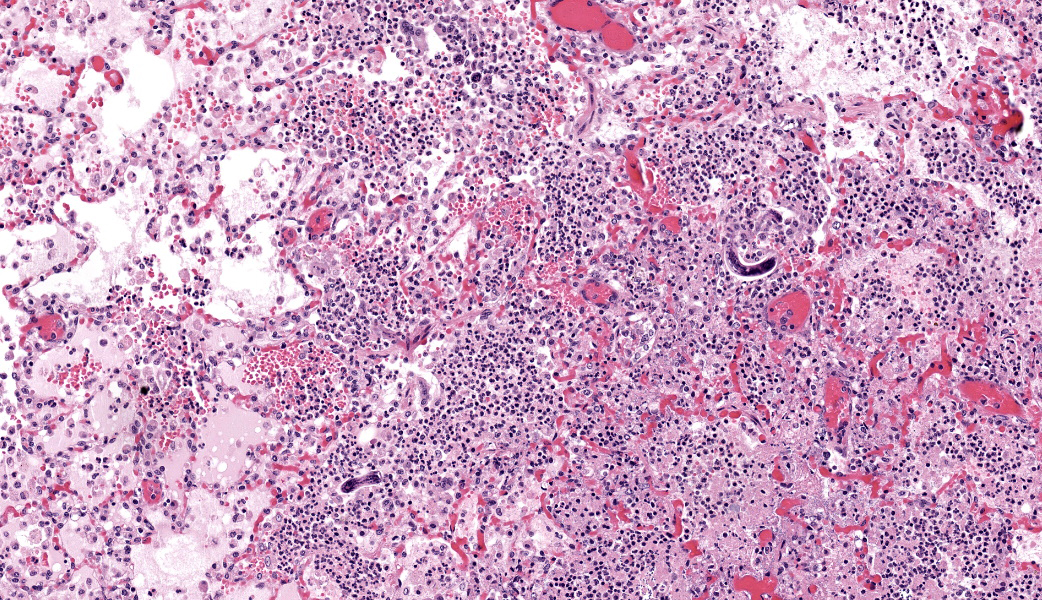
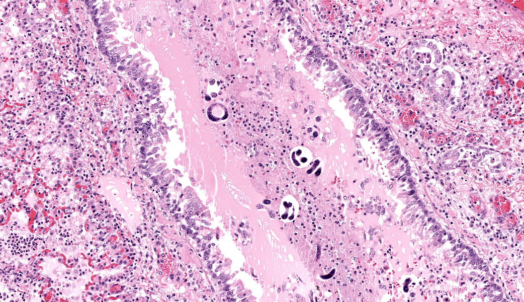
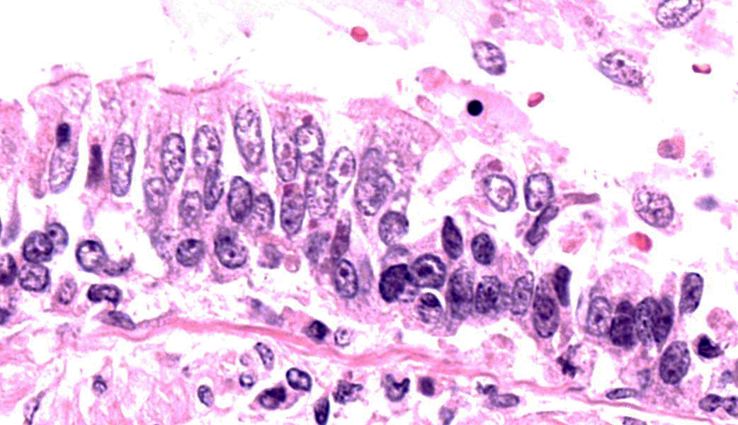
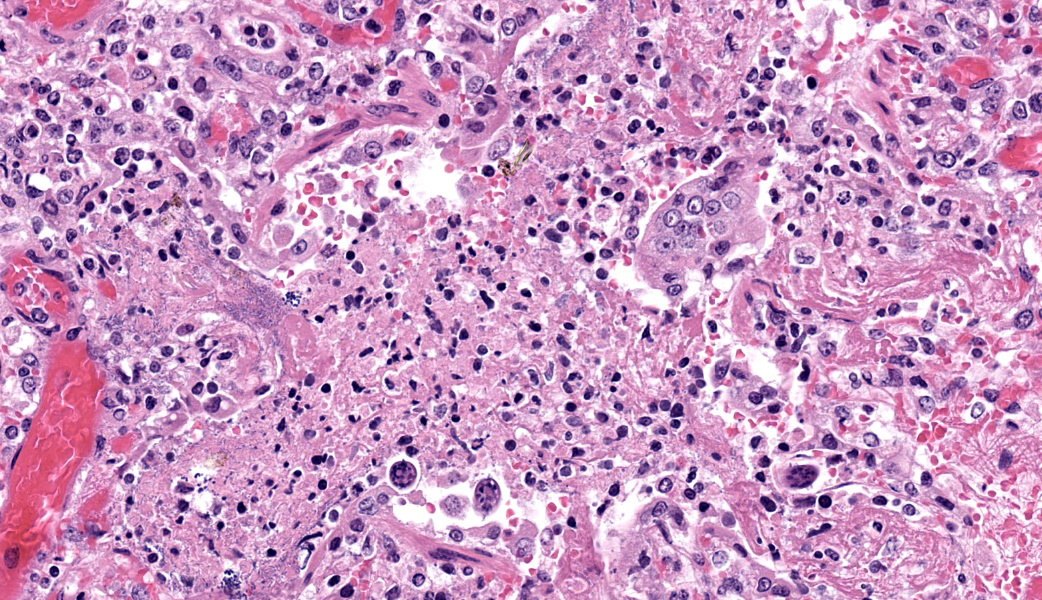
Pulmonary architecture is diffusely mildly to severely distorted by necrosis and inflammatory infiltrates. Bronchiolar submucosa is markedly expanded by dense infiltrates of neutrophils and macrophages, which often infiltrate surrounding alveoli. Diffusely, alveolar septa are undergoing fibrinoid necrosis, and protein-rich edema, sometimes with fibrin strands, fills alveoli. The walls of small arteries are infiltrated by neutrophils and macrophages and segments are undergoing fibrinoid necrosis. There are mild to severe hemorrhages scattered throughout the lung. There are large numbers of slender rod-type bacteria throughout most of the examined section, occasionally forming dense colonies that line vessels, alveolar walls, or bronchial and bronchiolar epithelium. There are smaller numbers of coccoid bacteria arranged in doublets or clusters, with small colonies generally found in close proximity to each other. Where bronchial and bronchiolar epithelium is present, there are moderate numbers of eosinophilic intracytoplasmic and rarely intranuclear inclusion bodies ranging from 2-6um diameter. Segmental bronchi are surrounded by marked interstitial edema, and there is mild to marked pleural edema and hemorrhage. Some sections have moderate numbers of larval nematodes which are 12-20um in diameter and up to 100um long, and lack a body cavity. These parasites have a slender serrated cuticle 1-2um thick and lateral alae. They are rarely abutted by multinucleated giant cells, and there are occasional elongate hyperchromatic basophilic structures suggestive of degenerating nematodes.
Contributor's Morphologic Diagnosis:
Lung: Pneumonia, bronchointerstitial, necrotizing, acute, diffuse, severe, with growth of multiple bacterial species, and eosinophilic intracytoplasmic and intranuclear viral inclusions in epithelia, etiology consistent with canine distemper virus; and regional pulmonary larval nematodiasis
Contributor's Comment:
Canine distemper virus (CDV) is an enveloped, negative sense, single-stranded RNA virus in the family Paramyxoviridae, genus Morbillivirus.3 The virus causes disease in both wild and domestic canids, as well as many other carnivores including seals, raccoons, weasels and the domestic ferret, skunks, and large cats such as lions and leopards; occasional outbreaks in populations of endangered large cats can have devastating population-level effect.1,5,7,8,11,18 Dogs and raccoons are the most common reservoir populations. Other closely related viruses include phocine distemper virus and the morbilliviruses that cause disease in cetaceans, measles virus, Hendra virus that infects horses and humans, and peste des petits ruminants viruses.3 Rinderpest, another morbillivirus that causes severe to fatal disease in cattle, was eradicated in 2011 with the help of vaccines.20
Initial replication of CDV occurs in tissue macrophages. The virus then spreads to tonsils and bronchial lymph nodes, spleen, lymphoid follicles in the lamina propria of the stomach and small intestines, other lymph nodes, and Kupffer cells; fever and leukopenia ensue. The virus is spread hematogenously to epithelia and to the central nervous system; once the virus begins replicating in epithelia, shedding ensues. Virus is most abundant in respiratory secretions and is commonly spread by droplets, but it can also be isolated from urine, and can be spread transplacentally. Shedding ceases once the immune system controls the virus, but can persist in some animals up to 90 days. Approximately 25-75% of dogs infected with CDV will have a strong humoral and cell-mediated immune response. Their signs may be entirely subclinical or limited to a brief fever, and the virus is cleared within two weeks of infection. Where dogs fail to develop a strong immune response, initial clinical signs are conjunctivitis and fever, followed by vomiting and diarrhea, anorexia, cough, increased respiratory effort, and secondary infections.3 Some patients may then resolve the CDV infection, while others develop neurologic signs, which are almost always progressive. Neurologic signs include ataxia, tremors, myoclonus, seizures, coma, and death.2
Canine distemper virulence is associated with immunosuppression and neurotropism. Viral H protein binds to CD150 (signaling lymphocyte activation molecule), which is found on dendritic cells and activated monocytes, as well as both B- and T-cells. Early infection in monocytes and dendritic cells may interfere with normal antigen presentation, and upregulation of CD150 in infected animals may further enhance spread of the virus amongst lymphocytes.14,17 CD4 (+) lymphocytes are first infected and lysed6, and viral depletion combined with lack of cytokine stimuli from CD4 (+) cells combine to markedly reduce B-cells as well. Severe immunosuppression predisposes infected animals to secondary viral and bacterial infections, as with the bacterial pneumonia seen in this skunk.2
One to three weeks after recovery from systemic illness, some dogs develop neurologic disease. Virus is spread both anterograde up the axon and via circulating lymphocytes. Once in the brain, replication in the ependymal and choroid plexus epithelium allows virus to circulate in the CSF.16 Acute encephalitis is a result of direct viral replication and injury in the central nervous system, with minimal inflammation. Virus is first detected in astrocyte foot processes and then neurons, and occasionally neuronal necrosis can lead to foci of polioencephalomalacia.13 Historically, CDV infection has been thought of as a demyelinating disease. Though oligodendrocytes are seldom infected, infection of surrounding astrocytes and microglia are thought to lead to oligodendrocyte dysfunction. However, other studies have demonstrated axonopath prior to demyelination.12 Whether axonopathy is due primarily to neuronal infection or to bystander damage from infected astrocytes and microglia is uncertain. In patients that survive distemper virus infection, with or without associated neural signs, a replication-deficient form of the virus may persist in the brain and can re-emerge years later to cause neural signs in a condition termed "Old Dog Encephalitis".3
Suspicion for canine distemper virus infection is based on noting clinical signs in a susceptible animal. Antemortem testing includes serologic assays and PCR, and observation of distemper inclusions in circulating erythrocytes. Common findings on histology of tissues collected post-mortem are atrophy of lymph nodes, splenic white pulp, and other lymphoid organs; and during the systemic phase of disease, secondary bacterial infection may be present in the lungs and gastrointestinal tract. Lesions in the brain are usually leukoencephalomalacia with marked spongiosis. Mononuclear infiltrate is variable; there may be digestion chambers and large numbers of gitter cells. Caracteristic eosinophilic cytoplasmic viral inclusions can be found in lungs, brain, adrenal gland, gastric mucosa, and urinary bladder to varying degrees in any infected carnivore.4 Nuclear inclusions are more common in neurons. If inclusions are not obvious, immunohistochemistry for the virus is elucidative.3
At necropsy, this skunk had severe cranioventral pneumonia. Lung submitted for aerobic culture grew a Pseudomonas; this bacterium may have inhibited growth of other organisms seen histologically. Bacterial infection was secondary to severe canine distemper virus infection. Inclusions are readily apparent in the lung, but were confirmed with immunohistochemistry. The skunk tested negative for rabies on DFA, and distemper inclusions were absent from the brain: as the skunk was still in the systemic phase of disease, intracranial infection may have been yet to be established. Regional infection of the lung by larval nematodes was an incidental finding; these may be Filaroides milksi or Crenosoma mephiditis.
Contributing Institution:
University of Connecticut Department of Pathobiology and Veterinary Sciencehttp://www.patho.uconn.edu/
JPC Morphologic Diagnosis:
Lung: Pneumonia, bronchointerstitial, necrotizing and neutrophilic, subacute, diffuse, marked, with bronchiolar epithelial hyperplasia, viral syncytia, intraepithelial and intrahistiocytic intracytoplasmic and intranuclear viral inclusions, larval nematodes, and mixed bacteria.
JPC Comment:
What an excellent write-up by this contributor! Dr. Holder set the stage for this case with a quick skunk taxonomy review. Their scientific name, Mephitis mephitis, basically translates to "stinky stinky." As if that wasn't enough, their family name, Mephitidae, also means "stinky." Taxonomists really wanted to make sure no one missed the point there. With that poignantly olfactory context established, discussion advanced.
The lung sections showcased classic morbillivirus pathology, including syncytia in both epithelial cells and macrophages. However, because this skunk also had a secondary bacterial pneumonia, participants found themselves squinting at multinucleated cells wondering, "Is this a viral syncytial cell or just an enthusiastic giant cell macrophage"? Dr. Holder pointed out that using viral inclusions when visible within multinucleated cells can help tip the pathologist one way or the other, but she encouraged participants to accept that, without IHCs, not every multinucleated Joe on a slide is going to confess to its villain origin story.
One of the most interesting features in this case was the prominence of pulmonary megakaryocytes, which appear as large, basophilic, "ink blot"' cells lounging around in the septal capillaries like they own the place. One participant pointed out that the lungs have their own megakaryocytes separate from those in the bone marrow and are a major site of platelet production in many mammalian species. Pulmonary megakaryocytes have important immune functions, as well. In humans, pulmonary megakaryocytes can become significantly more noticeable in severe disease states, and, in SARS-CoV-2 infections, they're considered a poor prognostic indicator.15
Regarding the bacterial infection that set up shop after morbillivirus did its damage and immunosuppressed this skunk, there were two distinct bacterial morphologies within areas of necrotic airways. Because tissue preservation was good in this case, these organisms were interpreted as true pathogens rather than postmortem freeloaders.
The group briefly discussed the nematodes present in the lung. Some larvae had lateral alae, prompting speculation about Baylisascaris columnaris, but the overall appearance favored metastrongyles as discussed by the contributor. Dr. Holder summed it up with characteristic flair by proclaiming, "A wild animal without parasites is a stadium without fans."
Dr. Holder shared that she is convinced if we ever get a zombie apocalypse, it's going to be due to a morbillivirus. She wrapped up this case discussion with morbillivirus immunopathology, including lymphotropism, immunosuppression, and "immune amnesia." Morbilliviruses target lymphocytes via CD150 (SLAM). When a dendritic cell gets infected and traffics to regional lymph nodes, it can transmit virus to memory T cells, which are subsequently destroyed. Memory B cells are also depleted, and morbilliviruses can wipe out up to 70% of preexisting immune memory cells.10 The result is a long-lasting vulnerability to other infections and reduced vaccine effectiveness.10 Bummer
References:
- Appel MJ, Yates RA, Foley GL, Bernstein JJ, Santinelli S, Spielman LH, Miller LD, Arp LH, Anderson M, Barr M, Pearce-Kelling S, Summers BA. Canine distemper virus epizootic in lions, tigers, and leopards in North America. J. Vet. Diagn. Invest. 1994; 6(3): 277-288
- Beineke, A, Puff, C, Seehuesen, F, Baumgärtner. Pathogenesis and immunopathology of systemic and nervous canine distemper. Vet Immunol Immunopathol. 2009; 127(1-2): 1-18.
- Greene, CE and Appel, MJ. Infectious Diseases of the Dog and Cat. 3rd Ed. Philadelphia, PA: Elsevier; 2006: 25-4
- Greene, RG. A comparative study of distemper inclusions. Am J Epidemiol. 1939; 2: 73-87
- Habermann, RT, Herman, CM, Williams Jr., FP. Distemper in raccoons and foxes suspected of having rabies. JAVMA. 1958; 132(1): 31-35.
- Iwatsuki, K, Okita, M, Ochikubo, F, Gemma, T, Shin, YS, Miyashita, N, Mikami, T, Kai, C. Immunohistochemical analysis of the lymphoid organs of dogs naturally infected with canine distemper virus. J. Comp. Pathol. 1995; 113(2): 185-190
- Kennedy, S, Kuiken, T, Jepson, PD, Deaville, R, Forsyth, M, Barrett, T, van de Bildt, MWG, Osterhaus, ADME, Eybatov, T, Duck, C, Kydyrmanov, A, Mitrofanov, I, Wilson, S. Mass die-off of Caspian seals caused by canine distemper virus. Emerg. Infect. Dis. 2000; 6(6): 637-639.
- Kilham, L, Habermann, RT, Herman, CM. Jaundice and bilirubinemia as manifestation of canine distemper in raccoons and ferrets. Am J Vet Res. 1956; 17(62): 144-148.
- Lemp, C, Spitzbarth, I, Puff, C, Cana, A, Kegler, K, Techangamsuwan, S, Baumgärtner, W, Seehusen F. New aspects of the pathogenesis of canine distemper leukoencephalitis. Virus. 2014; 6(7): 2571-2601.
- Morales GB, Munoz MA. Immune amnesia induced by measles and its effects on concurrent epidemics. J R Soc Interface. 2021;18(179):20210153.
- Roelke-Parker ME, Munson L, Packer C, Kock R, Cleaveland S, Carpenter M, O'Brien SJ, Pospischil A, Hofmann-Lehmann R, Lutz H, Mwamengele GL, Mgasa MN, Machange GA, Summers BA, Appel MJ. A canine distemper virus epidemic in Serengeti lions (Panthera leo). Nature. 1996; 379:441-445.
- Seehusen, F, Baumgärtner, W. Axonal Pathology and Loss Precede Demyelination and Accompany Chronic Lesions in a Spontaneously Occurring Animal Model of Multiple Sclerosis Brain Pathol. 2010; 20(3): 551-559
- Summers, BA, Greisen, HA, Appel, MJG. Early events in canine distemper encephalomyelitis. Acta Neuropathol.1979; 46(1-2): 1-10.
- Tatsuo, H, Ono, N, Yanagi, Y. Morbilliviruses use signaling lymphocyte activation molecules (CD150) as cellular receptors. J. Virol. 2001; 75(13): 5842-5850.
- Vendevelde, M, Zurbriggen, A, Higgins, RJ, Palmer, D. Spread and distribution of viral antigen in nervous canine distemper. Acta Neuropathol. 1985; 67(3-4): 211-218.
- Wenzlow, N, Plattet, P, Wittek, R, Zubriggen, A, Grone, A. Immunohistochemical demonstration of the putative canine distemper virus receptor CD150 in dogs with and without distemper. Vet Pathol. 2007; 44(6): 943-948.
- Valdivia-Mazeyra MF, Salas C, Nieves-Alonso JM, et al. Increased number of pulmonary megakaryocytes in COVID-19 patients with diffuse alveolar damage: an autopsy study with clinical correlation and review of the literature. Virchows Arch. 2021;478(3):487-496
- Woolf, A, Gremillion-Smith, C, Evans, RH. Evidence of canine distemper virus infection in skunks negative for antibody against rabies virus. JAVMärtner, W. Phenotypical characterization of T and B cell areas in lymphoid tissues of dogs with spontaneous distemper. Vet Immunol Immunopathol. 2000; 73(1): 83-98.
- Yadugiri, VT. Rinderpest: The fall of a virus. Curr Sci. 2011; 101(8): 990-993.