Signalment:
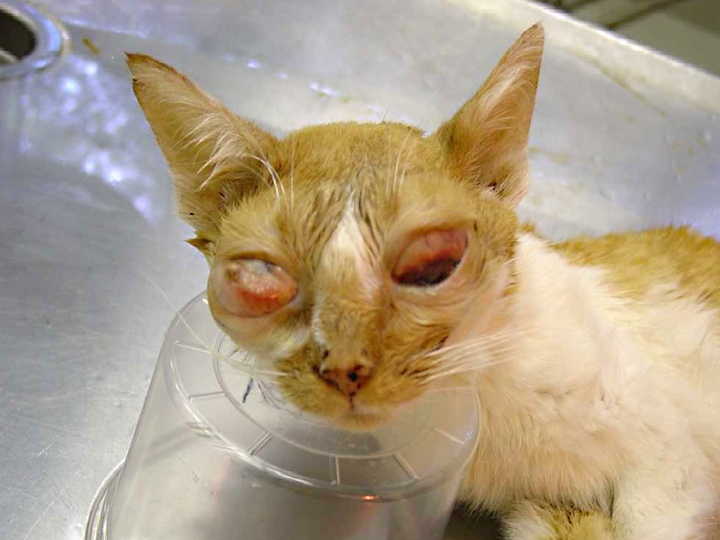
5-year-old spayed female domestic shorthair cat, Felis domesticus, feline.Five-year-old spayed female cat whose pupils began to dilate and ears to twitch after application of Bio Spot for cats on 05/27/2009. Cat returned to clinic with bilateral enlarged eyeballs with normal tonometry on 7/7/09. Conjunctiva slightly enlarged and oozy. Treated with antimicrobial eye ointment and dexamethasone. 7/23/09 and began treatment with metacam, doxycyclin, prednisone, antirobe, and strongid. Vaccination status unknown. Lesions progressively worsened over next few months. Animal euthanized and submitted for necropsy 09/08/09.
Additional History after diagnosis was made:
The owner moved into her present premises several months ago. The previous owner used one of the stalls in the barn as a chicken coop. Owner reported that all her animals (horses, dogs, and cats) began to lose weight with no response to deworming attempts. All other animals, except this cat, appeared to slowly recover on their own. At time of euthanasia all other animals were in good condition, even the other cats which had been treated with Bio Spot for cats.
Gross Description:
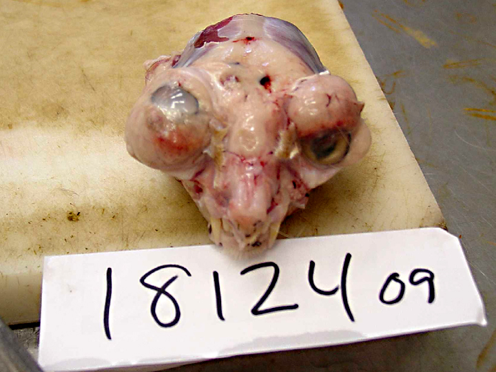
The cat is extremely cachectic. All bones can be easily palpated and the skin moves easily over the bones due to a scant amount of subcutaneous adipose tissue. There is no fat around the kidney, in the mesentery, or in the coronary groove. The eyes bulge forward. A portion of the cloudy cornea can be seen on the left eye. The sclera of both eyes is composed of bulging proliferative white tissue. Similar tissue is found in the frontal sinus, bulging through the calvarium at the bridge of the nose and extending down, around, and behind both eyes.
Histopathologic Description:
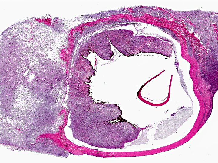
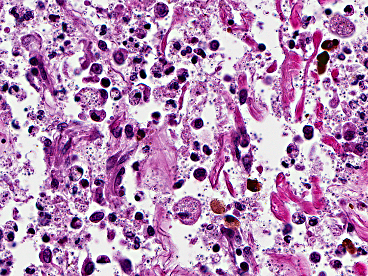
Cross sections of the eyes reveal that all layers of the globe, including the iris, portions of the cornea, and portions of the eyelids are effaced by sheets of macrophages distended with
Histoplasma capsulatum and necrotic debris. The tissue removed from the nasal cavity and frontal sinus are found to be distorted in a histologically similar manner.
Morphologic Diagnosis:
Chronic, severe, bilateral granulomatous panophthalmitis and blepharitis with intracellular
Histoplasma capsulatum, feline.
Lab Results:
07/23/09: CBC, Chemistry: WNL; FeLV/FIV negative.Â
Toxoplasmosa negative.Â
Hemobartonella positive.
Condition:
Histoplasma capsulatum
Contributor Comment:
Dogs are very susceptible hosts for histoplasmosis and they are considered sentinel animals.(3,4) However, while cats appear less susceptible to the disease, ocular manifestations of histoplasmosis are more commonly encountered in the cat.(4,5)
Histoplasma capsulatum is a soil-borne dimorphic fungus.(2) It grows best in moist soil containing nitrogen-rich organic matter such as bird or bat feces. The fungus can grow in moist soil associated with house plants, so even indoor-only cats can be at risk. Similar to blastomycosis, the disease is acquired by inhalation of fungal microconidia. The incubation period is 12 to 16 days. The microconidia in the lung convert to the yeast form which is phagocytized by macrophages, where they undergo further intracellular replication. The organism is then disseminated via the lymphatic and circulatory system. Occasionally, histoplasmosis infects the eye causing conjunctivitis, granulomatous blepharitis, granulomatous chorioretinitis, retinal detachment, and optic neuritis.
Histoplasmosis is an important zoonotic disease. People do not contract the disease from animals, but may contract the disease if they are exposed to the same environmental source; consequently, anyone in the household experiencing respiratory disease or nonspecific clinical symptoms should consult a physician as soon as possible. Laboratory workers can also contract the disease from fungal cultures containing mycelial growth of
Histoplasmosis capsulatum.
JPC Diagnosis:
Eye: Panophthalmitis, pyogranulomatous, focally extensive, severe, with numerous intrahistiocytic and extracellular yeasts consistent with
Histoplasma capsulatum.
Conference Comment:
Other common systemic mycoses in animals in North America can typically be readily differentiated from histoplasmosis in tissue section.Â
Blastomyces dermatitis, the most frequently reported cause of intraocular mycosis in dogs(6), appear as in tissue as 5-15 μm round nonencapsulated yeast with a 1 μm thick wall and broad-based budding. Yeasts of
Cryptococcus neoformans have a thick capsule and often form a soap bubble appearance, frequently extend from brain infection via the optic nerves and generally cause little tissue reaction. Spherules of
Coccidioides immitis and
C. posadasii, which are generally the most destructive and the most limited in geographical distribution, range from 20-200 μm in diameter and contain endospores.(1) Protozoal endophthalmitis caused by
Toxoplasma gondii, Encephalitozoon spp., Neospora spp., or
Leishmania spp. is more difficult to differentiate histologically and may require additional diagnostic modalities such as immunohistochemistry and electron microscopy.(6)
Toxoplasma gondii causes necrotizing granulomatous or lymphoplasmacytic ophthalmitis with tachyzoites in intracellular pseudocysts or cysts.Â
Neospora caninum may or may not reside in a parasitophorous vacuole and requires EM to distinguish it from Toxoplasma. The amastigote stage of
Leishmania spp. are present in macrophages and are 2-4 μm in diameter with a rod-shaped kinetoplast that is perpendicular to the nucleus.Â
Encephalitozoon cuniculiare intracellular, gram-positive, 1-2 μm protozoa present in large (up to 120 μm) pseudocysts and may induce periarteritis within the uvea and retina. Of these protozoal conditions, only
Toxoplasma gondii and
Neospora caninum specifically cause intraocular lesions and should be considered in the differential diagnosis for this case(6).
References:
1. Caswell JL, Williams KJ. Respiratory system. In: Maxie MG, ed.Â
Jubb, Kennedy, and Palmer's Pathology of Domestic Animals 5th ed. vol. 1, Philadelphia, PA: Saunders Elsevier; 2007:642.
2. Green CE. Histoplasmosis. In: Greene CE, ed.Â
Infectious Diseases of the Dog and Cat. ,3rd ed. St. Louis, MO: Elsevier Saunders; 2006;577-584.
3. Gwin RM, Makley TA, Wyman M, et al. Multifocal ocular histoplasmosis in a dog and a cat.Â
J Am Vet Med Assoc. 1980;176:638-642.
4. Percy DH. Feline histoplasmosis with ocular involvement.Â
Vet Pathol. 1981;18:163-169.
5. Stiles J. Ocular Infections. In
Infectious Diseases of the Dog and Cat, , 3rd ed., pp. 974-991. Elsevier Saunders, St. Louis, Missouri, 2006.
6. Wilcock BP. Eye and ear. In: Greene CE, Maxie MG, eds.Â
Jubb, Kennedy, and Palmer's Pathology of Domestic Animals. 5th ed. Vol. 1. Philadelphia, PA: Saunders Elsevier; 2007:502-3.