Signalment:
Gross Description:
Histopathologic Description:
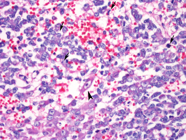
Sections of the tumor mass consist of sheets of small, polygonal to stellate cells with scant cytoplasm and dense, central nuclei within a fibrovascular stroma. Islands of large, polygonal cells resembling neurons are scattered irregularly within lobules. Similar foci are seen in the liver, but the spleen is filled primarily with the small cells.
The small cells observed in all sections are consistent with neuroblasts, whereas the larger neuron-like cells resemble ganglion cells. The histologic features are consistent with ganglioneuroblastoma, a rare tumor arising from the sympathetic ganglia. This one was malignant and sent metastases to the liver and spleen. Metastases were not found in the lung.
Morphologic Diagnosis:
Lab Results:
Reticulocyte counts were 8.1% one week prior to euthanasia.
Bacterial culture of the liver taken at necropsy was negative.
Condition:
Contributor Comment:
Clinical signs will vary depending on the location of the tumor. In all of the previously cited cases, clinical signs were limited to the effects of the space occupying mass on the local tissue. This dog was presented for non-specific lethargy, pain, and recumbency that was most likely due to severe compression of the heart and lungs. The cause of the anemia and thrombocytopenia in this case was not identified.
Immunohistochemical markers used in previous studies4-7 demonstrate consistent staining of ganglion-like cells with neurofilament protein (NFP), but variable patterns of staining with markers such as S100, synaptophysin, GFAP, and NSE. Case reports are few therefore a pattern of immunoreactivity is not clearly established for the dog. Studies of human neuroblastic tumors8 report variable staining for these and other markers.
JPC Diagnosis:
Conference Comment:
In this case, the predominant cell type is neuroblastic. There are scattered areas containing poorly differentiated ganglion cells that lack Nissl substance. Following the conference the case was reviewed in consultation with pathologists in the AFIP Department of Soft Tissue Pathology. Their diagnosis, based on the human classification of the International Neuroblastoma Pathology Committee (INPC), was neuroblastoma (Schwannian stroma-poor), differentiating subtype, with ganglion cells. The designation Schwannian stroma-poor indicates neuroblastic cells forming groups and nests without or with limited Schwannian proliferation. In the differentiating subtype, Schwannian stromal development containing mature and maturing ganglion cells comprise less that 50% of the neoplasm. We made our diagnosis based on the predominance of neuroblastic cells and absence of mature ganglion cells.
References:
2. Koestner A, Higgins RJ: Tumors of the endocrine system. In: Tumors in Domestic Animals, ed. Meuten DJ, 4th ed., pp. 715-716, 736-737. Iowa State University Press, Ames, IA, 2002
3. Koestner A, Bilzer T, Fatzer R, Schulman FY, Summers BA, Van Winkle TJ: Histological classification of tumors of the nervous system of domestic animals. In: WHO International Histological Classification of Tumors of Domestic Animals, ed. Schulman FY, pp. 1338. Armed Forces Institute of Pathology, Washington, DC, 1999.
4. Kuwamura M, Kotera T, Yamate J, Kotani T, Aoki M, Hori A: Cerebral ganglioneuroblastoma in a golden retriever dog. Vet Pathol 41:282-284, 2004
5. Mattix ME, Mattix RJ, Williams BH, Ribas JL, Wilhelmsen CL: Olfactory ganglioneuroblastoma in a dog, a light, ultrastructural, and immunohistochemical study. Vet Pathol 31:262265, 1994
6. Nakamura K, Ochiai K, Kadosawa T, Kimura T, Umemura T: Canine ganglioneuroblastoma in the oral mucosa. J Comp Pathol 130:205-208, 2004
7. Schulz KS, Steele KE, Saunders GK, Smith MM, Moon ML: Thoracic ganglioneuroblastoma in a dog. Vet Pathol 31:716-718, 1994
8. Shimada H, Ambros IM, Dehner LP, Hata J, Joshi VV, Roaki B: Terminology and morphologic criteria of neuroblastic tumors. Cancer 86:349-363, 1999
9. Zachary JF: Nervous System. In: Pathologic Basis of Veterinary Disease, eds. McGavin MD, Zachary JF, 4th ed., pp. 948-949. Elsevier, St. Louis, MO, 2007