Signalment:
Adult male wild house mouse,
Mus musculusFound dead.
Gross Description:
The mouse was 17.5 g and had minimal to no adipose stores. There were multiple hairless patches along the dorsum and face. Bilaterally, the margins of the pinnae were irregular and lacerated. The right pinna contained a 5 x 3 x 3 mm tan, multinodular skin mass. There were similar 1 mm diameter tan nodules within the hairless patches of the dorsum and face. The thoracic cavity contained a moderate amount of dark red hemorrhage.
Histopathologic Description:
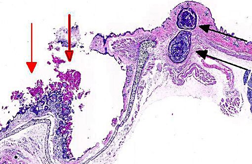
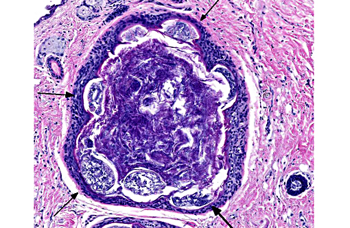
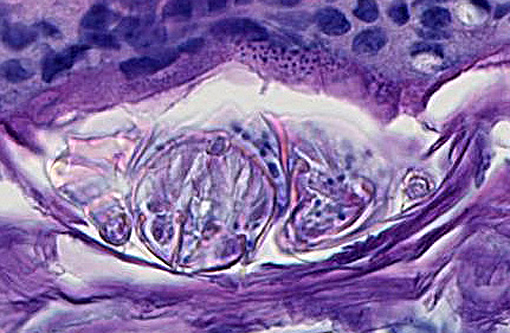
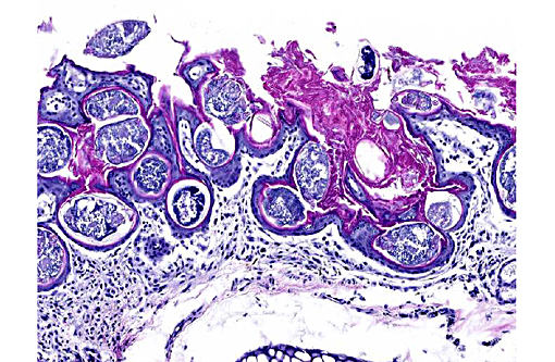
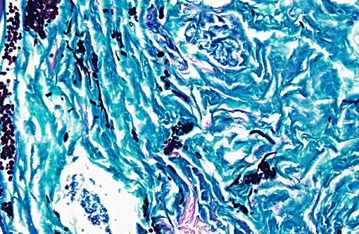
Hair follicles of the face, pinnae, and dorsum are multifocally dilated and contain abundant arthropods (mites) embedded in hyperkeratotic and hyperplastic follicular epithelium. Arthropods are 80 - 110 microns in diameter with a chitinous exoskeleton, multiple jointed appendages, skeletal muscle, and a reproductive tract containing basophilic material. Similar mites are embedded in hyperkeratotic surface epithelium. There are multifocal 40 - 60 micron diameter basophilic eggs. Rare sections contain subcutaneous inflammation characterized by lymphocytes, plasma cells, and neutrophils and hemorrhage. Rarely, follicles also contain clusters of 3 - 5 micron round to oval structures that are periodic acid-Schiff positive (yeasts).
Morphologic Diagnosis:
Haired skin (face, pinnae, and dorsum): Severe follicular plugging, hyperkeratosis, and hyperplasia with intrafollicular and superficial epidermal mites (etiology:
Psorergates simplex)
Lab Results:
None
Condition:
Psoregates simplex
Contributor Comment:
The gross and histologic presentation in this case is typical of the follicular mite,
Psorergates simplex. The characteristic gross lesion caused by this mite is numerous 2 mm tan to white cystic dermal nodules.(1,2,4,5) The nodules, also described as nests and pouches,(3) are most obvious when the skin is reflected back during postmortem examination.(1,2,4) Lesions most commonly occur in the loose skin of the neck, back, trunk, shoulders, and abdomen, but can occur anywhere, including the face and legs. The cysts resemble comedones and histologically are characterized by dilated follicles plugged with abundant mites and keratin debris.(1,5) Inflammation is usually minimal but will occur around ruptured follicles (furunculosis). The presence of mites in hair follicles of the pinnae, as was seen in this case, is a less common presentation.(1) Nodules can occur on either or both sides of the pinna and may need to be differentiated from notoedric ear mange,(1) in which the lesion tends to be more superficial and proliferative with mites embedded in the stratum corneum.(4)
Notoedres sp. are also larger mites (250 - 400 microns long), whereas
P. simplex are 90 - 150 microns long.(1)
Infection with
P. simplex is uncommon in wild and pet mice and rare to absent in laboratory mice. The complete life cycle of
P. simplex is unknown.(1,5) The mites are transmitted by direct contact(1) and gravid females enter the hair follicles to form nests which expand the follicles by internal pressure.(1,2) All stages of the mite life cycle (eggs, larvae, nymphs, and adults) can be found within hair follicles.(1,5)
Death of this wild house mouse was attributed to a suspected traumatic event which caused dermal hemorrhage and hemothorax. The mites were an incidental finding. We receive few wild mice at our laboratory but in our experience
P. simplex is uncommon. Rare yeasts in the follicles are most consistent with
Malassezia sp. and most likely represent a secondary infection.
JPC Diagnosis:
1. Haired skin: Comedones, multiple, with infundibular adult mites and eggs.Â
2. Haired skin: Infundibular fungal arthrospores (presumptive) and hyphae.Â
3. Pinna: Otitis externa, hyperkeratotic and lymphohistiocytic, diffuse, moderate, with infundibular adult mites and eggs.
Conference Comment:
For an institution with thousands of animals comprising hundreds of different species, this case serves as a reminder of the importance in monitoring the health of wildlife pests in addition to exhibit animal population.Â
Psorergates simplex mites were once prevalent in laboratory mice but are now only readily recognized among wild and pet mice.(5) While little is known regarding its pathogenesis, most skin mites of mice are directly transmissible which may pose a risk to some zoo inhabitants. Conference participants discussed the finding of some sections of mites which appeared to be larger (200-300 um) and found superficial to the epidermis in some slides. These larger mites often had a striated cuticle not observed among the intrafollicular mite sections, leading many to speculate on the presence of a second species. Several species of mites are relatively common in mice, including
Myobia musculi, Myocoptes musculinis, and
Radfordia affinis.(5) Additionally, the contributor mentions
Notoedres sp. which are much larger and more superficial in histologic sections. Any of these are a possibility, as all lack distinguishing morphologic characteristics. In fact, of all genres of mites, only
Sarcoptes sp. (with dorsal cuticular spines) and
Demodex sp. (with elongated abdomen and closely apposed appendages) can be readily identified on histologic section by their morphology alone.(3)
Myobia sp. are the most clinically significant mites which cause a hypersensitivity reaction while
Myocoptes sp. is most common.(5)
We also observed the 3-5 um spores present often in conjunction with mites in dilated follicles. These are PAS- and GMS-positive, which also revealed few fungal hyphae in the same location. Upon histochemical staining, we are unable to determine the specific species of this fungus though we do not believe this morphology is consistent with
Malassezia sp.; their location within the follicles as well as hyphal formation is consistent with a dermatophyte. The lack of inflammation associated with the dilated follicles was curious, as neither the mites nor fungi seemed to elicit a response from the host. In some slides, sections of ear pinna were identified which appeared to be the only area where lymphocytes and macrophages were recruited. We elected to include a third diagnosis for this location, though it is worth mentioning the mites seemed to be concentrated in larger numbers in these sections.Â
References:
1. Baker DG. Parasites of Rats and Mice. In: Flynns Parasites of Laboratory Animals. 2nd ed. Ames, IA: Blackwell Publishing Professional: 2007: 366-367.
2. Flynn RJ, Jaroslow BN. Nidification of a mite (Psorergates simplex Tyrell, 1883: Myobiidae) in the skin of mice. J Parasitology. 1956;42:49-52.
3. Gardiner CH, Poynton SL. An Atlas of Metazoan Parasites in Animal Tissues. Washington, DC: Armed Forces Institute of Pathology; 1999:56-58.Â
4. Izdebska JN, Fryderyk S. New for the fauna of Poland species of Psorergates spp. with the data of occurrence of mites from Psorergatidae family (Acari, Prostigmata) in native mammals. Annals Parasitology. 2012;58:19-22.
5. Percy DH, Barthold SW. Mouse. In: Pathology of Laboratory Rodents and Rabbits. 3rd ed. Ames, IA: Blackwell Publishing Professional: 2008: 85-87.