Signalment:
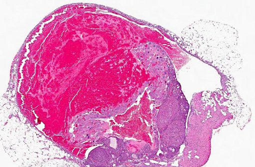
Gross Description:
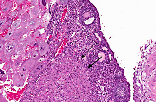
Histopathologic Description:
Morphologic Diagnosis:
Condition:
Contributor Comment:
The choriocarcinoma is a malignant neoplasm of trophoblastic cells, which in women often has widespread metastases. Choriocarcinoma is one of the rarest ovarian tumors in women, estimated to occur in only 1 in 369,000 women.3 Most choriocarcinomas in women occur in the uterus, post pregnancy. These tumors are also rare in animals, although they have been reported in rhesus and cynomolgus macaques, rabbits, a cow, dogs, mice and rats.(1,2,3,4,6,7) Choriocarcinomas may arise in the uterus, ovary or testis.Â
The incidence of ovarian tumors in mice varies with the strain.(2,4) Ovarian tumors are also more common in older mice (> 18 months of age). In mice, ovarian neoplasms are more common in the B6C3Fl mouse, but ovarian choriocarcinoma is only 1% of ovarian tumors.(4) The most common ovarian tumors in B6C3F1 mice are epithelial, granulosa cell tumors and teratomas.(1) In our laboratory, we have identified six ovarian choriocarcinomas in the last 10 years, all in sentinel mice as an incidental finding during health monitoring.
JPC Diagnosis:
Conference Comment:
References:
2. Alison RH, Lewis DJ, Montgomery CA. Ovarian choriocarcinoma in the mouse. Vet Pathol. 1987;24:226-230.
3. Farman CA, Benirschke K, Horner M, Lappin P. Ovarian choriocarcinoma in a rhesus monkey associated with elevated serum chorionic gonadotropin levels. Vet Pathol. 2005;42:226-229.
4. Frith CH, Evans MG. Spontaneous ovarian choriocarcinoma, yolk sac carcinoma, and teratoma in B6C3F1 mice: a case report. Toxicol Pathol. 1993;21:91-98.
5. Horn LC. Intermediate Trophoblastic Tumors and Tumor-like Lesions-the Clinicopathologic Aspects. IAP 2006 Annual Congress. Symposium #41, section 3. Accessed online at http://www.uscap.org/index.htm?iap2006/symp41-3.htm on 3 February 2013.Â
6. Kaufmann-Bart M, Fischer I. Choriocarcinoma with metastasis in a rabbit (Oryctolagus cuniculi). Vet Pathol. 2008;45:77-79.
7. Marbaix E, Defrere S, Ho Minh Duc K, Lousse JC, Dehoux JP. Nongestational malignant placental site trophoblastic tumor of the ovary in a 4-year-old Rhesus Monkey. Vet Pathol. 2008;45:375-378.Â
8. Pirak M, Waner T, Abramovici A, Scolnik M, Nyska A. Histologic and immunohistochemical study of a spontaneous choriocarcinoma in a male Sprague Dawley rat. Vet Pathol. 1991;28:93-95.