Signalment:
[Follow-up by phone conversation: The referring veterinarian was contacted by phone 6 months after biopsy submission. The veterinarian reported that all masses that had abruptly appeared regressed and were completely gone by 2-3 months. The veterinarian reported that the rabbit was currently healthy.]
Gross Description:
Histopathologic Description:
Morphologic Diagnosis:
Condition:
Contributor Comment:
The lesions caused by SFV in rabbits were first described by Dr. R.E. Shope, a physician and virologist, in 1932.(8,9) SFV is transmitted to rabbits through biting insect and arthropod vectors such as mosquitoes and fleas.(1,5) Tumors can arise from each bite site, resulting in one to several masses appearing on the patient.(5) Lesions are most often located on the head and legs.(5) The disease exhibits seasonality with most cases reported in the fall (September-November).(5) Lesions typically reach maximum size between 7-12 days after inoculation, which can account for the relatively rapid onset noticed by owners as in this case. Afterwards, lesions will regress over 1-1.5 months leaving the host immune.(5) Infection by SFV in neonatal or very young rapids can lead to fatal infections with disease resembling myxomatosis.(1,5)
Von Bomhard et al. performed a retrospective study on cutaneous lesions in rabbits, separating lesions into virus-induced and non-virus-induced tumors.(11) Shope fibroma was the most common virally induced tumor followed by Shope papilloma (Papovavirus). Overall, trichoblastomas and collagenous hamartomas were the most common cutaneous tumors reported from this study. Interestingly, this study reported a sex predilection for mesenchymal tumors in rabbits, with mesenchymal proliferations occurring significantly more in male rabbits than females.Â
JPC Diagnosis:
Conference Comment:
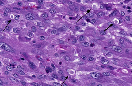
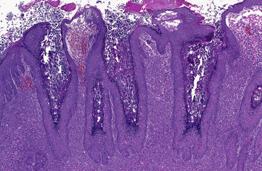
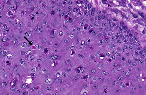
The fibroblast proliferation is unusual among the poxviruses, which are generally epitheliotropic in nature. Viral replication within epithelial cells causes ballooning degeneration and necrosis, with sites of replication observable histologically as small, basophilic, intracytoplasmic inclusions designated as type B inclusions or Guarnieri bodies, while the larger eosinophilic inclusions as observed in this rabbit are those of type A which occur later in the replication cycle.(7)
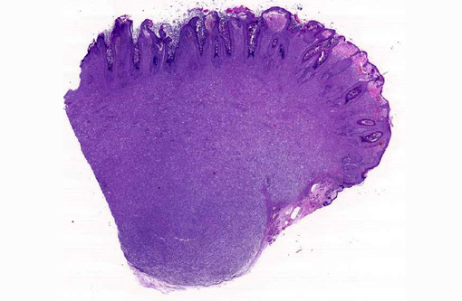
Specific to the members of the Leporipoxvirus family (Shope fibroma virus, squirrel fibroma virus and myxoma virus), the proliferation of fibroblasts expanding the dermis is the more prominent histologic lesion. In SFV, poxviral inclusions are also found in these mesenchymal cells as observed in this case. In contrast, myxoma virus infections induce a more mucinous matrix separating fibroblasts which lack viral inclusions (but are seen in overlying epithelial cells).(6) Poxviruses are known to express proteins with similar mechanisms of action as epidermal growth factor(2), and it is logical to conclude members of the Leporipoxvirus family may similarly produce fibroblast growth factors, though this has not been demonstrated as research on these niche viruses is limited.Â
Through the discovery that pretreatment with RANTES (CCL5) to cells in culture could inhibit infection of myxoma virus, researchers uncovered its affinity for the chemokine receptor CCR5, in addition to CCR1 and CXCR4.(4) This is a noteworthy feature of HIV and SIV infections, in which individuals have been identified with a natural resistance to infection due to a mutation in the CCR5 gene.(10) These chemokine receptors are specific to white blood cells, which alludes to the immunosuppressive nature of HIV infections.(9) What this means, if anything, for poxviruses and specifically the behavior of the Leporipoxvirus family is still undetermined.Â
References:
1. Dalmat HT. Arthropod transmission of rabbit fibromatosis (Shope). J Hyg 1959;57:1-30.Â
2. Ginn PE, Mansell JL, Rakich PM. Skin and appendages. In: Maxie MG ed. Jubb, Kennedy, and Palmers Pathology of Domestic Animals. 5th ed. Vol. 1. Philadelphia, PA: Elsevier Saunders; 2007:664-674.
3. Jones TC, Hunt RD, King NW. Veterinary Pathology. Baltimore, MD: Lippincott Williams & Wilkins; 1997:208-209.
4. Lalani AS, Masters J, Zeng W, et al. Use of chemokine receptors by poxviruses. Science. 1999;286:1968-1971.
5. Meredith AL. Viral skin diseases of the rabbit. Vet Clin North Am Exot Anim Pract. 2013;16:705-714.Â
6. Percy DH, Barthold S. Pathology of Laboratory Rodents and Rabbits. 3rd ed. Ames, IA: Blackwell Publishing; 2007:257-258.
7. Pfeffer M, Meyer H. Poxvirus diagnostics. In: Mercer AA, Schmidt A, Weber O, eds. Poxviruses. Basel, Switzerland: Birkh+�-�user Verlag; 2007:359.
8. Pulley LT, Shively JN. Naturally occurring infectious fibroma in the domestic rabbit. Vet Pathol. 1973;10:509-519.
9. Shope RE. A transmissible tumor-like condition in rabbits. J Exp Med. 1932;56:793-802.Â
10. Silva E, Stumpf MP. HIV and the CCR5-32 resistance allele. Micro Let. 2004;214(1):1-12.
11. Von Bomhard W, Goldschmidt MH, Shofer FS, Perl L, Rosenthal KL, Mauldin EA. Cutaneous neoplasms in pet rabbits: a retrospective study. Vet Pathol 2007;44:579-588.
12. Willer DO, McFadden G, Evans DH. The complete genome sequence of shope (rabbit) fibroma virus. Virology. 1999;264:319-343.