Signalment:
10-year-old, castrated male, border collie cross dog (
Canis familiaris).One-month history of a mass on the ventral abdomen. The mass had doubled in size over the last few
weeks. It was freely moveable within the subcutaneous layers of the skin and measured approximately 3 x 3 cm at
the time of removal.
Gross Description:
Received a 3 x 2.5 cm ellipse of haired skin containing a firm, pale 2 cm diameter subcutaneous
mass.
Histopathologic Description:
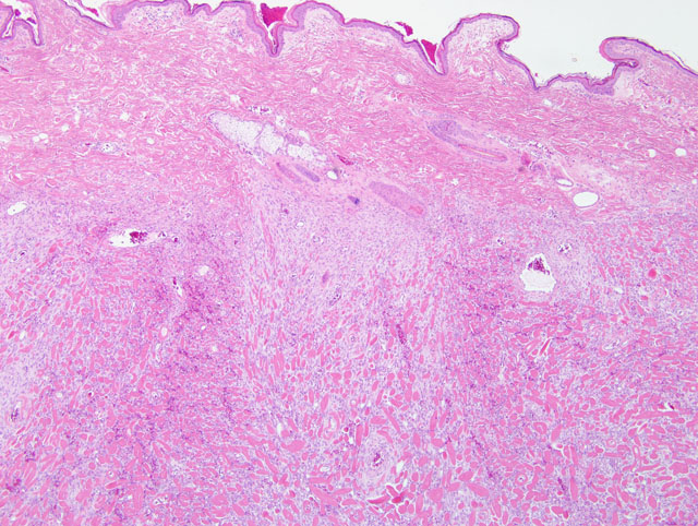
Haired skin: Beneath the superficial dermis is an unencapsulated, well demarcated,
moderately cellular, focally infiltrative neoplastic mass. Neoplastic cells are arranged in thin streams dissecting
between large thick bundles of well defined eosinophilic homogenous (hyalinized) birefringent material (collagen)
within a fine fibrovascular stroma. In one location, neoplastic cells form interlacing bundles and streams with small
embedded collagen bundles. Cells are spindloid with indistinct cell borders, a scant amount of finely fibrillar
eosinophilic cytoplasm with an elongate central nucleus with finely stippled chromatin and indistinct nucleoli.
There is three-fold anisocytosis and anisokaryosis. The mitotic rate is low with an average of 0.1 mitotic figures per
400x HPF. Neoplastic cells extend to one margin of the biopsy. Small areas of hemorrhage are scattered throughout
the tumor and around the tumor base. Small numbers of lymphocytes and plasma cells surround small vessels
around the margins of the tumor.
Morphologic Diagnosis:
Keloid fibrosarcoma.
Condition:
Keloidal fibrosarcoma
Contributor Comment:
Keloid fibrosarcomas are an uncommon variant of fibrosarcomas, distinct from the other
forms of fibrosarcoma and other collagen-rich masses due to the presence of thick bands of hyalinized collagen.(3)
These bands of hyalinized collagen can also be seen in cytologic preparations.(2) Keloidal tumors are infrequently
reported in dogs and have not been reported in other domestic animal species. They appear somewhat similar
histologically to keloids, hypertrophic scars and keloid dermatofibromas in humans.(2) The one published
retrospective study of keloidal tumors in dogs suggests that the presence of macrophages within the tumors may
indicate that these tumors are reactive inflammatory lesions, rather than true neoplasms.(3)
Differentiation of keloid fibromasarcomas from keloidal fibromas is based on the presence of portions of the tumors
that are composed largely of thickly packed neoplastic cells with only a small amount of fibrovascular stroma and
small numbers of hyalinized fibers.(3) These areas are reportedly more common in the deeper margins of the tumors
as is the case in this example. It was suggested that keloidal fibrosarcomas may represent a malignant
transformation of keloid fibromas; however, no difference in prognosis has been demonstrated between keloid
fibromas or keloid fibrosarcomas.
The neoplastic cells in keloidal tumors of dogs are vimentin positive and smooth muscle actin negative and are
interpreted to be fibroblasts in contradistinction to human keloidal tumors which are comprised of myofibroblasts.
(3)
JPC Diagnosis:
Haired skin and subcutis: Fibrosarcoma, low grade (keloidal).
Conference Comment:
The large bundles of hyalinized collagen are a distinctive histologic feature in this case,
and in dogs are generally limited to keloidal fibromas and fibrosarcomas and mast cell tumors with keloidal change.
(1) Differentiation between keloidal fibromas and keloidal fibrosarcomas is based on the presence of interlacing
fascicles of neoplastic cells and/or infiltration in the malignant variant. Since keloidal fibrosarcomas may arise from
keloidal fibromas, the malignant characteristics may be present only in a small portion of the tumor.(3) Keloidal
fibrosarcomas also may contain cellular atypia and have an increased mitotic rate.(1) The differential diagnosis
includes dermatofibroma and peripheral nerve sheath tumor (PNST). In dermatofibromas, and occasionally in
PNSTs, the spindle cells are separated by enlarged, but non-hyalinized, collagen bundles. Additionally,
dermatofibromas usually contain inflammation and overlying epithelial hyperplasia, while PNSTs have a neural
pattern, demonstrate variation in cellularity, and frequently contain a myxomatous matrix.(1)
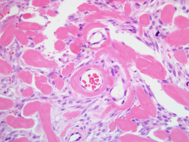
Conference participants also noted the presence of small capillaries within the large, hyalinized collagen fibers and
areas of microhemorrhage; these changes have been described previously.(3)
References:
1. Gross TL, Ihrke PJ, Walder EJ, Affolter VK: Skin Diseases of the Dog and Cat: Clinical and Histopathologic
Diagnosis, 2nd ed., pp. 721-722. Blackwell Publishing, Ames, IA, 2005
2. Little LK, Goldschmidt M: Cytologic appearance of a keloidal fibrosarcoma in a dog. Vet Clin Pathol
36:364-367, 2007
3. Mikaelian I, Gross TL: Keloidal fibromas and fibrosaromas in the dog. Vet Pathol
39:149-153, 2002