Signalment:
10-month-old female BALB/c,
Mus musculus.The mouse was group housed with other females in an individually ventilated cage (IVC) system. No experimental procedures had been performed on the mouse as she was part of a cohort being allowed to age prior to study commencement. The technicians noted a mass on the side of the face/neck, culled the animal and removed tissues for histological assessment.
Gross Description:
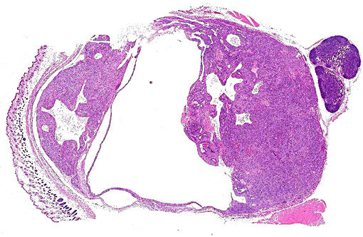
A piece of haired skin with a 10 mm diameter cavitated, fluid filled mass was received in formalin. The mass had a multifocally thin wall (less than 1 mm fixed tissue thickness) but in other foci the wall of the cavitation was expanded by beige homogeneous tissue up to 5 mm in thickness. The fluid contained within the cavitated structure was clear. Thoracic and abdominal viscera were also submitted fixed in formalin and no other abnormalities were detected.
Histopathologic Description:
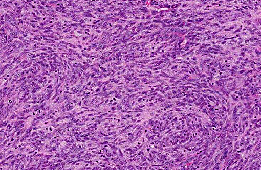
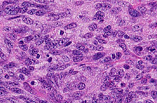
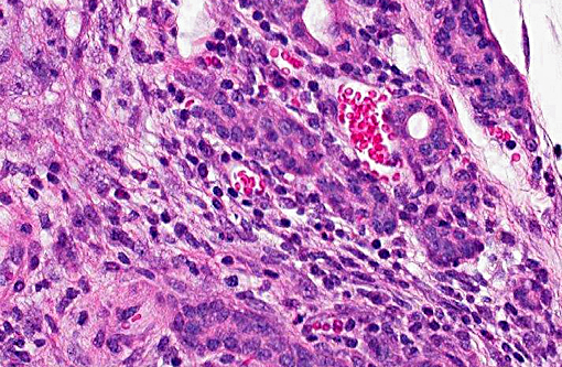
Within the subcutis of the haired skin, adjacent to the salivary tissue and lymph node, is an unencapsulated, but well-circumscribed, neoplastic mass, which is bluntly infiltrative in some tissue planes, and moderately densely cellular. The neoplastic cells are arranged in heterogeneous patterns including poorly defined clusters, occasionally in tubular structures, and also multifocally in sheets and broad bundles and streams, supported by a multifocally dense collagenous stroma. The cells are arranged around variably sized, but frequently large, cavitations and in some foci the cells palisade around the spaces. The neoplastic cells are pleomorphic polygonal to spindle-shaped, and are moderately sized, with indistinct cell margins and a moderate amount of eosinophilic cytoplasm. The nuclei are frequently oval and exhibit lightly stippled to clumped chromatin and a variable number of variably prominent nucleoli. The mitotic rate is high at 35 mitoses per 10 high power fields. There are foci of necrosis characterized by nuclear karyorrhexis and accumulations of amorphous eosinophilic material. Multifocal aggregates of small to moderate numbers of lymphocytes, with lesser numbers of plasma cells, are present multifocally. The cavitations within the mass multifocally contain strands of pale eosinophilic material and there are multifocal groupings of small numbers of macrophages, some of which contain grey to amphophilic intracytoplasmic material.
Morphologic Diagnosis:
Haired skin on side of face/ neck: Myoepithelioma
Condition:
Myoepithelioma
Contributor Comment:
In the planes of tissue examined, the lesion is consistent with a myoepithelial carcinoma arising from the adjacent salivary tissue (not present on all sections). Alternative differential diagnoses based on the site and gross morphology included a Harderian gland cystadenoma, but the histological appearance of the specimen was most consistent with a tumor arising from the myoepithelial cells of the salivary gland. Myoepitheliomas generally exhibit a low mitotic rate,(6) but this example exhibited frequent mitoses and thus classification as a myoepithelial carcinoma was preferred. The cavitations exhibited by this mass are considered characteristic and are frequently the result of necrosis leading to the formation of cyst-like structures.(4) Had immunohistochemical staining been undertaken, myoepitheliomas stain positively for keratins 5 and 14, reflecting their myoepithelial origin.(6)
Although myoepithelial tumors are infrequent in most strains of mice, they are relatively more common in BALB/c mice, especially females, such as in the present case. They most frequently arise from the submaxillary and parotid salivary glands and present as swellings of the subcutaneous tissue of the ventral neck.(4) A retrospective study of 142 myoepitheliomas determined that these tumors also occur spontaneously in several other inbred strains of laboratory mice including A/HeJ, A/J, LLC.A/Ckc and NOD/Lt.(6) Recently, salivary carcinomas exhibiting myoepithelial and basal cell differentiation have been reported in the Justy mutant mouse strain (background: C3HeB/FeJ) which bears a recessive mutation in the Gon4l gene that regulates gene expression during development.(5)
Myoepithelial carcinomas are rare in humans and in the domestic species(3) although there are at least two reports of myoepithelial carcinoma (synonym: malignant myoepithelioma) in dogs.(1,2)
JPC Diagnosis:
Parotid gland: Myoepithelioma
Conference Comment:
This is a rarely reported neoplasm in domestic species (other than the mouse), and without distinguishing features, its specific histogenesis is often left to immunohistochemistry. In this particular case, we ran pancytokeratin, vimentin and smooth muscle actin, with only pancytokeratin yielding positive staining. Myoepithelial cells are modified epithelial cells with contractile properties.(6) They exist in glandular tissue, such as salivary and mammary glands, and function by forcing secretions into and through a duct system with their contraction.(6) Myoepithelium stains positive for both epithelial and muscle markers; however, their staining seems to be more variable compared to the apparent high specificity of CK5 and CK14 as mentioned by the contributor.(1,5,6)
The question of malignancy was debated in this case, as some pointed out the well-circumscribed appearance and uniform cell population. We agree with the contributor, that the high mitotic rate and areas of necrosis are reason to worry, but elected to avoid a malignant modifier without definitive evidence such as vascular invasion.Â
References:
1. Faustino AM, Pereira PD. A salivary malignant myoepithelioma in a dog. Vet J. 2007:173:223-226.
2. Gorlin RJ, Barron CN, Chaudhry AP, et al. The oral and pharyngeal pathology of domestic animals: a study of 487 cases. Am J Vet Res. 1959:20:1032-1061.
3. Head KW, Else RW, Dubielzig RR. Tumors of the alimentary tract. In: Meuten D, ed. Tumors in Domestic Animals. 4th ed. Ames, Iowa: Iowa State Press; 2002:415.Â
4. Percy DH, Barthold SW. Mouse. In: Percy DH, Barthold SW, eds. Pathology of Laboratory Rodents and Rabbits. 3rd ed. Oxford, UK: Blackwell publishing; 2007:120-121.
5. Simons AL, Lu P, Gibson-Corley KN, et al. The Justy mutant mouse strain produces a spontaneous murine model of salivary gland cancer with myoepithelial and basal cell differentiation. Lab Invest. 2013:93:711-719.
6. Sundberg JP, Hanson CA, Roop DR, et al. Myoepitheliomas in inbred laboratory mice. Vet Pathol. 1991:28:313-323.