Joint Pathology Center
Veterinary Pathology Services
Wednesday Slide Conference
2017-2018
Conference 10
November 29th, 2017
CASE IV: 2014 Case 1 (JPC 4050142).
Signalment: Adult, female, breed unspecified, Bos taurus, bovine.
History: The bovine was presented for slaughter at a USDA Food Safety Inspection Service (FSIS) inspected slaughter facility and passed antemortem inspection. The carcass was retained for further diagnostic testing after green, caseous-appearing lesions were observed in the liver and portal lymph nodes during postmortem inspection.
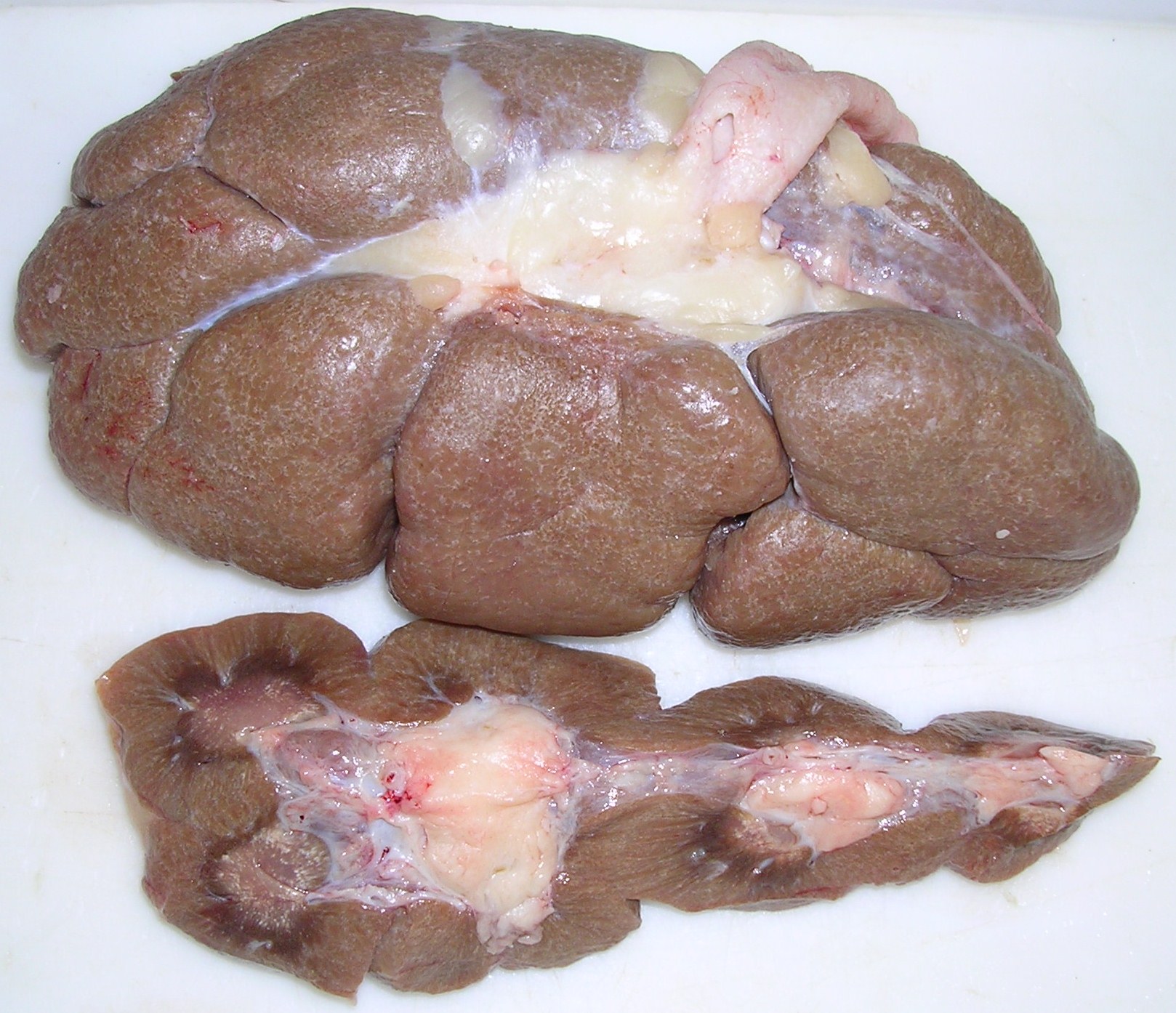
Gross Pathology: Green, caseous-appearing lesions in the liver and portal lymph nodes.
Laboratory results:
None provided.
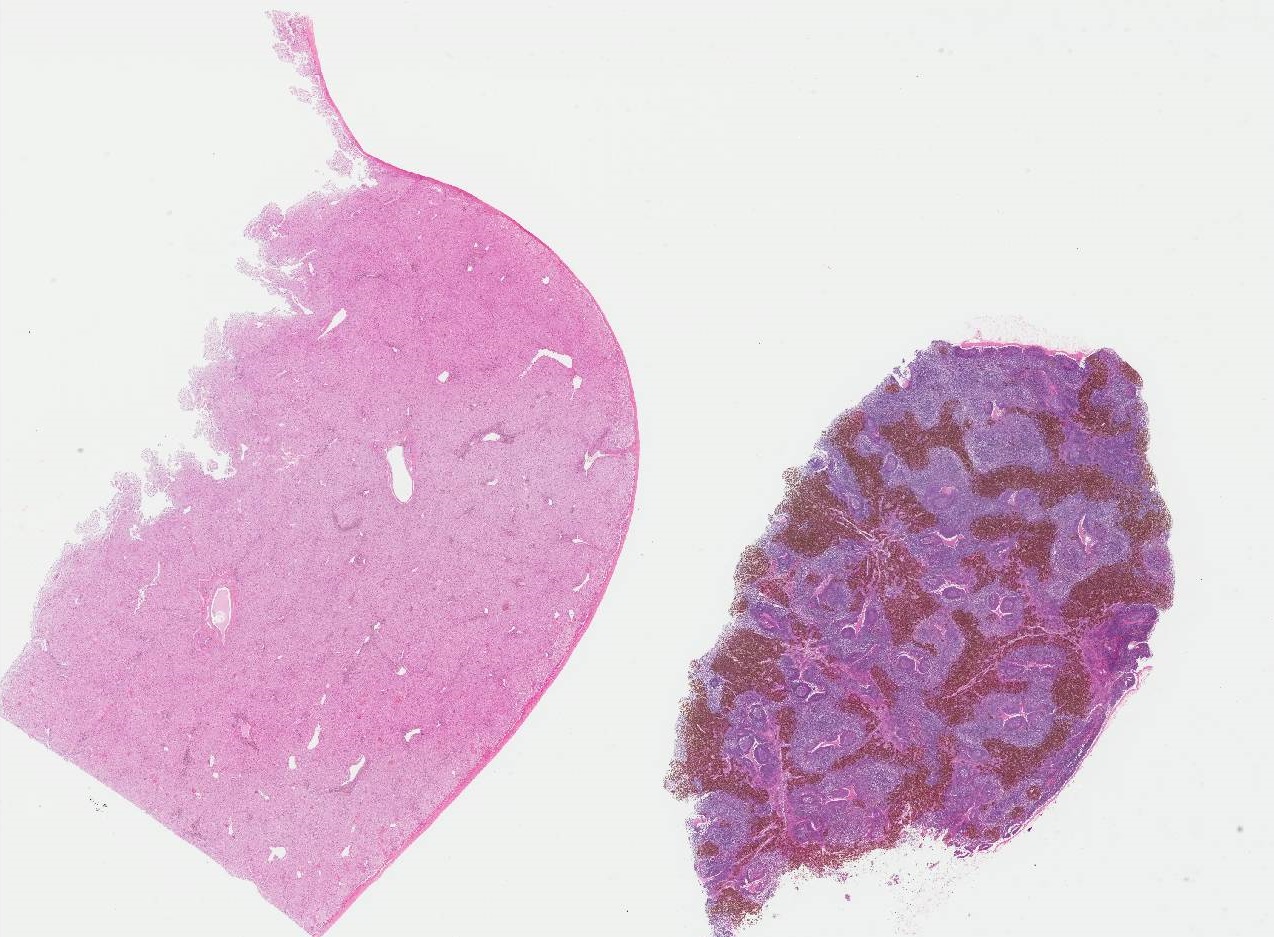
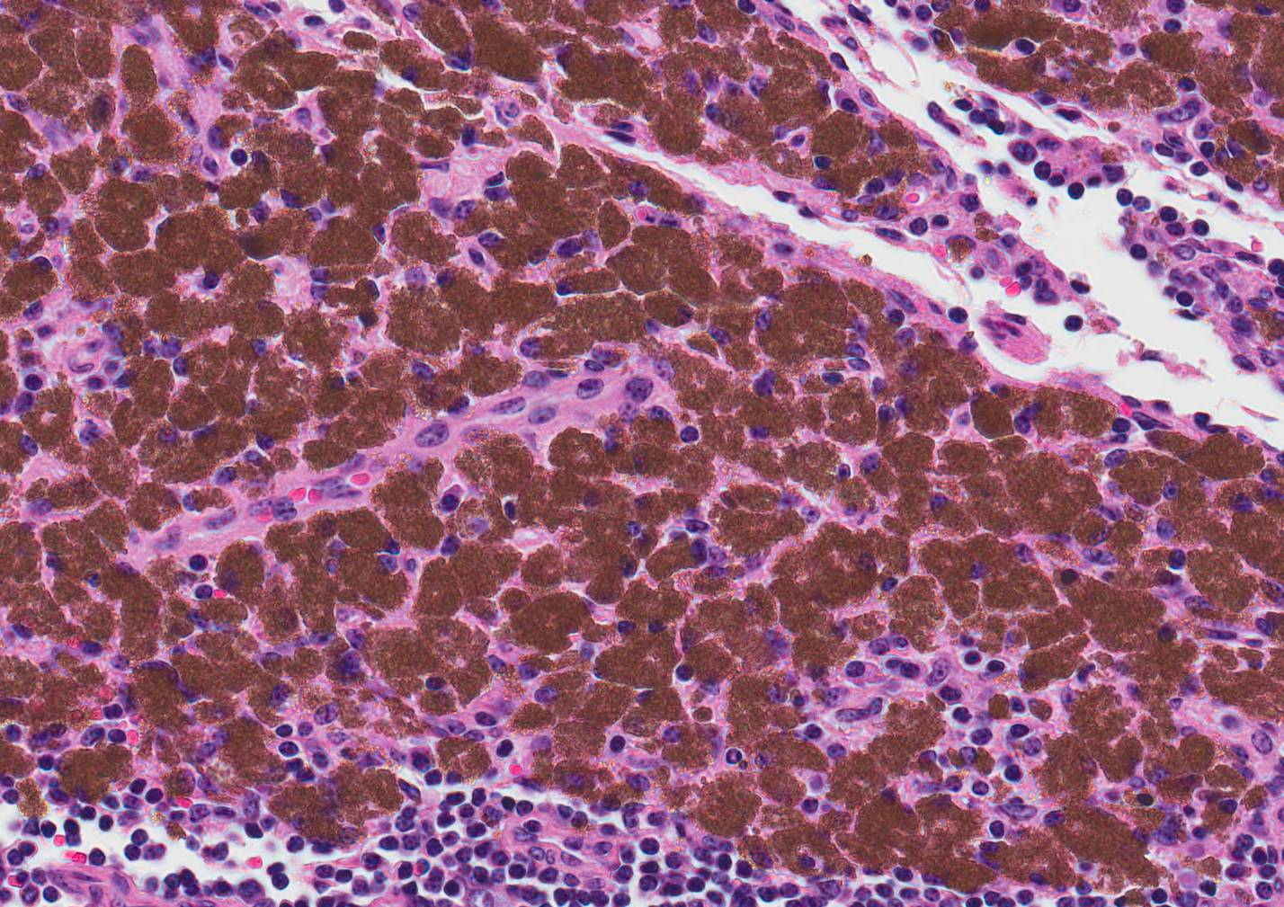
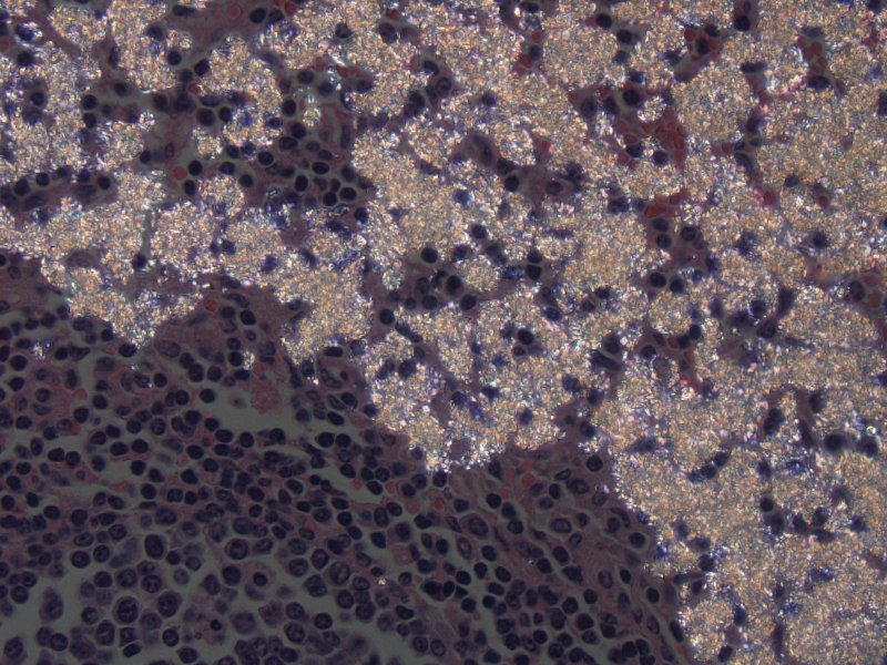
Microscopic Description: Liver: Predominantly in portal areas, macrophages contain intracytoplasmic light green to light tan crystalline material which is birefringent when viewed under polarized light. Individual crystals are irregular to rod-shaped and range in size from <1µm to approximately 3μm in length and aggregates can be >100µm in diameter. Small numbers of crystals are found within scattered hepatocytes. Occasionally surrounding the affected cells are clusters of free erythrocytes that obscure the parenchyma. Sinusoids in the adjacent areas are often congested. There is variable subcapsular hemorrhage present.
Lymph node: Cortical and medullary sinuses are expanded by moderate to large numbers of macrophages engorged with intracytoplasmic material identical to that described in the liver.
Contributor’s Morphologic Diagnosis:
- Liver: Accumulation of refractile material consistent with 2,8-dihydroxyadenine, Bos taurus, bovine.
- Lymph node: Cortical and medullary histiocytic accumulation of refractile material consistent with 2,8-dihydroxyadenine.
Contributor’s Comment: 2,8-dihydroxyadeninosis or “green liver disease” is a rare condition of bovines that is caused by the tissue deposition of 2,8-dihydroxyadenine (2,8-DHA), an insoluble green crystalline material. Accumulation of 2,8-DHA is most commonly seen in the liver and hepatic lymph nodes, but has also been reported in the kidney, and the renal, mediastinal, intercostal, and gastric lymph nodes.7 The crystals appears as a light green to light tan mottling on the liver surface and parenchyma. Larger coalescing foci of 2,8-DHA are present in the hepatic lymph nodes. Renal lesions, if present, consist of light green mottling or streaking on the surface and on cut section or as concretions in the renal pelvis. The condition in bovines was initially reported about 20 years ago after unusual green lesions were observed during postmortem examination at federally inspected abattoirs. Although the condition is rare, FSIS personnel are trained to recognize it since even rare conditions can be observed during the postmortem exam of the 30 million head of cattle that undergo federal inspection in the US every year.9
Bovines with 2,8-dihydroxyadeninosis are thought to have an enzyme deficiency affecting purine catabolism. Normally, adenine is phosphorylated to adenine monophosphate in the first step of adenine catabolism. In the absence of the enzyme adenine phosphoribosyltransferase (APRT), adenine may instead be hydroxylated by the enzyme xanthine oxidase to 2,8-DHA, which precipitates in tissues as an insoluble crystal.7 The light green to light tan color of the precipitate is retained throughout processing, embedding, and staining.
Deposits of 2,8-DHA in the renal tubules and lower urinary tract have been reported in humans, dogs, and APRT knockout mice. Uroliths and tubular casts can block the urinary tract, and lead to renal failure in these species.5
Other causes of grossly green discoloration in bovine tissues include green algal lymphadenitis (chlorellosis),4 exogenous green pigment in lymph nodes draining tattoo ink from skin,6 eosinophilic myositis,10 green liver cell adenomas,1,3 and bile imbibition. Green discoloration has been noted in injection site lesions in cuts of beef in modified-atmosphere packages.8
JPC Diagnosis:
- Lymph node, medullary sinus: Sinus histiocytosis with intrahistiocytic birefringent pigment, breed unspecified (Bos Taurus), bovine.
- Lymph node: Reactive lymphoid hyperplasia, diffuse, moderate.
- Liver, portal areas: Histiocytosis, multifocal, mild with abundant intrahistiocytic birefringent pigment.
Conference Comment: An extremely rare condition, 2,8 dihydroxyadenine accumulation in cattle, has only been reported on once in veterinary literature,7 but results in striking gross and microscopic lesions that are worth mentioning. Adenine is a purine that is normally found in all tissues of the body (as it serves as one of the bases for the nucleic acids that form DNA and RNA) and is converted to adenylate by an enzyme called adenine phosphoribosyltransferase. If this enzyme is lacking, adenine is excreted in the urine (resulting in crystalluria) or oxidized by xanthine oxidase to form 2,8-dihydroxyadenine. This substance is not soluble in water and at the body’s pH precipitates to form crystals. Grossly, these crystals give tissues a light green hue and are most prominent around the portal triads of the liver and medullary sinuses of lymph nodes. When lymph nodes are incised, the surface of the knife becomes covered in a green, mucoid material. Rarely, there are light-green urinary calculi found within the renal pelvis or anywhere throughout the entirety of the lower urinary tract.2 In this study7, the most remarkable microscopic changes were in the portal triads of the liver, which were so enlarged by crystals, macrophages, and multinucleated giant cells that they comprised up to 30 percent of the hepatic mass. Crystal laden macrophages were also present in the medullary sinuses of regional lymph nodes. To date, the pathogenesis of 2,8 dihydroxyadenine accumulation has not been ascertained.
Contributing Institution:
National Centers for Animal Health, Ames, IA
www.ars.usda.gov/main/site_main.htm?modecode=36-25-30-00
References:
- Chu HH, Moon WS. Beta-catenin activated hepatocellular adenoma. Clin Mol Hepatol. 2013;19: 185-189.
- Cianciolo RE, Mohr FC. Urinary system. In: Maxie MG, ed. Jubb, Kennedy, and Palmer's Pathology of Domestic Animals. 2. 6th ed. St. Louis, MO: Elsevier; 2016:429.
- De Kock G, Fourie P. Green liver cell adenoma in a bovine. Digitized by the University of Pretoria, Library Services.
- Hafner S, Brown CC, Zhang J. Green algal peritonitis in 2 cows. Vet Pathol. 2013;50:256-259.
- Houston DM, Moore AE, Mendonca SZ, Taylor JA. 2,8-Dihydroxyadenine uroliths in a dog. J Am Vet Med Assoc. 2012;241:1348-1352.
- Ladds PW. A Colour Atlas of Lymph Node Pathology in Cattle. Ames, IA: Iowa State University Press; 1986.
- McCaskey PC, Rigsby WE, Hinton DM, Friedlander L, Hurst VJ. Accumulation of 2,8 dihydroxyadenine in bovine liver, kidneys, and lymph nodes. Veterinary Pathology Online. 1991;28:99-109.
- Roeber DL, Belk KE, Engle TE, Field TG, et al. The effect of vitamin E supplementation on discoloration of injection-site lesions in retail cuts and the greening reaction observed in injection-site lesions in muscles of the chuck. J Anim Sci. 2003;81:1885-1894.
- USDA-NASS: Livestock Slaughter 2012 Summary; 2013.
- Vangeel L, Houf K, Geldhof P, De Preter K. Different Sarcocystis are present in bovine eosinophilic myositis. Vet Parasitol. 2013;197:543-548.