Joint Pathology Center
Veterinary Pathology Services
Wednesday Slide Conference
2019-2020
Conference 11
20 November 2019
Charles
W. Bradley, VMD, DACVP
Assistant Professor, Pathobiology
University of Pennsylvania School of Veterinary Medicine
4005 MJR-VHUP
3900 Delancey Street
Philapdelphia, PA, 19104
CASE IV: 847/19 (JPC 4137402).
Signalment: 17-year-old, neutered female, Domestic Shorthair (Felis catus), feline
History: The cat has been clinically diagnosed with intermittent eczema of the dorsal and ventral neck areas with alopecia and mild pruritus over the past two years.
Treatment consisted of glucocorticoids (prednisolone), but only slight improvement could be achieved. A strict diet over one year and additional treatment with the Janus kinase inhibitor Oclacitinib, commonly used to treat atopic and allergic dermatitis, and with an anthelmintic drug (Moxidectin) had no effect on the clinical picture.
Gross Pathology: Two skin biopsies from areas with alopecia and crusting measuring 0.8 x 0.7 x 0.4 cm and 0.8 x 0.6 x 0.4 cm respectively, were fixed in formalin and submitted.
Laboratory results:
Cytological examination of a superficial skin lesion identified few neutrophils but no evidence of bacterial or fungal structures. Microbiological and a fungal culture were negative, too.
Microscopic Description:
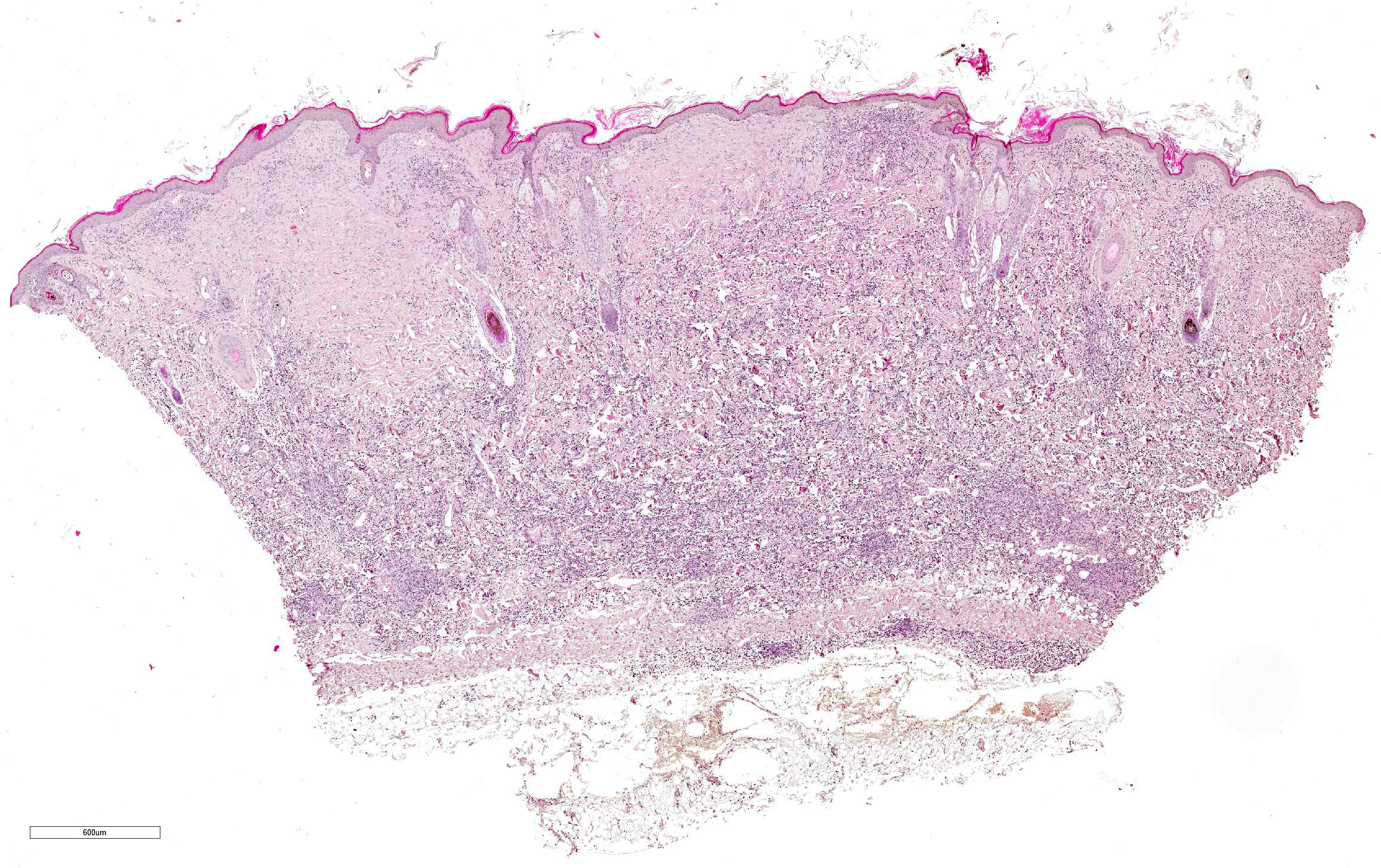
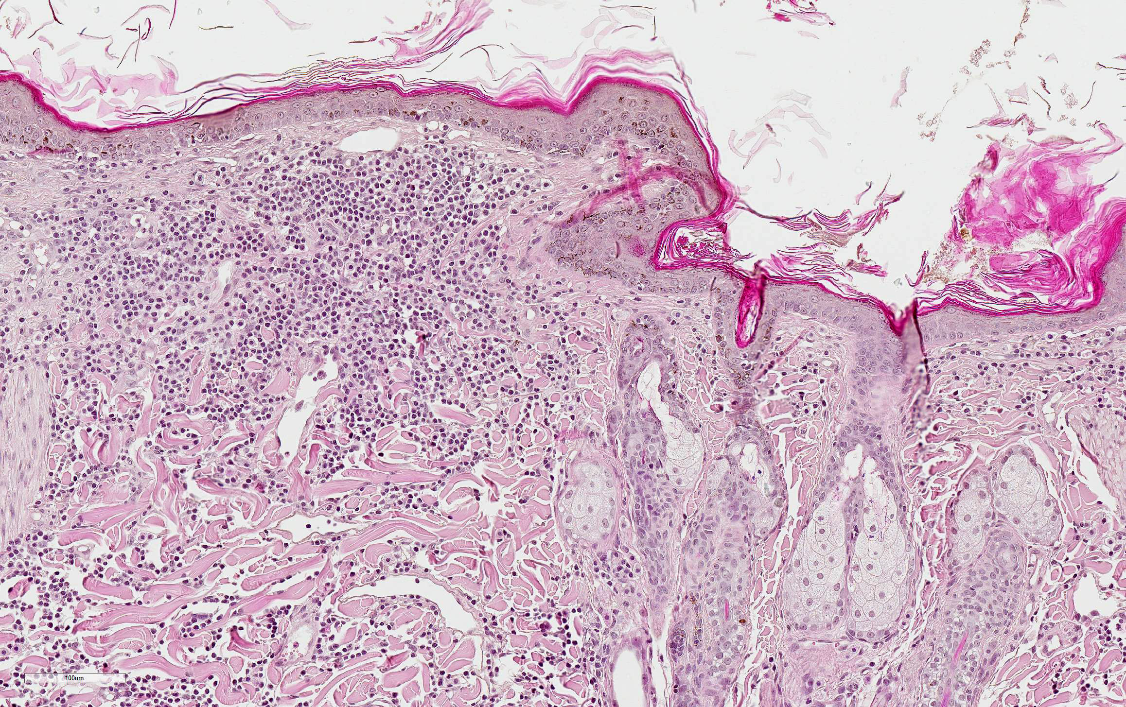
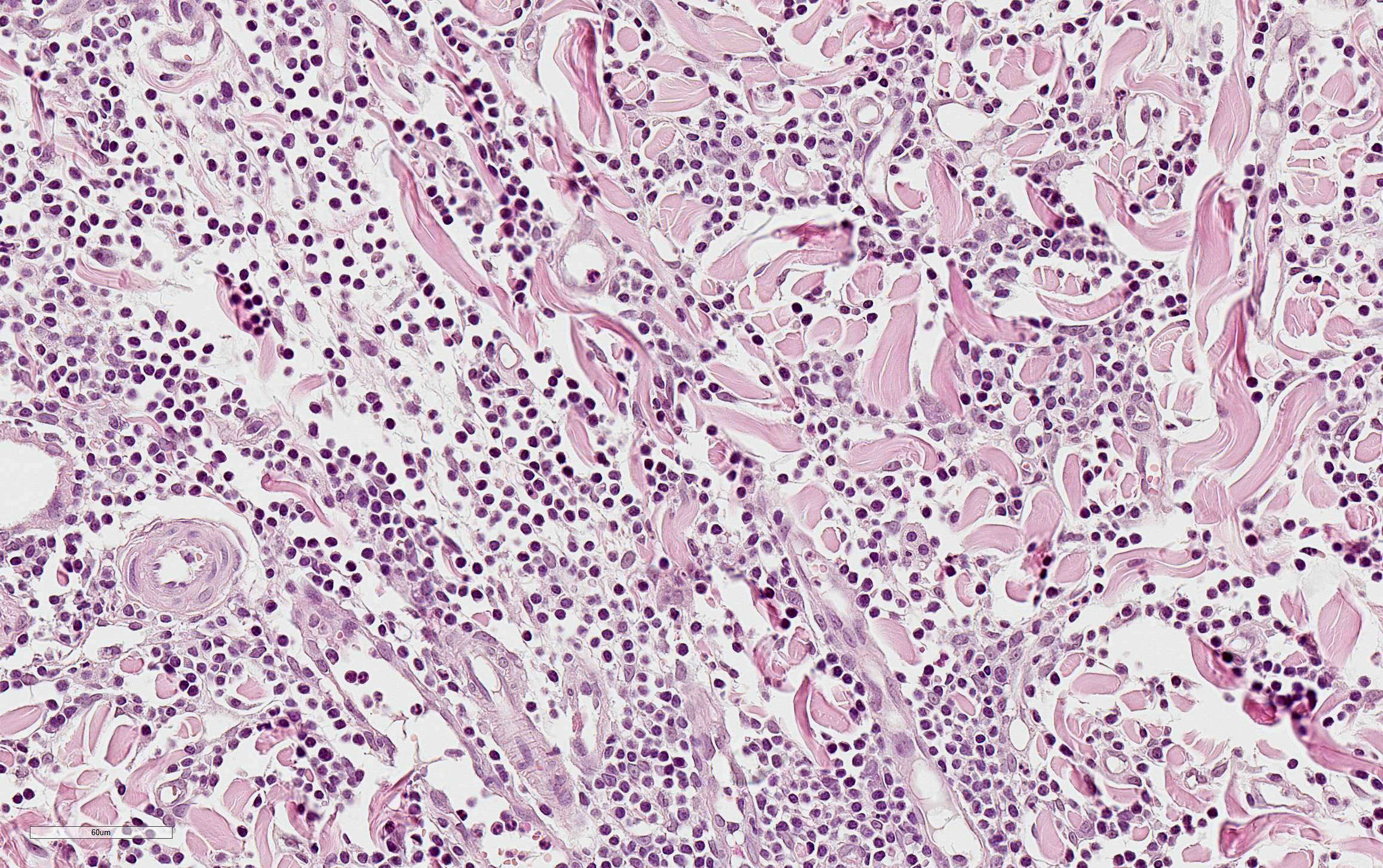
Haired skin: Multifocally to coalescent the dermis is loosely infiltrated by mostly uniform, small round cells with scant or only minimal pale amphophilic cytoplasm and dark round to oval nuclei with condensed chromatin consistent with mature lymphocytes. The infiltrate abuts the epidermal and follicular epithelia but infiltrates them only occasionally.
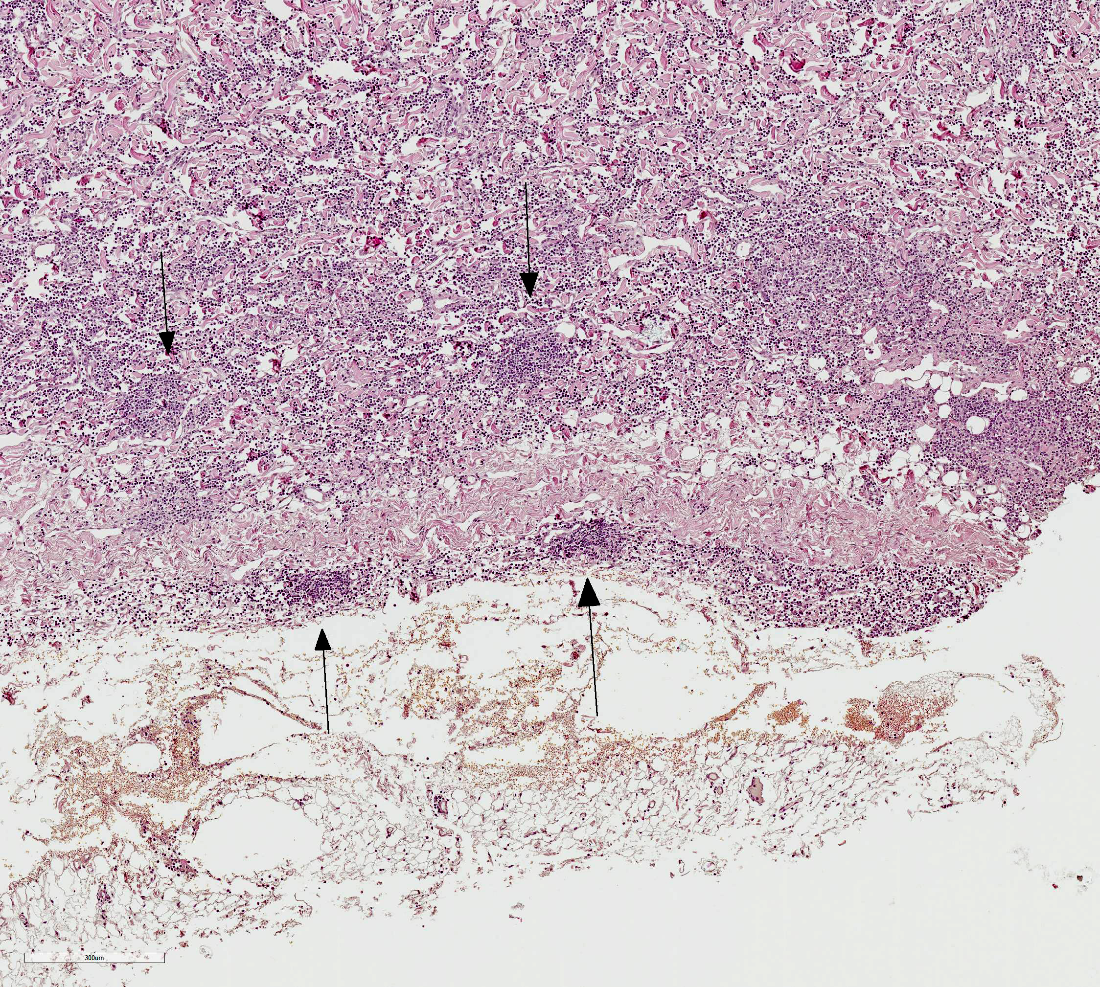
The deep dermis contains perivascular to nodular aggregates of tightly packed mature lymphocytes with almost no cytoplasm and dense basophilic nuclei. Mitotic figures were not seen.
Small numbers of mast cells, plasma cells and neutrophils are loosely intermingled. The epidermis shows mild to moderate epidermal hyperplasia and mild orthokeratotic hyperkeratosis.
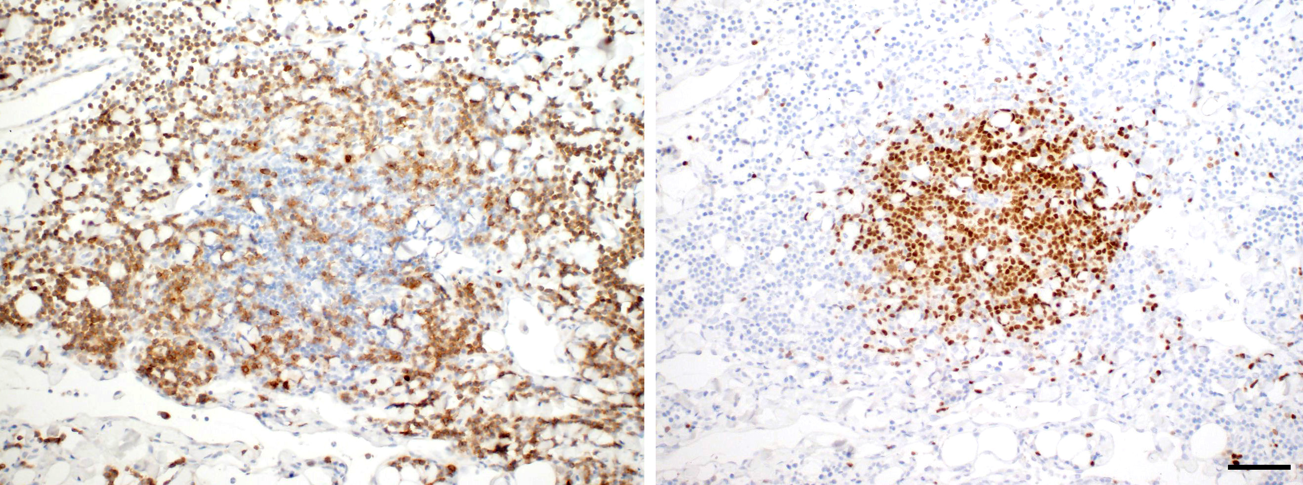
Immunohistochemistry : identified almost all cells in the superficial and deep dermis as CD3-positive T cells. The small, tightly packed aggregates in the deep dermis consisted of PAX5-positive B cells.
Contributor Morphologic Diagnosis:
Haired skin: cutaneous lymphocytosis, moderate to severe, diffuse
Contributor Comment: Cutaneous lymphocytosis is an uncommon disease and has been described in cats, dogs and humans 1,2,11. The condition is most often seen in older cats, without a breed predisposition, but a slight predominance in females. The etiology is unknown.2
The typical clinical presentation is characterized by acute onset with slow progression.2 Cutaneous lymphocytosis is mostly a solitary lesion showing alopecia, erythema and scaling, with or without crusting or ulceration. Pruritus can be seen in more than half of the cases, most commonly affecting the lateral thorax but also various other body sites (legs, pinnae, flank, neck, abdomen, hip, digit and planum nasale for instance).2 It is a relatively benign disease with rare occasions of systemic illness in some cats and dogs associated with anorexia, weight loss and well-differentiated T-cell infiltrates in internal organs.1,2 In most of the cases, treatment with glucocorticoids resulted in marked improvement of the clinical outcome.2
Cutaneous lymphocytosis in cats and dogs resembles cutaneous pseudolymphoma (lymphocytoma cutis, cutaneous lymphoid hyperplasia) in humans, a reactive proliferation of well-differentiated T- or B-cells in the skin of humans. In most cases, hypersensivity reaction to various antigenic stimuli has been described (such as arthropod bites, vaccination, tattoo dyes, drugs and contactants).6,7,10
In human cases, malignant transformation has been observed in some cases with systemic lymphoid involvement including enlarged mesenteric lymph nodes, infiltration of the liver, pancreas, stomach, kidney and heart by T lymphocytes and B cell infiltration of the small intestine.4,8,11
Histologically, cutaneous lymphocytosis in dogs and cats is characterized by proliferation of well differentiated CD3-positive T cells in the superficial and deep dermis and in most cases also nodular aggregates of CD79-positve B-cells in the dermis.2,4,11 In some cases, a T-cell receptor gamma gene rearrangement (TCRγ) can be verified suggesting that this condition is a low-grade or indolent T cell lymphoma.3
As a differential for cutaneous lymphocytosis, cutaneous T cell lymphoma must be considered. Cutaneous histiocytosis may be difficult to clinically or histologically differentiate from well-differentiated cutaneous malignant lymphoma with predominance of mature T-cells. However, histopathology of cutaneous T cell lymphomas often identifies epitheliotropic lymphoma characterized by a diffuse superficial or follicular epitheliotropic infiltrates of medium sized to large lymphocytes / lymphoblasts and in some cases additional Pautrierâs intraepidermal aggregates. In non-epitheliotropic lymphomas a deep dermal and subcutaneous infiltration of small to blastic lymphoid cells (bottom heavy configuration) with a variable mitotic activity can be seen.9
Contributing Institution:
Department of Veterinary Pathology, Freie Universität Berlin http://www.vetmed.fu-berlin.de/en/einrichtungen/institute/we12/index.html
JPC Diagnosis: Haired skin: Lymphocytosis, diffuse, marked, with mild acanthosis and orthokeratotic hyperkeratosis.
JPC
Comment: The contributor
has provided an excellent review of this uncommon entity in the dog and cat.
This particular specimen appears to very closely match descriptions in the
veterinary literature. Immunohistochemical stains run at the JPC revealed the
infiltrate to be overwhelmingly populated by CD-3 positive T-cells, with
scattered aggregates of CD-20 and Pax-5 positive B cells in the deeper regions
of the dermis.
In Gross et al, while the condition share most features in dogs and cats, cases
in the dog often demonstrate a Grenz or lymphocyte-free zone in the superficial
dermis at the dermo-epidermal junction.5
The attendees found it of note that a PubMed search for Dr. Nicolas Kluger7 (cited below) yielded 77 publications on dermatologic conditions involving tattoos.
The moderator pointed out that in the second reference by Gilbert at all, 14 of the 20 cases showed clonality, suggesting that this would be better called an indolent cutaneous lymphoma, although this term and cutaneous lymphocytosis are largely considered synonymous in the veterinary literature at this time.
In cats as well, these lesions tend to progress over time, and eventually often appear in the viscera, also suggesting that these cases may represent lymphoma rather than an inflammatory lesion.
References:
1. Affolter VK, Gross TL, Moore PF. Indolent cutaneous T-cell lymphoma presenting cutaneous lymphocytosis in dogs. Vet Dermatol. 2009 Oct 20;5- 6:577-585.
2. Gilbert S, Affolter VK, Gross TL, et al. Clinical, morphological and immunohistochemical characterization of cutaneous lymphocytosis in 23 cats. Vet Dermatol. 2004 Feb 15;1:3-12.
3. Gilbert S, Affolter VK, Schmidt P, et al. Clonality studies of feline cutaneous lymphocytosis. Vet Dermatol. 2004 Aug 15;S1:24.
4. Gilliam AC, Wood GS. Cutaneous lymphoid hyperplasia. Semin Cutan Med Surg. 2000 Jun 19;2:133-141.
5. Gross TL. Ihrke PJ, Walder EJ, Affolter VK. Lympocytic tumors. In: Skin Disesaes of the dog and cat: clinical and histopathologic diagnosis, 2nd Ed. Oxford UK Backwell Science 2003; .. 872-875.
6. Hartsock RJ. Postvaccinal lymphadenitis. Hyperplasia of lymphoid tissue that stimulates malignant lymphomas. Cancer. 1968 Apr 21;4:632- 649.
7. Kluger N, Vermeulen C, Moguelet P, et al. Cutaneous lymphoid hyperplasia (pseudolymphoma) in tattoos: a case series of seven patients. J Eur Acad Dermatol Venereol. 2010 Feb 24;2:208-213.
8. Lackey JN, Xia Y, Cho S, et al. Cutaneous lymphoid hyperplasia: a case report and brief review of the literature. Cutis. 2007 Jun 79;6:445-448.
9. Pariser MS, Gram DW. Feline cutaneous lymphocytosis: case report and summary of the literature. J Feline Med Surg. 2014 Sep 16;9:758-763.
10. Rijlaarsdam JU, Willemze R. Cutaneous pseudolymphoma: classification and differential diagnosis. Seminars in Dermatology. 1994 Sep 13;3:187-196.\
11. Rook KA. Canaine and Feline Cuatneous epitheliotropic lymphoa and cutaneous lymphocytosis. Vet Clin Small Anim 2019: 49:67-81.
12. Wood GS. Inflammatory diseases that stimulate lymphomas: cutaneous pseudolymphomas. In: Goldsmith AL, Katz SI, Gilchrest BA, et al. eds. Fitzpatrickâs dermatology in general medicine. New York, NY: McGraw Hill Medical; 2008:1402-1408.