Signalment:
Gross Description:
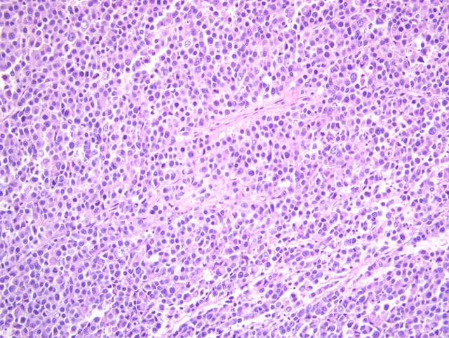
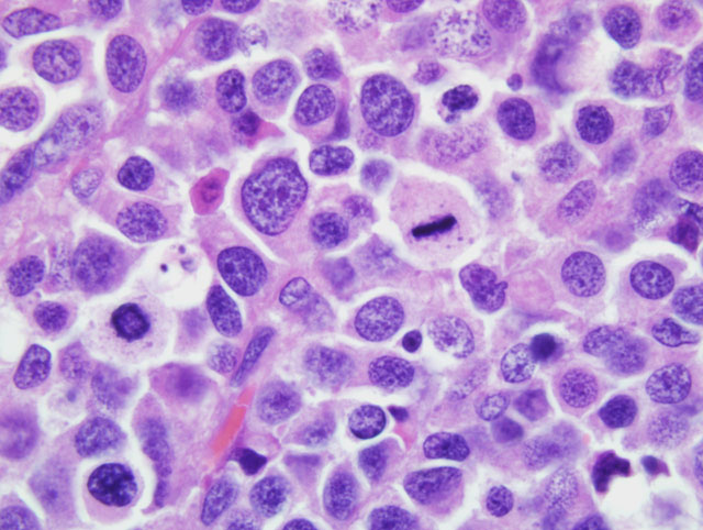
Histopathologic Description:
Morphologic Diagnosis:
Condition:
Contributor Comment:
JPC Diagnosis:
Conference Comment:
Conference participants reviewed the primary ovarian tumors of domestic species, among which the dysgerminoma is rare. As classified by the World Health Organization, these are summarized below:(3,4)
| Primary Ovarian Tumors of Domestic animals | ||
|---|---|---|
| Category | Notes | |
| Tumors of the surface coelomic epithelium | Papillary adenoma; papillary cystadenoma |
Common only in the bitch; arise from the coelomic mesothelium forming surface epithelium or subsurface epithelial structures (SES); form papillary projections covered by ciliated cuboidal to columnar cells, with or without glandular or cystic cavity formation |
| Papillary adenocarcinoma | Common only in the bitch; malignant counterpart of papillary adenomas; larger and extends through ovarian bursa; fronds may dislodge and cause metastatic implantations (carcinomatosis) and ascites) | |
| Rete adenoma | Rare; reported only in the bitch; arise from the tubular network of rete; differentiate from papillary adenomas by location in the tubal extremity of the ovarian medulla (vs. surface epithelium for the latter) | |
| Mesenchymal tumors | Hemangioma | Most common ovarian tumor of the sow; rare in the cow, mare, and bitch |
| Leiomyoma | Rare; reported in the bitch, queen, and sow; arise from smooth muscle of the mesovarium | |
| Sex cord-stromal (gonadostromal) tumors | Granulosa (granulosatheca) cell tumor; thecoma (theca cell tumor); interstitial cell tumor (luteoma, lipid cell tumor, steroid cell tumor) | Most common ovarian tumor of the cow and mare; slightly less common than epithelial tumors of the ovary in the bitch; infrequent in the queen; arise from granulosa and theca interna cells (and their luteinized counterparts); may produce estrogens or androgens; produce inhibin in the mare with contralateral ovarian atrophy; rarely metastasize; distinctive Call-Exner bodies are present histologically |
| Germ cell tumors | Dysgerminoma | Uncommon; arise from germ cells before differentiation; may metastasize or spread locally but biological behavior largely unknown due to low incidence in domestic species; histologically indistinguishable from the much more common testicular seminoma |
| Teratoma | Rare; arise from totipotential germ cells that have undergone somatic differentiation; consist of two or more of the three embryonic layers (i.e. endoderm, mesoderm, ectoderm); usually well-differentiated and benign | |
| Embryonal carcinoma | Arise from embryonic multipotential cells capable of further differentiation; variable histologic pattern |
References:
2. Jackson ML, Mills JHL, Fowler JD: Ovarian dysgerminoma in a bitch. Can Vet J 26:285-287, 1985
3. Kennedy PC, Cullen JM, Edwards JF, Goldschmidt MH, Larsen S, Munson L, Nielsen S: Histological Classification of Tumors of the Genital System of Domestic Animals, 2nd series, vol. IV, ed. Schulman YF, pp. 24-28. Armed Forces Institute of Pathology (in cooperation with the ARP and the WHO Collaborating Center for Worldwide Reference on Comparative Oncology), Washington, DC, 20306
4. Schlafer DH, Miller RB: Female genital system. In: Jubb, Kennedy, and Palmer's Pathology of Domestic Animals, ed. Maxie MG, 5th ed., vol. 3, pp. 450-454. Saunders Elsevier, Philadelphia, PA, 2007