Signalment:
Gross Description:
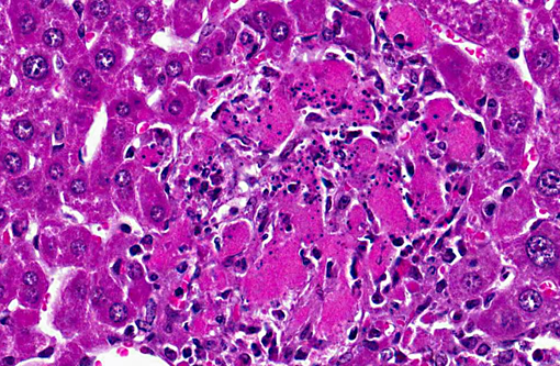
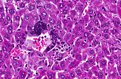
Histopathologic Description:
Morphologic Diagnosis:
Lab Results:
Condition:
Contributor Comment:
Mouse hepatitis virus is a single-stranded, enveloped RNA virus of the family Coronaviridae. Approximately 25 strains or isolates of MHV have been described. Isolates can be divided into two biotypes with varying degrees of overlap: the respiratory (polytropic) and enterotropic strains.(3) Respiratory strains of MHV are extremely contagious and are transmitted primarily via aerosol, direct contact, and fomites. Once inhaled, the virus replicates in the nasal mucosa and subsequently spreads via the blood and lymphatics through cervical and mesenteric lymph nodes to multiple tissues.(1) Dissemination is more likely with more virulent strains of virus in infant mice without maternal antibody and in immunocompromised (particularly nude) mice. MHV has high morbidity and mortality in these groups. Immunocompetent mice may clear the infection in five to seven days post-infection with no carrier status. BALB/c mice are generally quite susceptible to MHV, whereas SJL mice are remarkably resistant. The spike protein (SP), responsible for viral entry, is a major determinant of tropism and virulence.(2) SP binds carcinoembryonic antigen-related cell adhesion molecule 1, CEACAM- 1 on either the cell surface or in endosomes. SP also mediates cell-to-cell fusion. The allelic form of CEACAM in a particular strain of mouse is a determinate of susceptibility. Strains that express CEACAM-1α such as Balb/C are susceptible. Strains that express CEACAM -1β such as SJL, are resistant. (2) Disease severity is determined by thrombosis and coagulation necrosis due to induction of procoagulant activity by macrophages in susceptible mice.(5) T cell function is important for viral clearance depending on both CD8 activity and antibody and cytokine induction. (2)
Enterotropic strains of MHV selectively infect intestinal mucosal epithelium, with minimal to no dissemination to other organs, even in immunodeficient mice.(4) All stages and strains of mice are susceptible to enterotropic MHV infection, including SJL mice, which are resistant to polytropic MHV.(5) The severity of intestinal disease is associated with age-related intestinal mucosal proliferative kinetics, thus severe disease occurs only in infant mice.(4) Even in nude or SCID mice, disease can be minimal; however hyperplastic colitis has been reported in nude mice5. Recovery from enterotropic MHV is T-cell dependent; persistent infections can occur in immunodeficient mice. (3)
Diagnosis is usually made via serology using an ELISA, but MHV may also be isolated in susceptible tissue culture cells. Differential diagnoses include Salmonellosis, Tyzzers disease and reovirus infection. (5) Respiratory syncytial virus forms syncytial cells similar to MHV, but these cells are restricted to the pulmonary parenchyma.Â
JPC Diagnosis:
Conference Comment:
References:
2. Bender SJ, Weiss SR. Pathogenesis of murine coronavirus in the central nervous system. J Neuroimm Pharm. 2010; 5(3): 336-354.
3. Compton SR, Barthold SW, Smith AL. The cellular and molecular pathogenesis of coronaviruses. J Lab Animals. 1993; 43: 15-28.
4. Homberger FR. Enterotropic mouse hepatitis virus. J Lab Anim. 1997; 31(2): 97-115.
5. Percy DH, Barthold SW. Coronavirus infection: Mouse Hepatitis Virus. In: Pathology of Laboratory Rodents and Rabbits. 3rd ed. Ames, IA: Blackwell Publishing; 2007: 31-36.
6. Saif LJ. Coroniviridae. In: MacLachlan NJ, Dobovi EJ, eds. Fenners Veterinary Virology. 4th ed. London,UK: Elsevier; 2011:404-406.