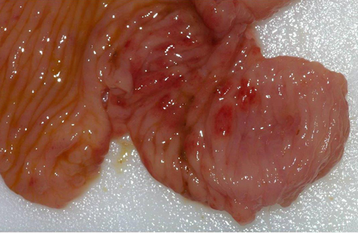
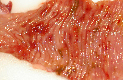
Signalment:  

1.5-year-old male non-castrated colony beagle, dog (Canis familiars).The dog was received from the provider when it was eight months old. Until the onset of clinical signs, it was never part of a toxicology study. One month before euthanasia, this dog presented with bloody stool, which was refractory to metronidazole and fenbendazole. Colonoscopy revealed multiple erosive lesions in distal colon. 


Gross Description:  

Body condition was thin. There were small amounts of adipose tissue in the subcutis and abdomen. Mesenteric and submandibular lymph nodes were slightly enlarged and red. The cecum had numerous, sometimes coalescing, 0.3 to 0.6 cm diameter erosions-ulcers in the mucosa. Similar findings were observed in the distal colon, 5 to 12 cm cranial to the anus, where the mucosa was thickened, reddened and numerous 0.3 to 0.8 cm erosions-ulcers were present. 


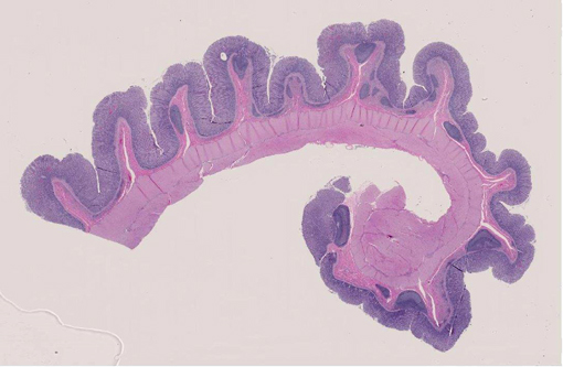
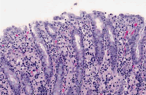
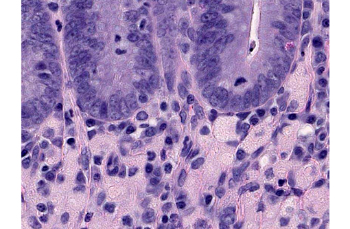
Histopathologic Description:

Multifocally expanding the mucosa of the colon, separating mucosal crypts and extending into the submucosa, there is a dense inflammatory infiltrate composed of large numbers of macrophages and fewer neutrophils, and occasional lymphocytes and plasma cells. These macrophages have abundant eosinophilic cytoplasm and occasionally contain pale eosinophilic, variably sized vacuoles, bacillary bacteria or cellular debris in the cytoplasm. Along the mucosal surface, there are multiple erosions of the epithelium, with the presence of numerous bacillary bacteria, and associated with necrotic cellular debris in the mucosal and apical lamina propria. Colonic crypts are often dilated with desquamated and necrotic cellular debris, and lined by flattened epithelium. Additionally, there are focal hemorrhages at the apical portion of the mucosa. Numerous foamy macrophages are separating the basal crypts from the muscularis mucosa and are present in the submucosa, surrounding mucosa associated lymphoid tissue. Frequently in the crypts, there are numerous spirochetes.

In the cecum (not submitted), findings are similar as described above. 

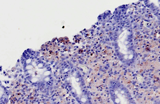
Macrophages infiltrating the colonic mucosa and submucosa are PAS-positive (data not shown). Immunohistochemical stains using polyclonal anti-Escherichia coli antibody highlighted numerous bacillary bacteria present free in the upper lamina propria and in the cytoplasm of macrophages.


Morphologic Diagnosis:  

Colon and cecum: Marked, multifocal to coalescing, chronic histiocytic typhlocolitis, with intralesional Escherichia coli.


Condition:  

Canine histiocytic ulcerative colitis


Contributor Comment:  

Canine histiocytic ulcerative colitis is an idiopathic, probably multifactorial, inflammatory condition of the cecum, colon and rectum of dogs. This condition was first described by Van Kruiningen et al. (1965),(9) after observing chronic hemorrhagic diarrhea in a Boxer colony, which was characterized as a granulomatous colitis with resemblance to Whipples disease. Subsequent reports led to the use of the term of canine histiocytic ulcerative colitis (CHUC), which has since gained general acceptance.(6) CHUC has been distinctively recognized in Boxers and the related French bulldogs. Nevertheless, this condition has also been described in a Doberman pinscher, a Mastiff and one Alaskan malamute.(7) Here, we report the first case of CHUC in a colony Beagle dog. 

Grossly, the colon of affected dogs is thickened, folded and perhaps dilated, with focal areas of scarring. On the mucosa, the lesion varies from red, frequently raised areas to patchy or coalescing ulcers. Histologically, it is characterized by a dense inflammatory infiltrate in the mucosa and submucosa, composed mainly by large numbers of foamy, PAS-positive macrophages which contain phagocytized necrotic cellular debris and bacteria. Additionally, there is distortion of the normal glandular architecture and decreased numbers of goblet cells. Ulceration, which does not progress beyond the submucosa, arises from the superficial epithelial erosion and destruction of the basement membrane.(1) Resultant clinical signs consist of large bowel diarrhea, tenesmus, hematochezia and marked weight loss.(4)

The inflammatory infiltrate of the colonic mucosa and submucosa was further characterized in an immunohistochemical study. German et al (2000) found increased IgG plasma cells, plus PAS, CD3, L1 and MHC class II positive cells in the lamina propria, in conjunction with increased enterocyte MHC II class expression, and decreased goblet cell numbers in the epithelium. These findings are similar to those of human ulcerative colitis and might suggest a similar pathogenesis at a cellular level.(3)

The exact cause of this condition remains unknown, and a multi-factorial etiology is highly suspected. In an ultrastructural study, it was concluded that CHUC was probably caused by a lipid-rich, ribosome-rich, coccoid to coccobacillary organism that possesses a cell membrane and range from 100 to 500 nm in size. This agent was suspected to be a Chlamydia-like organism.(10) In more recent reports, the infectious agent hypothesis was again brought to attention, since dogs completely recover after treatment with enrofloxacin.(2,4) In one study, there was demonstration of E. coli in 100% of the examined colonic samples with a polyclonal antibody, strongly associating this organism to the disease. In the same study, there was a comparison of this entity with granulomatous leptomeningitis of Beagle dogs, and malakoplakia and xanthogranulomatous cholecystitis in man, in which E. coli has also been incriminated, together with abnormal lysosomal function.(11) In 2009, Mansfield et al. observed remission of CHUC in Boxer dogs in correlation with the eradication of invasive intramucosal E. coli, further supporting the causal involvement of E.coli in the development of the disease in susceptible Boxer dogs.(5)


JPC Diagnosis:  

Colon: Colitis, histiocytic, multifocal, moderate, with glandular hyperplasia.


Conference Comment:  

In addition to reviewing the contributors excellent summary of canine histiocytic ulcerative colitis (CHUC), conference participants discussed the use of the term histiocytic versus granulomatous, noting that histiocytic is preferred in this condition, as the lesions lack the epithelioid macrophages and multinucleated giant cell macrophages that are characteristic of granulomatous inflammation. The comparison between human inflammatory bowel disease (IBD), which encompasses both Crohns disease and ulcerative colitis, and CHUC is interesting. Human IBD is thought to be the result of a combination of defects affecting the hosts interaction with intestinal microbes as well as intestinal epithelial dysfunction and abnormal immune responses.(8) Additionally, in humans, Crohns disease is thought to be associated with a TH-1 response; whereas ulcerative colitis has been suggested to be a TH-2 mediated disease.(8) This may not be clear-cut, however, as polymorphisms of the IL-23 receptor have been shown to be protective against both Crohns disease and ulcerative colitis, suggesting there may also be involvement of a TH-17 response in both conditions. Furthermore, anti-TNF therapy has been effective in some human patients with ulcerative colitis, indicating that a TH-1 response may play a role in ulcerative colitis as well.(8) As the contributor notes, similar to IBD in humans, a multifactorial etiology is suspected in CHUC, although neither has yet to be fully elucidated. Participants noted some slide variation with some slides exhibiting crypt abscesses and/or crypt herniation absent in other slides.


References:

1. Brown CC, Baker DC, Barker IK. Alimentary system. In: Maxie MG, ed. Jubb, Kenedy and Palmers Pathology of Domestic Animals. 5th ed. Vol 2. Philadelphia, PA: Elsevier; 2007:112-114.
2. Davies DR, OHara AJ, Irwin PJ, Guilford WG. Successful management of histiocytic ulcerative colitis with enrofloxacin in two Boxer dogs. Aust Vet J. 2004;82:58-61.
3. German AJ, Hall EJ, Kelly DF, Watson ADJ, Day MJ. An immunohistochemical study of hisitiocytic ulcerative colitis of Boxer dogs. J Comp Path. 2000;122:163-175.
4. Hostutler RA, Luria BJ, Johnson SE, Weisbrode SE, Sherding RG, Jaeger JQ, et al. Antibiotic-responsive histiocytic ulcerative colitis in 9 dogs. J Vet Intern Med. 2004;18:499-504.
5. Mansfield CS, James FE, Craven M, Davies DR, OHara AJ, Nicholls PK, et al. Remission of Histioytic ulcerative colitis in boxer dogs correlates with eradication of invasive intramucosal Escherichia coli. J Vet Intern Med. 2009;23:964-969.
6. Sander CH, Langham RF. Canine histiocytic ulcerative colitis. A condition resembling Whipples disease, colonic histiocytosis and malakoplakia in man. Arch Pathol. 1968;85:94-100. 
7. Stokes JE, Kruger JM, Mullaney T, Holan K, Schall W. Histiocytic ulcerative colitis in three non-boxer dogs. J Am Anim Hosp Assoc. 2001;37:461-465. 
8. Turner JR. The Gastrointestinal tract. In: Kumar V, Abbas AK, Fausto N, Aster JC, eds. Robbins and Cotran Pathologic Basis of Disease. 8th ed. Philadelphia PA: Saunders Elsevier; 2010:807-813.
9. Van Kruiningen HJ, Montali RJ, Strandberg JD, Kirk RW. A granulomatous colitis of dogs with histologic resemblance to Whipples disease. Path Vet. 1965;2:521-544. 
10. Van Kruiningen HJ. The ultrastructure of macrophages in granulomatous colitis of Boxer dogs. Vet Pathol. 1975;12:446-459.
11. Van Kruiningen HJ, Civco IC, Cartun RW. The comparative importance of E. coli antigen in granulomatous colitis of Boxer dogs. APMIS. 2005;113:420-425.


Click the slide to view.


2-1. Cecum


2-2. Colon


2-3. Colon


2-4. Colon


2-5. Colon


2-6. Colon


Back | VP Home | Contact Us |