Signalment:  

A five-month-old male white-tailed deer (Odocoileus virginianus).The fourth fawn to die from the herd.


Gross Description:  

The cornea of the right eye was partially cloudy. There was a 1.5 cm area of hemorrhage in the skin of the right ear. There was subcutaneous yellow edema fluid in the ventral neck, surrounding the trachea and esophagus. The right half of the thorax contained a large blood clot. Dorsal to the lungs and heart there was a blood clot approximately 20 cm long that surrounded the aorta and was attached to the musculature ventral to the spinal column. There was acute hemorrhage in one of the adrenal glands and the left testicle. There were small multifocal hemorrhages in the right testicle. 


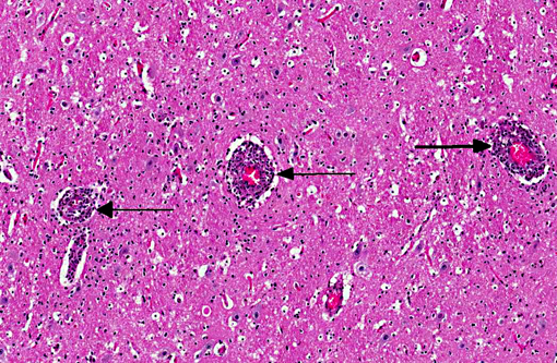
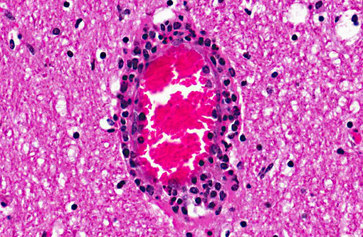
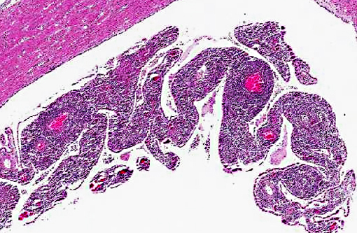
Histopathologic Description:

Cerebrum: Surrounding numerous meningeal and parenchymal vessels is an infiltrate composed of low to medium numbers of lymphocytes and neutrophils. The inflammatory cells often are equally present in the tunica media and tunica adventitia. There are variable numbers of macrophages, lymphocytes, and occasional plasma cells in the meninges. There are scattered glial cell infiltrates within the neuropil.

In most of the sections, there are several vessels in the leptomeninges that have fibrinoid necrosis of the vessel wall. Affected vessels often have adventitial to subintimal accumulation of neutrophils, lymphocytes, and macrophages.

In some sections, choroid plexus is present; it is heavily infiltrated by lymphocytes, plasma cells, macrophages, and neutrophils.


Morphologic Diagnosis:  


1. Cerebrum: Vasculitis and perivasculitis, necrotizing, lymphocytic, multifocal, moderate.
2. Cerebrum: Meningoencephalitis, lymphocytic, diffuse.


Lab Results:  

No anaerobic bacterial growth from lung, liver, or brain. Negative for epizootic hemorrhagic disease virus by PCR. PCR positive for Caprine Herpesvirus-2.


Condition:  

Malignant catarrhal fever, caprine herpesvirus-2


Contributor Comment:  

Gross and histologic lesions were consistent with malignant catarrhal fever (MCF), and infection with caprine herpesvirus 2 was confirmed by PCR testing. MCF occurs in ruminant species and is caused by several herpesviruses, an enveloped, linear, doubled-stranded DNA virus. These viruses are in the Rhadinovirus genus of the subfamily Gammaherpesvirinae. There are currently four known members of the MCF virus group: alcelaphine herpesvirus 1 (AlHV-1), ovine herpesvirus 2 (OvHV-2), caprine herpesvirus 2 (CpHV-2), and a gammaherpes virus found in white-tailed deer with no known reservoir (MCFV-WTD).(3,4) Infection of white-tailed deer with CpHV-2 has been previously reported.(3)

Typical gross changes associated with MCF include conjunctivitis, cutaneous exanthema, crusting, and alopecia, nasal discharge, oral and esophageal ulcerations, urinary mucosal hemorrhages, and lymphoid enlargement.(2) Mortality in susceptible species approaches 100%; however, in the natural host, infection is latent or inapparent with intermittent virus shedding. These viruses are difficult or impossible to isolate in cell culture.

The common histologic changes associated with MCF are lymphocytic perivasculitis and vasculitis with fibrinoid necrosis of medium sized arteries, and lymphoid hyperplasia.(2) Lesions are characterized by a proliferation of CD8+ T lymphocytes and tissue necrosis.(5,7) The mechanism of proliferation and vasculitis is unknown.(5)

MCF has been documented in white-tailed deer, along with other wild cervids, pigs, and cattle.(1,2,8) Raising bovids and deer around other small ruminants (sheep, goats) can be risky due to transmission of the MCF viruses from the host species that is not clinically ill. The incubation period is usually 2-10 weeks, but may on occasion be very much longer than this. Epizootic hemorrhagic disease (orbivirus) is a differential diagnosis for MCF in deer with acute hemorrhage.


JPC Diagnosis:  

Cerebrum: Arteritis and phlebitis, lymphoblastic and necrotizing, diffuse, moderate, with meningitis and choroid plexitis.


Conference Comment:  

The contributor provides an excellent summary of the gamma herpesviruses associated with malignant catarrhal fever in various species. In addition to the four members of the MCF virus group, there are several viruses that have been reported to cause MCF or MCF-like diseases in various species of hoofstock within zoological collections.(6) Recently, one such novel virus (MCFV-ibex) was identified as the etiologic agent for MCF observed in bongo antelope. In this outbreak, a Nubian ibex was found to be the source of the virus. The presentation of MCF in bongos differs from the classic presentation in domestic ruminants and deer in the following ways: The bongos developed necrotizing cholangiohepatitis, neutrophilic necrotizing myocarditis, and they lacked the typical erosive or ulcerative oronasal lesions and enteritis often seen in other ruminants with MCF.(6)


References:

1. Alcaraz A, Warren A, Jackson C, Gold J, McCoy M, Cheong SH, et al. Naturally occurring sheep-associated malignant catarrhal fever in North American pigs. J Vet Diagn Invest. 2009;21:250-253.
2. Brown CC, Baker DC, Barker IK. The Alimentary system. In: Maxie MG, ed. Jubb, Kennedy and Palmers Pathology of Domestic Animals, 5th ed. Vol 2. Philadelphia, PA: Elsevier Saunders; 2007:152-158.
3. Li H, Wunschmann A, Keller J, Hall DG, Crawford TB. Caprine herpesvirus-2associated malignant catarrhal fever in white-tailed deer (Odocoileus virginianus). J Vet Diagn Invest. 2003;15:4649.
4. Li H, Dyer N, Keller J, Crawford TB. Newly recognized herpesvirus causing malignant catarrhal fever in white-tailed deer (Odocoileus virginianus). J Clin Micro. 2000;38(4):13131318.
5. Dewals B, Boudry C, Farnir F, Drion PV, Vanderplasschen A. Malignant catarrhal fever induced by alcelaphine herpesvirus 1 is associated with proliferation of CD8+ T cells supporting a latent infection. PLoS ONE. 2008;3(2):e1627.
6. Gasper D, Barr B, Li H, Taus N, Peterson R, Benjamin G, et al. Ibex-associated malignant catarrhal fever−like disease in a group of bongo antelope (Tragelaphus eurycerus ). Vet Pathol. 2012:49;492. 
7. Russell GC, Stewart JP, Haig DM. Malignant catarrhal fever: A review. Vet J. 2009;179(3):324-35.
8. Vik+�-+ren T, Li H, Lillehaug A, Jonassen CM, B+�-�ckerman I, Handeland K. Malignant catarrhal fever in free-ranging cervids associated with OvHV-2 and CpHV-2 DNA. J Wildl Dis. 2006;42(4):797-807.


Click the slide to view.


3-1. Cerebrum at level of lateral ventricles


3-2. Cerebrum at level of lateral ventricles


3-3. Cerebrum at level of lateral ventricles


Back | VP Home | Contact Us |