Signalment:
Gross Description:
Histopathologic Description:
Morphologic Diagnosis:
Lab Results:
Condition:
Contributor Comment:
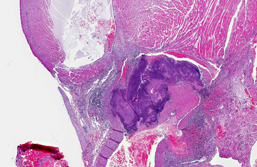
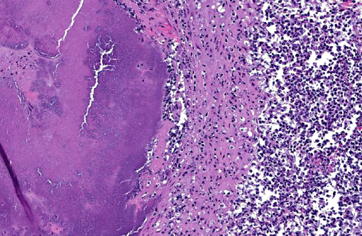
Bacterial endocarditis primary arises from adhesion of the microorganisms to the endocardium, leading to death of the endothelium and formation and adherence of a thrombus within which large colonies of bacteria proliferate. Such proliferative growths and thrombus are called vegetative endocarditis. Although not observed in the opossum, pieces of the vegetation may break free and circulate to other organs, causing septic infarcts or abscesses. It is not uncommon for valvular endocarditis to cause cardiac dysfunction leading to congestive heart failure.
JPC Diagnosis:
Conference Comment:
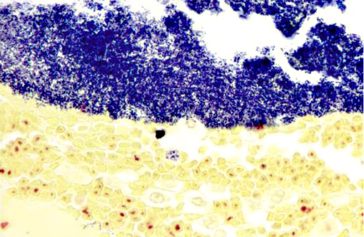
Spontaneous bacterial endocarditis in the Virginia opossum (Didelphis virginiana) is typically due to Streptococcus viridans or Staphylococcus aureus.(4) Although culture was not performed in this case, a tissue Gram stain reveals numerous intralesional Gram-positive cocci, supporting a similar etiology in this case. In research species, bacterial endocarditis has been associated with the use of vascular access ports and intravenous catheters. Vegetative valvular endocarditis is also seen in ruminants, swine, dogs, and rarely in cats and horses. Streptococcus sp., Staphylococcus sp. and E. coli are frequently implicated as the etiologic agents in many species. Additionally, Erysipelothrix rhusiopathiae is often isolated in pigs and (occasionally) dogs, while Bartonella sp. is more specific to the dog or the cat. Arcanobacterium pyogenes is a common pathogen in cattle and Actinobacillus equuli can occasionally cause valvular endocarditis in horses.(2) Regardless of the inciting cause, this condition can result in valve damage and the development of congestive heart failure, or detachment of the vegetations with subsequent embolic disease.(2) In this case, because the lesion is located in the left heart at the aortic valve, the kidney would be a likely anatomic location for secondary embolic lesions.
References:
2. Maxie MG, Robinson WF. Cardiovascular system. In: Maxie MG, ed. Jubb, Kennedy and Palmers Pathology of Domestic Animals. 5th ed. Vol. 3. Philadelphia, PA: Elsevier Saunders; 2007:27-29.
3. Samollow PB. The opossum genome: insights and opportunities from an alternative mammal. Genome Res. 2008;18(8):1199-1215.
4. Sherwood BF, Rowlands MD, Vakilzadeh J, LeMay JC. Experimental bacterial endocarditis in the Opossum (Didelphis virginiana). Am J Pathol. 1971;64(3):513-520.
5. Xie Q, Mackay S, Ullmann SL, Gilmore DP, Payne AP, Gray C. Postnatal development of Leydig cells in the opossum (Monodelphis domestica): an immunohistochemical and endocrinological study. Biol Reprod. 1998;58(3):664-669.