Joint Pathology Center
Veterinary Pathology Services
Wednesday Slide Conference
2019-2020
Conference 6
2 October, 2019
Dr.
Jeff Wolf, DVM
Senior Pathologist
Chief Scientific Officer/Pathology Manager
EPL, Inc.
Sterling, VA
CASE IV: WSC 18-19 #2 (JPC 4117006).
>
Signalment: 1-year-old, male, Puerto Rican crested toad (Peltophryne lemur)
History: The toad was found dead in its enclosure after an approximately several weeks long treatment for skin issues.
>
Gross Pathology: At necropsy, the toad was moderately desiccated and the skin of the ventrum and legs was diffusely brown and was easily peeled away from the body.
Laboratory results: N/A
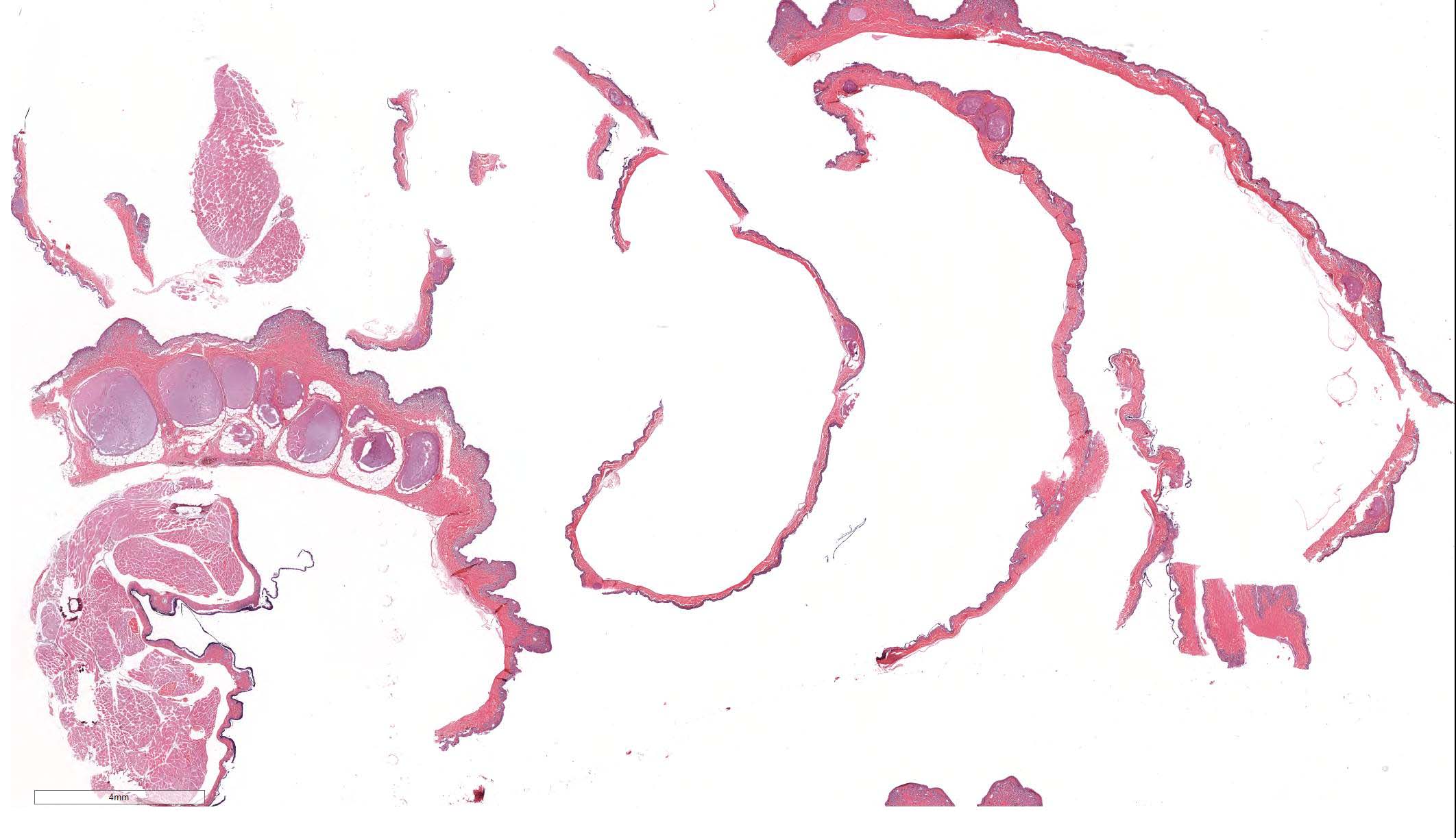
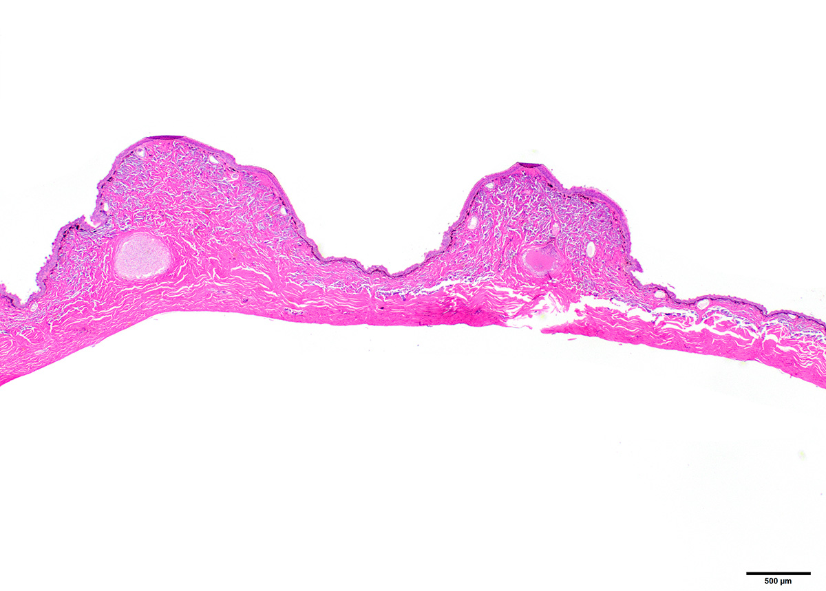
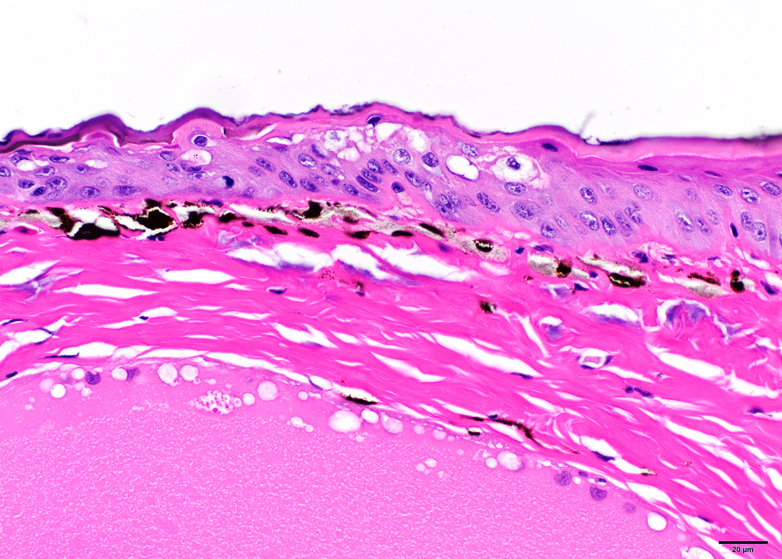
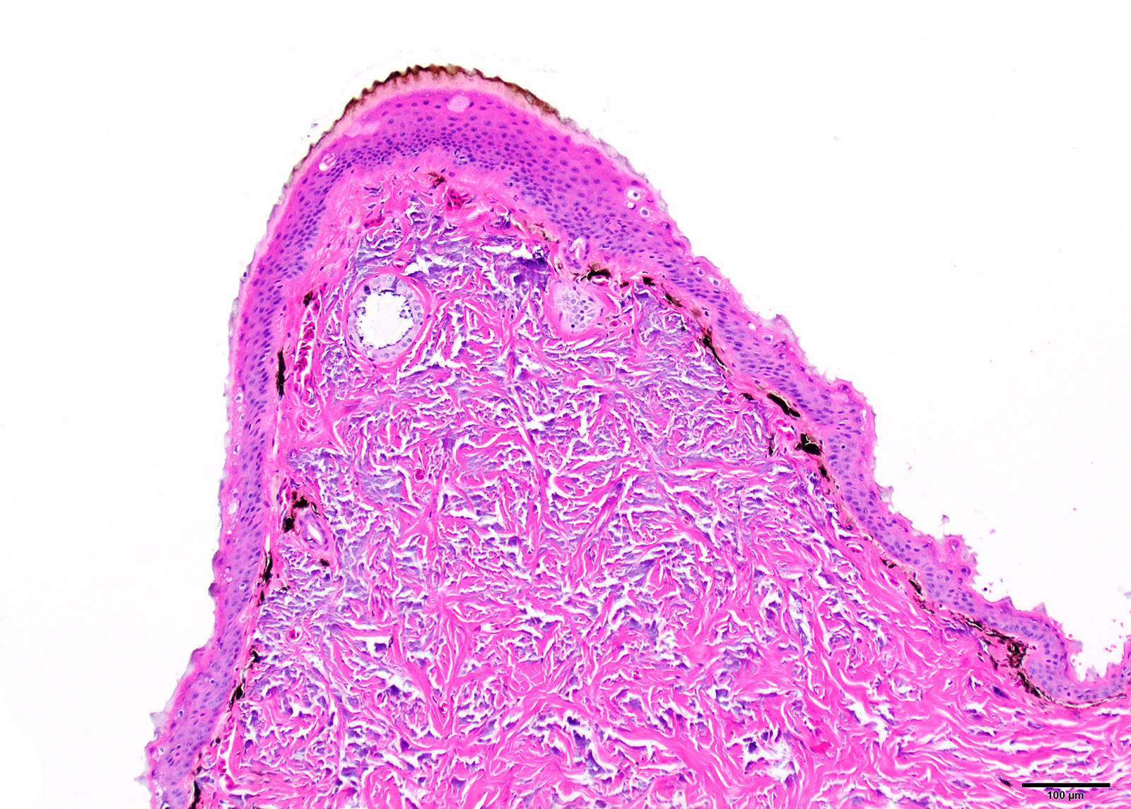
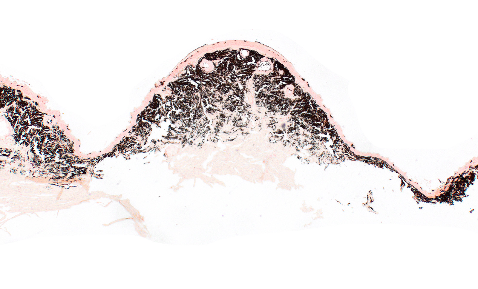
Microscopic Description:
There is multifocal to diffuse hyperplasia of the epidermis which is often covered by orthokeratotic hyperkeratosis. This keratin layer is often deep brown. Within the hyperplastic epidermis there are rare apoptotic keratinocytes and intracellular edema (spongiosis).There is a nearly diffuse layer of prominent melanocytes within the superficial dermis immediately adjacent to the basal layer of the epidermis. There is frequent thickening of the dermal Eberth-Katschenko (calcium) layer.
Contributor Morphologic Diagnosis:
Skin, Epidermal hyperplasia, orthokeratotic hyperkeratosis, spongiosis, rare keratinocyte apoptosis, and dermal melanocytosis, moderate, diffuse with thickened Eberth-Katschenko layer
Contributor Comment: The changes seen in the sections of skin from the submitted toad are consistent with the entity described as Brown Skin Disease of Puerto Rican Crested Toads (PRCT). Brown skin disease (BSD) is an idiopathic disease that has only been described in this species of toad. Grossly the disease is characterized by leathery, brown discoloration of the skin that can occur anywhere on the body with abnormal shedding of the skin (dysecdysis) particularly on the ventrum.1 Microscopically BSD is typically characterized by epithelial hyperplasia, spongiosis, orthokeratotic or parakeratotic hyperkeratosis with ’nuclear ghosts’ in keratinized epithelial cells, and occasional basal layer disorganization.1 Other features include single cell necrosis throughout the epithelium, erosion or ulceration, and thickening of the Eberth-Katschenko layer (a normal layer of mineralization present in the skin).1 See Crawshaw et al for excellent comparisons of normal and affected PRCT skin histology images.
Examination of many cases has failed to reveal a cause for the disease. Attempts at bacterial cultures have revealed growth of a number of different bacterial genuses including Aeromonas, Acinetobacter, and many others but these have been considered normal flora or contaminants and attempts to identify common fungi of toads including chytrid fungi have all been negative.1 In one study, supplementation of Vitamin A in 48 captive born PRCT failed to reduce the severity of disease when it developed.2 Further testing for bacterial or fungal organisms was not pursued in this case. Another PRCT was submitted from the same zoological collection and was similarly diagnosed with BSD. In that case bacterial culture of the skin revealed abundant growth of Providencia rettgeri, Acinetobacter sp., Pseudomonas putida, Citrobacter freundii, Enterococcus faecalis, Microbacterium sp., and Staphylococcus sp. (non-hemolytic). Fungal cultures of this second toad revealed unidentified yeast and further testing was not pursued.
Puerto Rican Crested Toads are a threatened species and have been listed as endangered by the International Union for Conservation of Nature (IUCN) since 1987.1 Since then a number of the species have been housed in a number of zoos in an attempt to save the species. Given the lack of a convincing infectious cause for this disease is it possible that a genetic component contributes to this disease. Evidence for this includes the initial case of BSD occurring in one male of a group of four toads that represented the last four of the Northern race of PRCT, six offspring from a pairing of this same group, and multiple animals from succeeding generations from this initial pairing.1 Despite this, cases of BSD have occurred in unrelated individuals from the Southern race housed at different zoos in close proximity to affected animals and on similar diets thus the true etiology remains elusive.1
Contributing Institution:
Kansas State Veterinary Diagnostic Laboratory, Department of Diagnostic Medicine/Pathobiology, www.ksvdl.org
JPC Diagnosis: Skin:
Epidermal hyperplasia and hyperkeratosis, diffuse, marked, with dermal
melanosis and mineralization.
JPC Comment: The
contributor has provided a concise review of this species-specific disease
which unfortunately is a problem in a threatened species. The contributor
describes the common finding of brown leathery skin in affected toads, which
does not apparently contain an increase in dermal melanocytes to explain the
coloration. Attempts to soak and remove affected skin from individuals have
been successful, but the disease recurs again soon after.1
A wide range of environmental factors (to include husbandry issues) and
infectious agents have been incriminated as causing skin lesions in anurans.
Once considering the problem of dysecdysis (abnormal skin shedding), low
humidity, low temperature, and poor nutrition have all been implicated in this
dysecdysis.2
Hypovitaminosis A has been particularly incriminated in amphibians with dysecdysis. Like all other species, retinoids must be obtained via the det in the form of provitamin A carotenoids or preformed Vitamin A. When absorbed, enterocytes hydrolyze dietary retinol esters or carotenoids to retinol where they are combined with dietary fat and cholesterol to form retinyl ested-laden chylomicons and resecreted. Dissolution of chylomicra in circulation allows the delivery of retinoids to peripheral tissues; remaining retinoids are taken up by the liver. In the skin, retinoids perform an important function in the regulation of gene transcription and affect processes of cellular proliferation, differentiation, and apoptosis.2
References:
1. Crawshaw G, Pienkowski M, Lentini, A, et al. Brown Skin Disease: A Syndrome of Dysecdysis in Puerto Rican Crested Toads (Peltophryne lemur). Zoo Bio. 2014. 33:558-564.
2. Dutton C, Lentini A, Berkvens, et al. The Effect of Supplementation with Vitamin A on Serum and Liver Concentrations in Puerto Rican Crested Toads (Peltophryne lemur) and its lack of Impact on Brown Skin Disease. Zoo Bio. 2014. 33: 553-557.