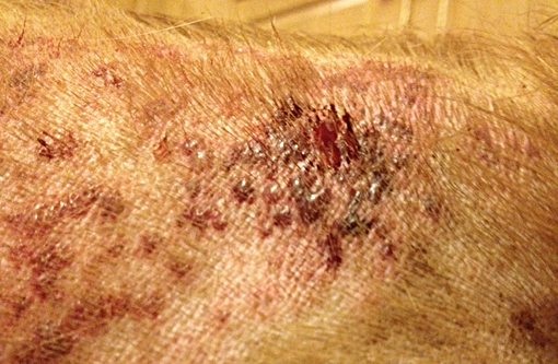
Signalment:  

1-year-old spayed female golden retriever (Canis familiars).The dog has an acute history of skin lesions that were noticed 12 to 14 hours prior to examination by a veterinary dermatologist. One week prior to presentation, the local veterinarian gave the dog a rabies vaccine, and no abnormalities were noted on physical exam. Three days prior to presentation, the dog was routinely bathed at a commercial grooming facility. On physical examination, the dog was febrile (104°F), lethargic, and painful and had a mild neutrophilia with left shift. Two 6-mm punch biopsies were taken from the dorsolumbar skin via local anesthesia. 


Gross Description:  

Coalescing erythematous to hemorrhagic papules and pustules on the dorsal midline.


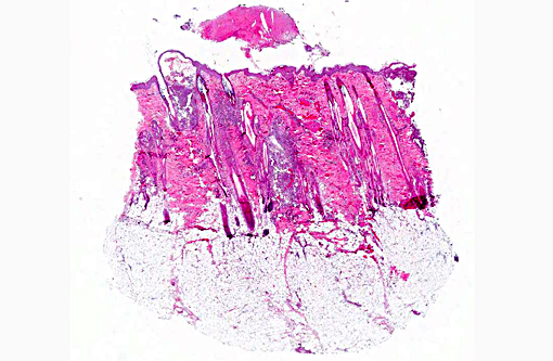
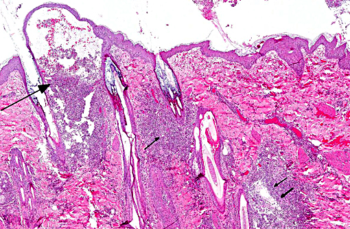
Histopathologic Description:

The main histologic change is multifocal outer root sheath necrosis with suppurative inflammation and acute hemorrhage in the superficial and mid-dermis. Dense localized aggregates of neutrophils and fewer macrophages surround adnexal units and are admixed with free keratin and erythrocytes. The suppurative aggregates form large subepidermal pustules. The epidermis is expanded by spongiosis with mild acan-thosis. There is mild superficial dermal edema. 


Morphologic Diagnosis:  

Furunculosis, necrosuppurative and hemorrhagic, acute, multifocal, severe. 


Condition:  

Post grooming furunculosis


Contributor Comment:  

The clinical and histologic features are typical of post-grooming furunculosis (PGF). This acute and severe inflammatory reaction is associated with the use of contaminated grooming products. While contaminated shampoos or conditioners are the most common causes, vigorous brushing (e.g. coat stripping), whirlpools, and contaminated surgical scrub have also been documented. Pseudomonas spp. is the most common isolate from the skin. In some cases, the bacteria can be cultured from both the skin and the affected product.

The clinical lesions typically arise on the dorsal midline as hemorrhagic pustules and crusts. The dogs may be febrile and very painful on thoracolumbar palpation. In dogs with a dense or thick haircoat (e.g. Golden retriever), the lesions may be missed on initial examination. In fact, some patients have been worked up for discospondylitis, back pain, tick borne diseases or pancreatitis.  In contrast to other causes of furunculosis (e.g. demodicosis, dermatophytosis, staphy-lococcal infection), PGF is acute. The outer root sheath is necrotic rather than hyperplastic and the rupture occurs in the superficial dermis. Furthermore, the inflammation is hemorrhagic and fibri-nosuppurative rather than pyogranulomatous. Bacteria may be difficult to discern on H&E and Gram stain as they are often small gram negative bacilli. 


JPC Diagnosis:  

Haired skin: Furunculosis, superficial, necrosuppurative and hemorrhagic, acute, multifocal to coalescing, marked.


Conference Comment:  

The presence of erector pili muscles allows localization of the section to skin of the back/dorsum. At subgross magnification, a distinctive histologic pattern is identified characterized by inflammation targeting the hair follicle in the superficial dermis. A coagulum of predominantly degenerate neutrophils, fibrin, hemorrhage and debris overlies the epidermis; folliculosebaceous units are infiltrated by neutrophils with fewer macrophages and plasma cells admixed with hemorrhage and edema within the superficial dermis and extending into the mid dermis. The infiltrate results in a mural folliculitis with progression to furunculosis. The inflammatory milieu associated with the corresponding furunculosis contains basophilic fragmented material, interpreted as keratin, along with free necrotic keratinocytes which can be differentiated from macrophages by the distinctive rounded cellular margins in the former. The moderator noted that in many inflammatory conditions targeting the hair follicle, the follicular epithelium is typically hyperplastic which is not a microscopic feature observed in cases of PGF. 

The superficial nature of the lesions, acute onset, hemorrhage and lesions isolated to the dorsal trunk can help differentiate this condition from other causes of folliculitis and furunculosis in dogs. The predilection for lesions over the dorsum in PGF is postulated to be associated with the nature of grooming or bathing activities that may concentrate brushing and shampooing efforts on this area. The dorsum also has increased hair density and hair shaft size. Cytologic features of impression smears may include neutrophils and macrophages with or without eosinophils, red blood cells, and intracellular or extracellular bacteria. Clinicopathologic abnormalities associated with this condition may include neutrophilia, with or without a left shift, monocytosis, lymphopenia and mild thrombocytopenia. These abnormalities, when considered with the presence of fever, may suggest a systemic inflammatory response.(1)

Pseudomonas aeruginosa, a gram negative bacillus, is the most common bacterial agent associated with post grooming furunculosis. Pseudomonas aeruginosa is a common bacterial contaminant associated with water and can survive in the presence of some disinfectants. Other bacteria, such as Serratia marcescens and Burkholderia cepacia, are associated with this condition. In the veterinary setting, S. marcescens has been implicated as a contaminant of intravenous catheters, and B. cepacia has been documented as a contaminant of ear cleaning solutions.(1)


References:

1. Baxter CG, Vogelnest LJ. Multifocal papular deep bacterial pyoderma in a Boxer dog caused by Pseudomonas aeruginosa. Aust Vet J. 2008;86:435439.

2. Cain C, Mauldin EA. Clinical and histopathologic features of dorsally oriented furunculosis in dogs following water immersion or exposure to grooming products. J Am Vet Med Assoc. 2015; 246(5):522-9.

3. Hillier A, Alcorn JR, Cole LK, et al. Pyoderma caused by Pseudomonas aeruginosa infection in dogs: 20 cases. Vet Dermatol. 2006;17:432439.

4. Ihrke PJ, Gross TL. Warning about postgrooming furunculosis. J Am Vet Med Assoc. 2006;229:10811082.


Click the slide to view.


4-1. Haired skin


4-2. Haired skin


4-3. Haired skin


Back | VP Home | Contact Us |