CASE 2: F930 VP19265N (4152714-00)
Signalment:
1-year 6-months-old, female, African hedgehog (Atelerix albiventris)
History:
The hedgehog showed ataxia and visited the animal hospital. At the time of the initial visit, the hedgehog was mildly thin and showed ataxia in the bilateral forelimb. Although corticosteroid, vitamin B complex, and vitamin E were continuously administrated, the symptoms progressed to paralysis, and the hedgehog was died two months later.
Gross Pathology:
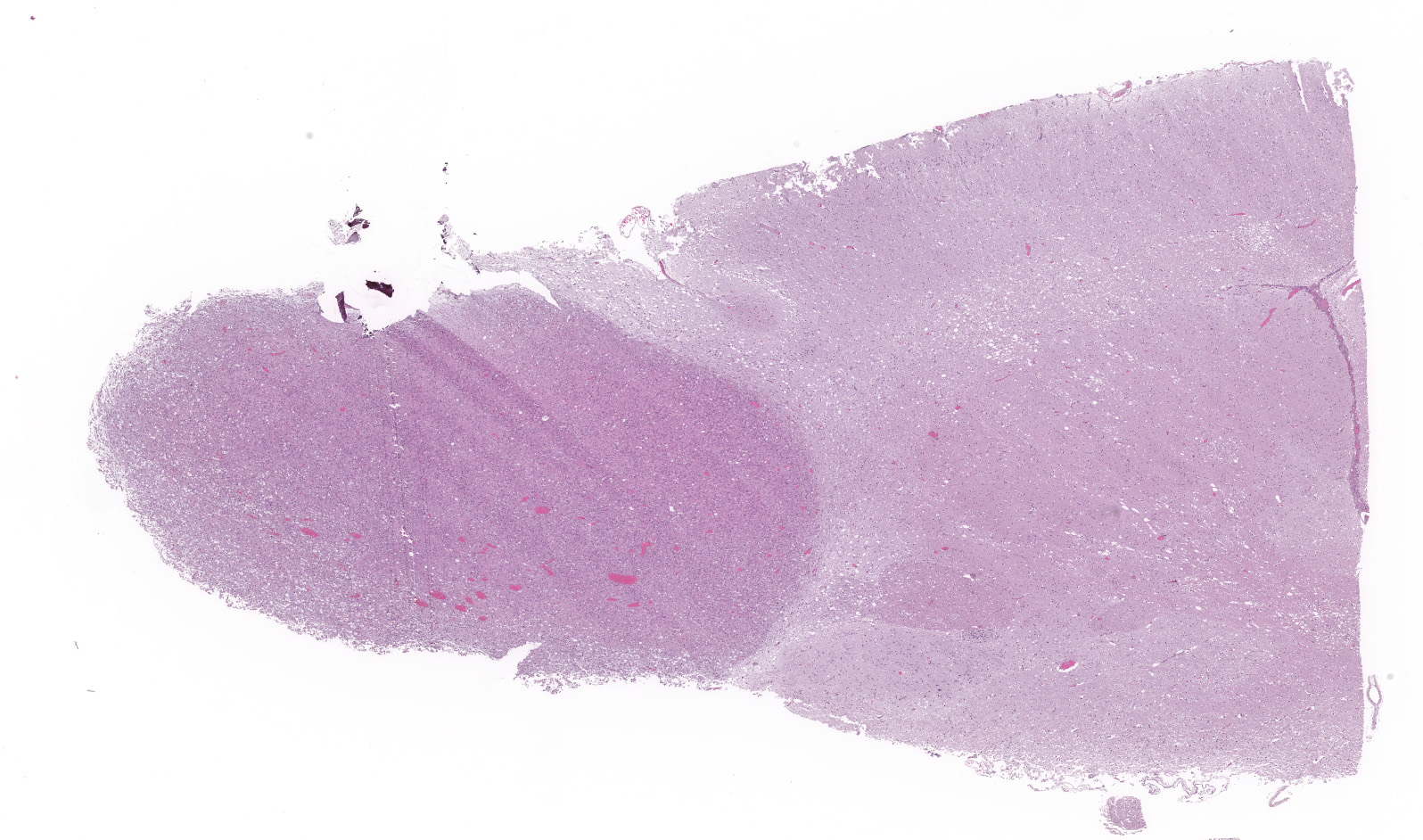
At necropsy, 3.5 mm soft, gelatinous, and grayish white mass was formed in rostral end of the right cerebral hemisphere. The liver was enlarged and discolored, and pulmonary edema was observed.
Laboratory results:
There were no abnormal findings on CBC, serum chemistry analysis, abdominal ultrasonography, and chest X-ray.
Microscopic description:
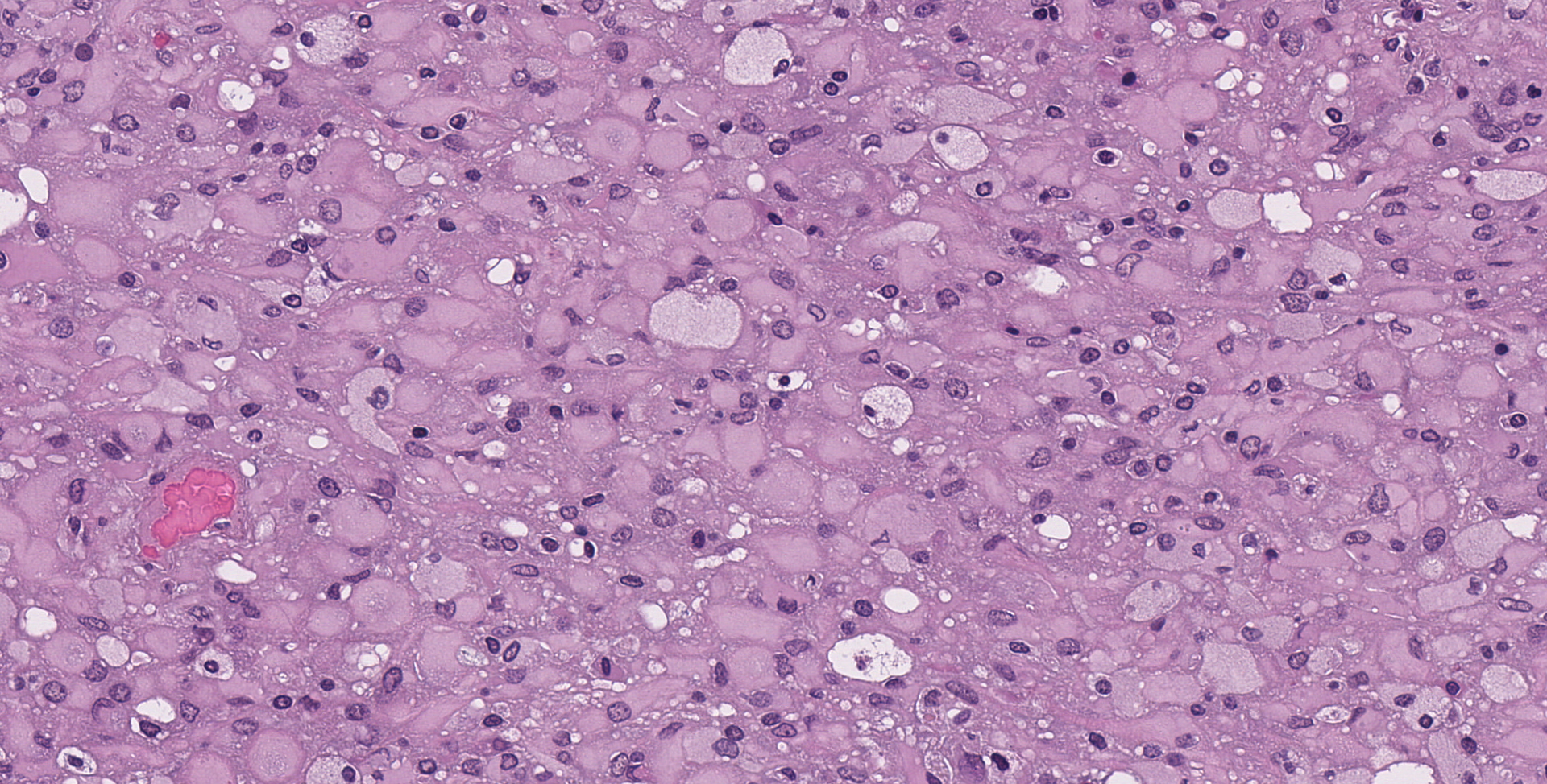
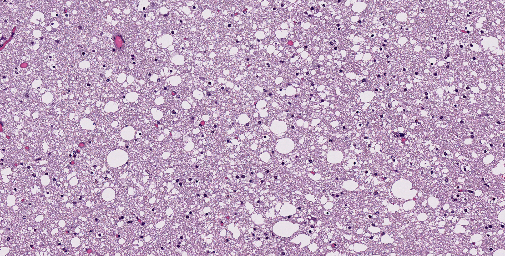
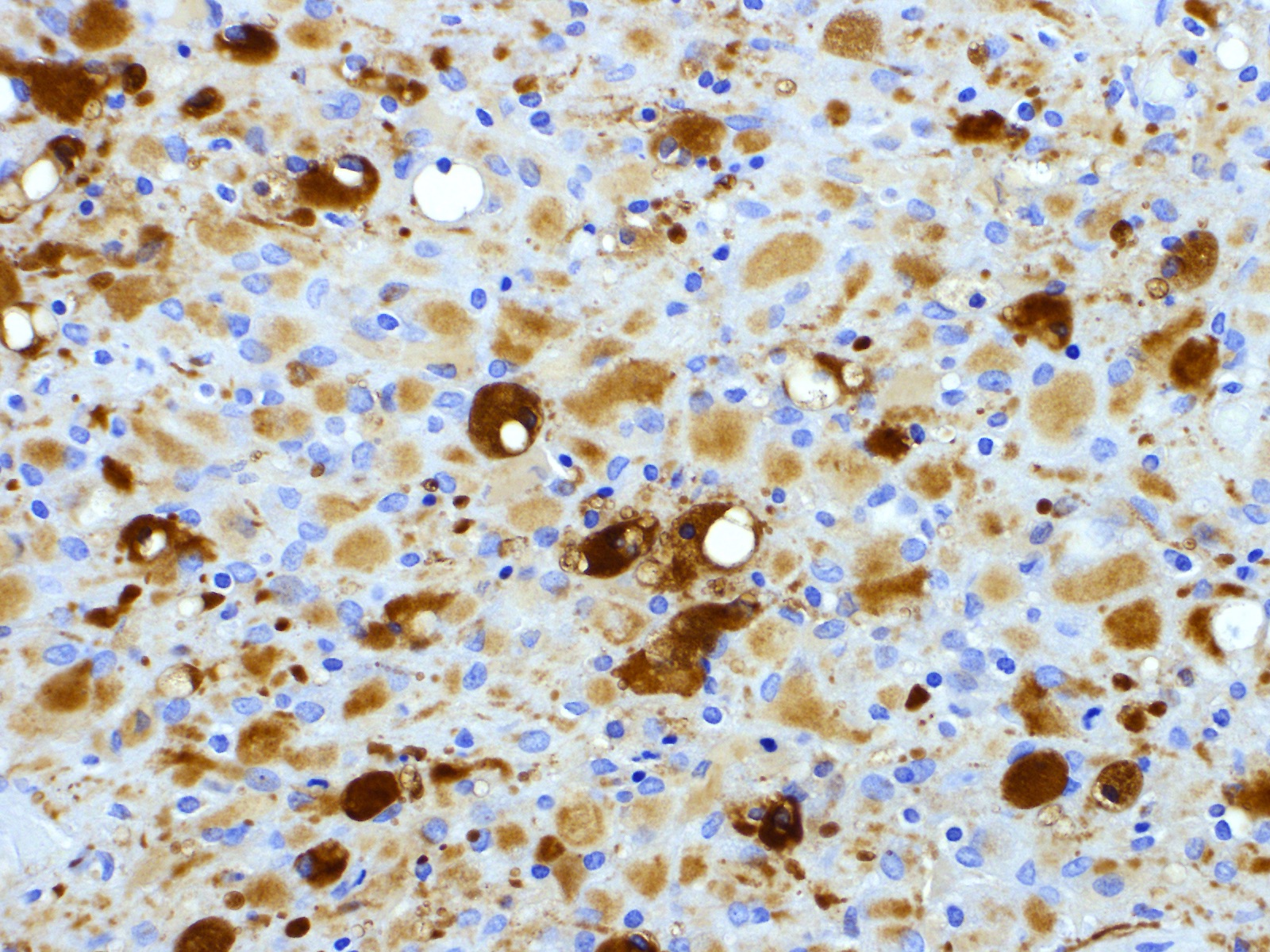
In the rostral end of the cerebrum, there is a well-demarcated, unencapsulated mass. The neoplasm is composed of diffusely growing round to angular cells. Neoplastic cells are 10-18 microns in diameter with glassy, abundant eosinophilic cytoplasm and have distinct cell borders. Nuclei are round to oval and eccentric, with finely stippled chromatin and indistinct nucleoli. Mitotic figures are rare. The neoplastic cells stained positively for GFAP, but negative with Olig2 and Iba-1.
Contributor's morphologic diagnosis:
Brain (cerebrum): Gemistocytic astrocytoma.
Contributor's comment:
Astrocytomas are a type of glioma that consist of neoplastic cells that are morphologically similar to astrocytes and occurs in the brain and spinal cord. This tumor is well known in dogs and is divided into three grades by World Health Organization (WHO) classification on tumors of domestic animals: low-grade (well-differentiated), medium-grade (anaplastic), and high-grade (glioblastoma) astrocytomas.5 Additionally, low-grade astrocytomas are classified into three subtypes: fibrillary, protoplasmic, and gemistocytic astrocytoma.5 Our case consistent with gemistocytic astrocytoma in terms of large, round to angular neoplastic cells with abundant eosinophilic cytoplasm.
In African hedgehogs, astrocytoma, oligodendroglioma, oligoastrocytoma, ganglioglioma, and histiocytic sarcoma have been reported as primary intracranial tumors.1, 3, 8-10 Muñoz-Gutiérrez et al. reported a series of primary central nervous system neoplasms in African hedgehogs, with primary central nervous system neoplasms identified in 12 of 762 hedgehogs.8 Of the 12 cases in their report, six were gangliogliomas, five were astrocytomas, and one was an oligodendroglioma, and all astrocytomas were gemistocytic subtype.8 Additionally, two cases of anaplastic astrocytoma have been reported.3, 9
Clinical symptoms such as progressive ataxia and paralysis suggestive of central nervous system manifestation should be differentiated from wobbly hedgehog syndrome (WHS) in African hedgehog. Histopathologically, WHS is characterized by bilaterally symmetrical myelin degeneration in the white matter.2 In our case, the onset of symptoms was at a young age of less than 2 years of age, and WHS was clinically suspected. However, WHS was clearly denied by confirmed the neoplastic lesion and the absence of myelin degeneration. Because some primary intracranial tumors do not form clear masses, a detailed pathological observation of the brain and spinal cord is needed to rule out WHS.
Contributing Institution:
Laboratory of Pathology
Faculty of Pharmaceutical Sciences
Setsunan University
45-1 Nagaotohge-cho, Hirakata
Osaka 573-0101, Japan
JPC diagnosis:
Cerebrum: Gemistocytic astrocytoma, low grade, African hedgehog, Erinaceinae.
JPC comment:
The contributor provides a brief summary of astrocytomas and Wobbly Hedgehog Syndrome. The 2016 WHO classification for human astrocytic neoplasms differs from the reference WHO classification in domestic animals, and now all diffuse human gliomas (astrocytic and not) are grouped together based on growth patterns and behaviors, and also the shared mutated isocitrate dehydrogenase (IDH) gene status. The new classification category includes WHO grade II and III astrocytic tumors, grade II and III oligodendrogliomas, grade II and III oligoastrocytomas, grade IV glioblastomas, and diffuse gliomas of childhood. While occasional gemistocytic astrocytes would be considered normal in a diffuse astrocytoma, there must be more than 20% gemistocytic astrocytes for the diagnosis of gemistocytic astrocytoma. As in this case, gemistocytic astrocytoma cells demonstrate strong cytoplasmic immunoreactivity to GFAP, and more than 80% of (human) cases exhibit nuclear immunoreactivity to p53.7
IDH mutation status provides important diagnostic and prognostic information for patients with diffuse gliomas. IDH status may also be associated with efficacy of individualized treatment, therapeutic vaccines, and anti-IDH treatment. While the gold standard for diagnosis of IDH status is immunohistochemistry (R132H antibody), recent computational pathology efforts have focused on making IDH status determination using H&E slides only. Deep learning models were developed using a training set of known IDH-mutated and IDH-wild type cases, and further augmented by Generative Adversarial Network (GAN) training, resulting in an accuracy of classification by IDH status of 85% using H&E alone. Adding an age variable to the model (< 55 years of age, 55 years of age or older) further improved the accuracy of classification to 96% in the older age group. This category of computational pathology may someday improve medical care and efficiency of resources.6
GFAP is usually considered a well conserved intermediate filament expressed in well differentiated, less malignant astrocytic neoplasms. However, GFAP expression has also recently been observed in radial glia of the developing (human) brain, and in adult neural stem cells, indicating the GFAP is also expressed in immature, less fully differentiated glial cells. While GFAP expression may not be correlated with astrocytoma of different grades of malignancy, the two isoforms of GFAP may be useful markers for this determination. Higher levels of GFAPd expression (alternative splice variant) relative to the normal isoform GFAPa appears to be correlated with higher grades of malignancy. Further research in veterinary species may be warranted in light of this human-centric work.12
The recently revised classification of canine gliomas attempts to simplify the categorization of these neoplasms and does not rely on stratification based on mutational status of previously identified susceptible genes in humans.4 While a very small sample size, it was once found that none of 25 sampled canine gliomas contained an IDH mutation, suggesting a potentially different pathogenesis than human gliomas.11 However, as was stated in the revised classification, "there is much to learn about the histogenesis and behavior of these tumors ? a more detailed system will depend on future molecular studies and more thorough clinical annotation."4
References:
1. Benneter SS, Summers BA, Schulz-Schaeffer WJ, Härtig W, Mollidor J, Schöniger S. Mixed Glioma (Oligoastrocytoma) in the brain of an African hedgehog (Atelerix albiventris). J Comp Pathol. 2014;151:420?424.
2. Díaz-Delgado J, Whitley DB, Storts RW, Heatley JJ, Hoppes 3, Porter BF. The Pathology of wobbly hedgehog syndrome. Vet Pathol. 2018;55:711?718.
3. Gibson CJ, Parry NM, Jakowski RM, Eshar D. Anaplastic astrocytoma in the spinal cord of an African pygmy hedgehog (Atelerix albiventris). Vet Pathol. 2008;45:934?938.
4. Koehler JW, Miller AD, Miller CR, et al. A revised diagnostic classification of canine glioma: towards validation of the canine glioma patient as a naturally occurring preclinical model for human glioma. J Neuropathol Exp Neurol. 2018;77(11):1039-1054.
5. Koestner A, Bilzer T, Fatzer R, Schulman FY, Summers BA, Van Winkle TJ: Astrocytic tumors. In: Histological classification of tumors of the nervous system of domestic animals, pp. 17?19. World Health Organization, Washington, DC, 1999.
6. Liu S, Shah Z, Sav A, et al. Isocitrate dehydrogenase (IDH) status prediction in histopathology images of gliomas using deep learning. Sci Rep. 10, 7733 (2020).
7. Louis DN, von Deimling A, Cavenee WK. Diffuse astrocytic and oligodendroglial tumours - Introduction. In: WHO Classification of Tumours of the Central Nervous System. Lyon, France: International Agency for Research on Cancer. 2016:15-23.
8. Muñoz-Gutiérrez JF, Garner MM, Kiupel M. Primary central nervous system neoplasms in African hedgehogs. J Vet Diagn Invest. 2018;30:715?720.
9. Nakata M, Miwa Y, Itou T, Uchida K, Nakayama H, Sakai T. Astrocytoma in an
African hedgehog (Atelerix albiventris) suspected wobbly hedgehog syndrome.
J Vet Med Sci. 2011;95:255?257.
10. Ogihara K, Suzuki K, Madarame H. Primary histiocytic sarcoma of the brain in an African hedgehog (Atelerix albiventris). J Comp Pathol. 2017;157:241?245.
11. Reitman ZJ, Olby NJ, Mariani CL, et al. IDH1 and IDH2 hotspot mutations are not found in canine glioma. Int J Cancer. 2010;127(1):245-246.
12. Van Bodegraven EJ, van Asperen JV, Robe PAJ, Hol EM. Importance of GFAP isoform-specific analysis in astrocytoma. Glia. 2019;67:1417-1433.