Signalment:
2-3 years, female, pony, equine,
Equus caballusThe case was part of an investigation in an animal cruelty case. The pony was cachectic and deteriorated. Due to poor body condition, severe elevated levels of urea and creatinine (azotaemia), the horse was euthanased. Multiple formalin-fixed fragments of right and left kidney were submitted for histopathology.
Gross Description:
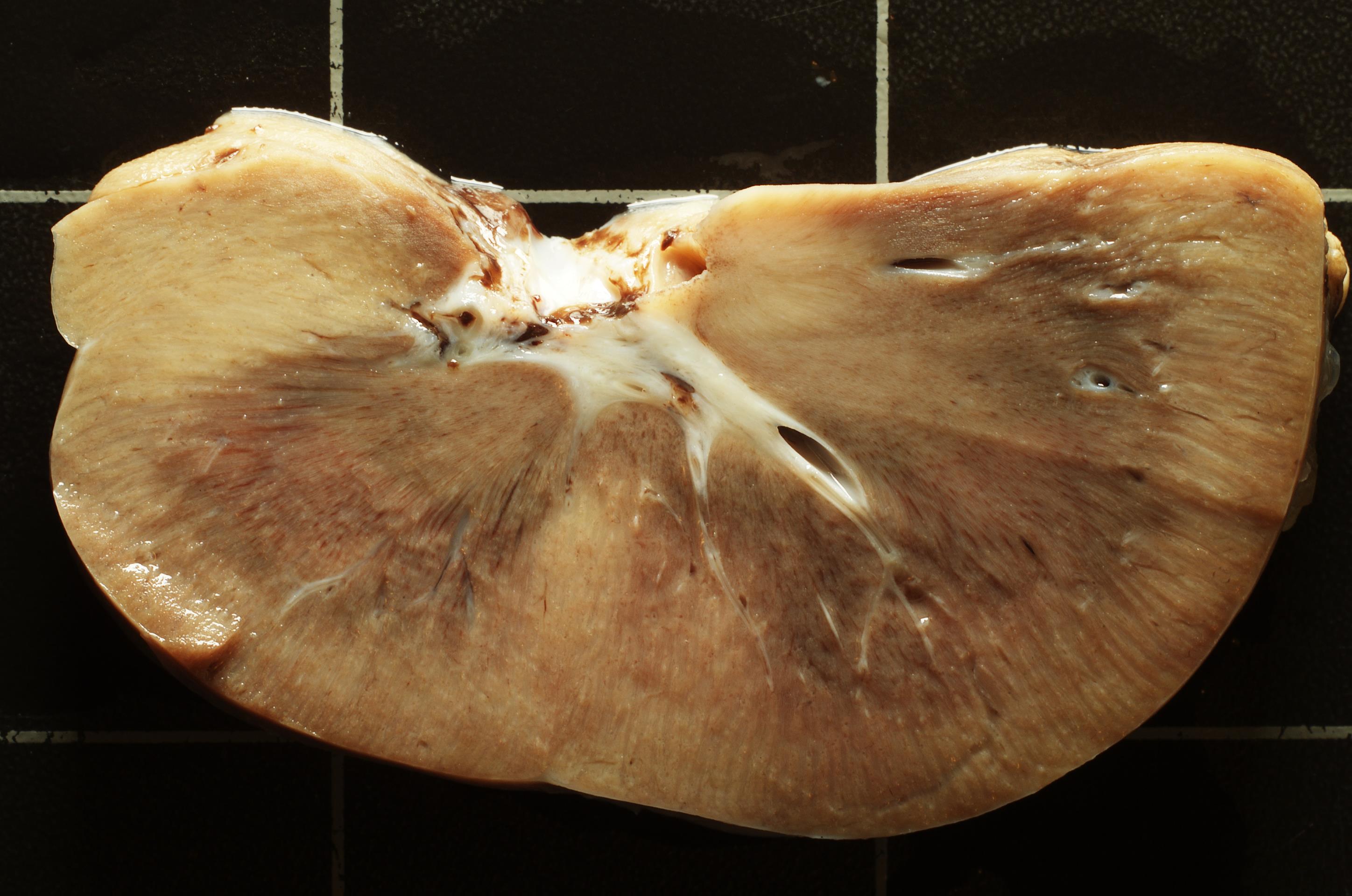
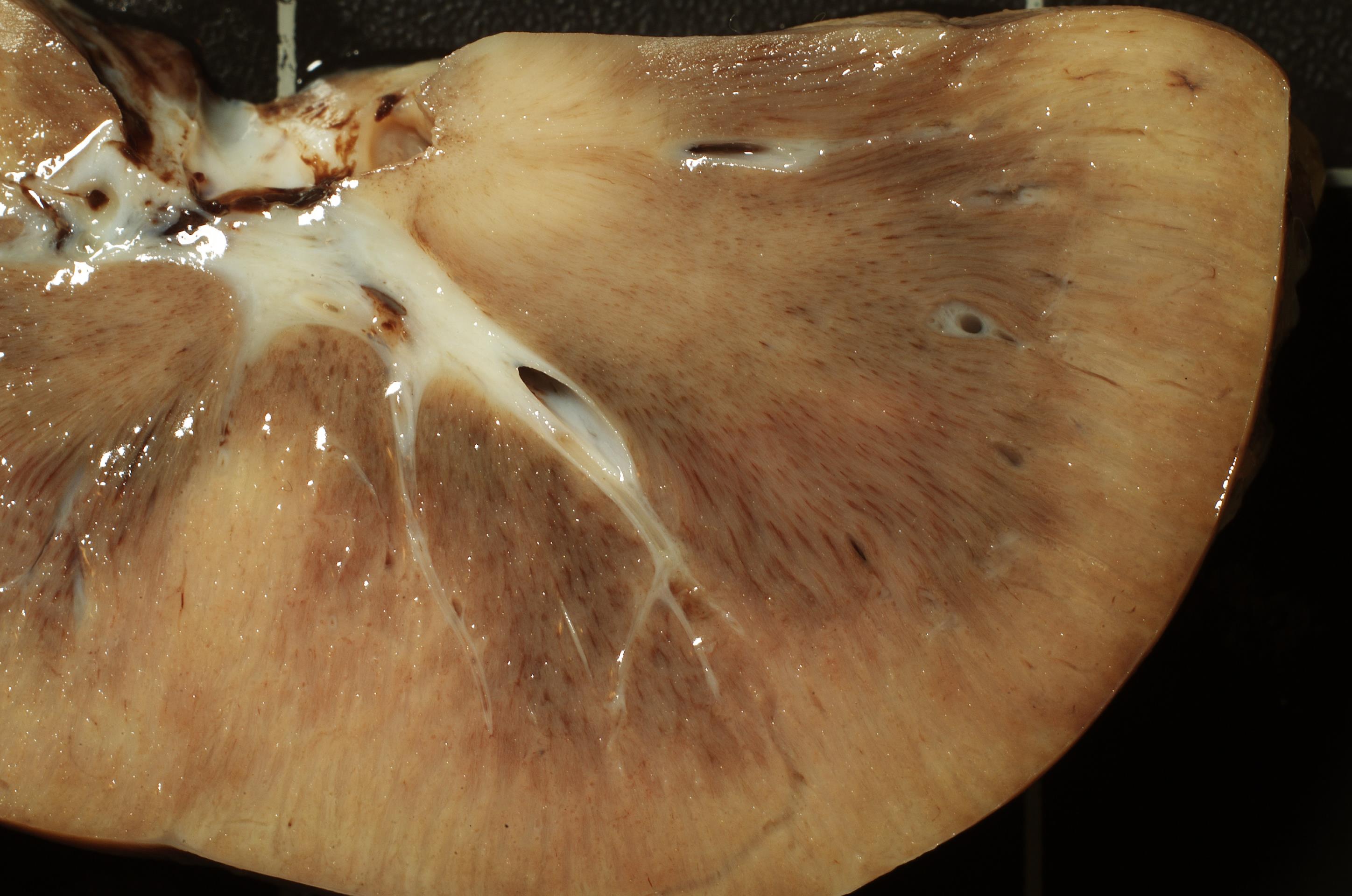
Kidney moderately firm, with prominent, pale beige glomerula, mild diffuse hyperaemia, moderate diffuse cortical and medullary interstitial fibrosis and focally indistinct cortico-medullary transition zone
(Fig. 2-1, 2-2)
Histopathologic Description:
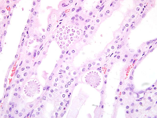
The renal architecture is preserved. Within the epithelium of proximal and distal tubular epithelia of the cortical and medullary zone, numerous intracytoplasmic apicomplexan coccidian trophrozoites and sporoblasts are seen. Few tubular epithelia are desquamated. Multifocal, mild to moderate tubular epithelial necrosis, intratubular, Von Kossa stain-positive mineralisation (calcification), and Von Kossa stain-negative, birefringent crystals (oxalate) are seen. Multifocally, distal tubules contain moderate to large amounts of cellular debris (desquamated, necrotic epithelia ?) and show mild dilation. There is focal mild glomerular fibrosis with glomerular synechia formation and multifocal mild to focally moderate interstitial fibrosis. A focal mild interstitial infiltration by lymphocytes, plasma cells and small numbers of eosinophils is seen.
Morphologic Diagnosis:
Glomerular and interstitial fibrosis, tubulonecrosis with (dystrophic) mineralisation, oxalate crystal formation, mixed cellular interstitial nephritis; diffuse, moderate (end-stage renal disease); with intracellular sporogenic and gametogenic stages of apicomplexan coccidian parasites, consistent with
Klossiella equi, (sporozoa, apicomplexa, coccidia) infection, kidney, horse,
E. caballus.
Condition:
Klossiela equi
Contributor Comment:
The parasitic genus
Klossiella belongs to the subphylum sporozoa which is characterised by intracellular life-cycle and an apical complex at some point during its development. The trophozoites have no cilia or flagella. The reproduction involves both asexual (schizogony) and sexual (gametogony) phases. Following gametogony a zygote is formed which divides to produce spores (sporogony). Klossiellidae can be found in the kidneys of equids, mice, guinea pigs, bats, opossums, and snakes.
2,7 Renal infections with
K. equi are often clinically inapparent. Mild infections, usually self-limiting, are followed by a predetermined cycle of development, with the production of infective stages that are shed in the host urine, and are thought to be ingested by another host. There are few reports on
K. equi-related nephritis in severely infected equids.
1,3 Both sporogenic and gametogenic stages of
Klossiella equi were identified in the kidneys. All stages developed in individual tubular epithelia cells. Schizonts were seen mostly in the proximal convoluted tubules, but also free within the lumen of a tubule. Macrogametocytes and microgametocytes were present in syzygy in the loop of Henle and collecting ducts. Sporont and budding sporont stages were also seen in the loop of Henle. All stages of development of sporoblasts were observed protruding into the tubular lumen. Sporocysts were identified rupturing out of the sporoblast membrane into the lumen of tubules. Renal tubules were greatly dilated and contained cellular debris. The tubulonecrosis and desquamation of tubular epithelia in this case most likely can be ascribed to the infection by
K. equi. Whether the additionally described chronic renal alterations are due to the parasitic infection or are a separate underlying pathomechanism, cannot be stated.
JPC Diagnosis:
1. Kidney, tubules: Degeneration and
necrosis, multifocal, moderate, with cellular casts and
protozoa
(Fig. 2-3), etiology consistent with Klossiella
equi, pony (Equus caballus), equine.
2. Kidney: Nephritis, interstitial, lymphoplasmacytic,
multifocal, moderate, with intratubular crystals.
Conference Comment:
The contributor gives an excellent
description of
Klossiella equi. Klossiella equi is the
only known coccidian parasite of the equine urinary tract,
with various stages of development located in the kidney.
3 The life cycle is not currently known, although
infection is presumed to occur via ingestion of infective
sporocysts that were shed in the urine.
6 It is also believed
that one schizont generation develops in the glomerular
endothelium and another in the proximal tubular epithelium,
with sporogony occurring in the epithelium of the
thick limb of Henle's loop.
4 Infection is thought to be an
incidental finding although it has been associated with
nephrosis and nephritis in immune-compromised individuals.
1,6 We agree with the contributor that it cannot be
determined if the interstitial nephritis and crystals are the
result of the
K. equi infection.
Ultrastructurally, developing sporoblasts are encased by a
bilaminated cell membrane composed of an overlying
thin granular layer, and an underlying dense inner layer.
1
We thank Dr. C. H. Gardiner, PhD, veterinary parasitology
consultant to the AFIP, for his review of this case.
References:
1. Anderson, W.I., Picut, C.A. and Georgi, M.E., 1988.Â
Klossiella equi induced tubular nephrosis and interstitial nephritis in a pony. Journal of Comparative Pathology 98(3): 363-366.
2. Gardiner CH, Fayer R, Dubey JP: An Atlas of Protozoan
Parasites in Animal Tissues, 2nd ed., pp. 61-62.
Armed Forces Institute of Pathology, Washington, DC,
1998
3. Karanja DNR, Ngatia TA, Wandera JG: Donkey
Klossiellosis in Kenya. Vet Parasitiol 53:1-5, 1994
4. Maxie MG, Newman SJ: Urinary system. In: Jubb,
Kennedy and Palmers Pathology of Domestic Animals,
ed. Maxie MG, 5th ed., vol. 2, pp. 498. Elsevier Saunders,
Philadelphia, PA, 2007
5. Newman SJ, Confer AW, Panciera RJ: Urinary system.
In: Pathologic Basis of Veterinary Disease, eds.
McGavin MD, Zachary JF, 4th ed., p. 657-658. Elsevier,
St. Louis, MO, 2007
6. Suedmeyer WK, Restis E, Beerntsen BT:
Klossiella
equi infection in a Hartmanns Mountain zebra (
Equus
zebra hartmannae). J Zoo Wildl Med 37:420-423, 2006
7. Taylor JL, Wagner JE, Kusewitt DF, Mann PE:
Klossiella parasites of animals: a literature review. Vet
Parasitol 5:137-144, 1979