Joint Pathology Center
Veterinary Pathology Services
Wednesday Slide Conference
2017-2018
Conference 13
January 3rd, 2018
CASE III: 719/16 (JPC 4095576).
Signalment: Adult, female, Cavia porcellus, guinea pig.
History: The animal stems from a large colony kept in a zoo and was euthanized due to an overall poor body condition. At necropsy the ocular lesion was an incidental finding.
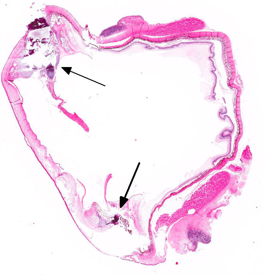
Gross Pathology: On external examination the left eye presented with an irregularly shaped, well-demarcated whitish-grey mass at the limbal region of the iris, which encircled the pupil entirely. On cut section there was a marked thickening of the ciliary body with a bone-like structure and projections extending into the anterior chamber. Otherwise the eye was unremarkable.
Laboratory results:
None provided.
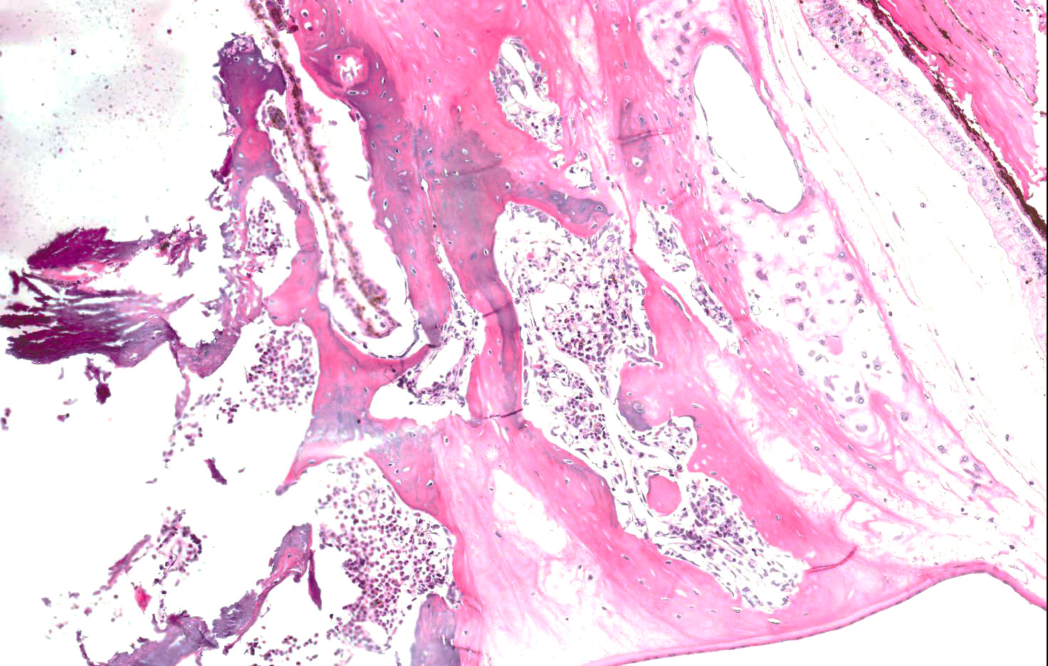
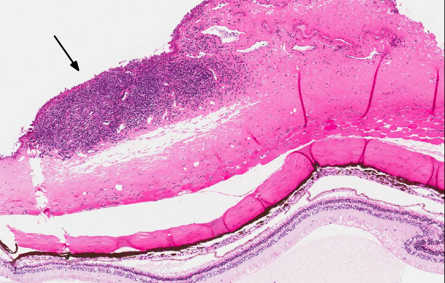
Microscopic Description: Eye: Expanding and replacing the stroma of the ciliary body and the iris is a proliferation of regularly formed lamellar bone containing multiple central cavities filled by hematopoietically active bone marrow. The bone is partially surrounded by a fine fibrous layer. Multifocally in the subepithelial connective tissue of the bulbar conjunctiva there are nodular infiltrates composed of large numbers of lymphocytes and few plasma cells.
Contributor’s Morphologic Diagnoses:
- Eye: Heterotopic bone formation of the ciliary body.
- Eye: Lymphoplasmacytic conjunctivitis, multifocal, chronic, mild.
Contributor’s Comment: Heterotopic bone formation, also known as osseous choristoma, is a sporadic, usually incidental finding in the eye of guinea pigs.5,10 Heterotopic bone formation of the ciliary body can be found in young as well as aged guinea pigs of both sexes and it might occur uni- or bilaterally.10,12 In general, affected animals don’t show any clinical symptoms, however, development of secondary open-angle glaucoma by blockage of the iridocorneal filtration angle has been reported with chronic exposure keratitis being the most notable associated sequelae.10 Other authors on the contrary found no elevation of the intraocular pressure inside eyes displaying heterotopic bone formation of varying degrees compared to unaffected eyes.8,12
The etiology of this condition has not been fully determined yet. Bone in the eye can arise within metaplastic or neoplastic processes particularly in phthisic eyes.5 In most affected guinea pigs, however, no previous ocular trauma or disease has been reported.5,10,12 Due to their slow-growing nature and well-differentiated appearance, they originally have been classified as choristoma, which is defined as histologically normal tissue arising in ectopic positions and considered a benign congenital neoplasm, therefore resulting of an incorrect embryogenesis.7
It has also been noted that the role of the ciliary epithelium in transporting and concentrating plasma ascorbic acid into the aqueous humor might be a pivotal factor for development since ascorbic acid positively influences trabecular bone formation by modifying the expression of several bone matrix genes in osteoblasts.1,13 Osseous metaplasia and mineralization are common findings in other organs of guinea pigs, especially the lungs.8
Similar ocular lesions have also been described in other species including dogs and also humans.6 In humans the location differs in that mainly choroidal structures are affected.5,11
JPC Diagnosis: Eye, anterior uvea: Osseous choristoma (heterotropic bone) with fibrovascular membrane formation, drainage angle occlusion, iris bombe, and diffuse mild retinal atrophy, Cavia porcellus, guinea pig.
Conference Comment: There have been several reports within the past 30 years detailing individual cases of osseous choristoma in guinea pigs with little additional information about this benign lesion which is considered an embryologic remnant that is left behind and grows as the animal ages.2, 3, 4, 5, 6, 10, 12
Bone is a normal feature of the eyes, particularly the sclera of birds and some reptiles, but not of the eye of mammals. Osseous metaplasia may result from trauma. Additionally, bone may develop as ossification within a pre-existing choroidal hemangioma.5 These causes can be ruled out by an otherwise normal ocular anatomy and no evidence of pre-existing vascular lesions or ocular trauma.
Osseous choristomas within the ciliary body appear microscopically as replacement and elevation of ciliary body tissue with mature bony spicules surrounded by a thin fibrous envelope. Frequently, the bone will contain vascular spaces and hematopoietically active bone marrow.5
Guinea pigs are particularly susceptible to ectopic mineralization, possibly related to the high calcium content of commercial guinea pig diets and alfalfa. In addition to heterotopic bone formation within the ciliary body of the eye, guinea pigs may develop ectopic ossification within the lungs and urolithiasis from high calcium within urine. Like rabbits, guinea pigs’ main mode of calcium homeostasis is renal excretion, thus, chronic renal disease predisposes animals to ectopic mineralization.9 Ectopic mineralization is different that an osseous choristoma, which is mature bone in an abnormal location.
In addition to the findings listed above by the contributor, conference attendees identified peripheral anterior synechiae, posterior synechiae, thinning of the sclera, and multifocal atrophy of the ganglion cell layer. Microscopic evidence for glaucoma was considered, and there was spirited discussion related to the cause, whether it was due to the pre-iridial fibrovascular membrane or compression of the trabecular meshwork by the osseous choristoma. Additional discussion centered on the presence of numerous lymphocytes within the conjunctiva and as to whether they represented true conjunctivitis or mucosal-associated lymphoid tissues which is commonly found at this site in older rodents.
Contributing Institution:
Institute of Veterinary Pathology
Faculty of Veterinary Medicine
LMU Munich
Veterinaerstr. 13
80539 Muenchen
http://www.patho.vetmed.uni-muenchen.de/index.html
References:
- Aghajanian P, Hall S, Wongworawat MD, Mohan S. The roles and mechanisms of actions of vitamin C in bone: new developments. J Bone Miner Res. 2015;30:1945-1955.
- Brooks DE, McCracken MD, Collins BR. Heterotopic bone formation in the ciliary body of an aged guinea pig. Lab Anim Sci. 1990;40(1):88-90.
- Cullen CL, Grahn BH, Wolfer J. Diagnostic ophthalmology. Right superficial corneal ulcer with secondary anterior uveitis and osseous choristoma in a guinea pig. Can Vet J. 2000;41(6):502-503.
- Donnelly TM, Brown C, Donnelly TM. Heterotopic bone in the eyes of a guinea pig: osseous choristoma of the ciliary body. Lab Anim (NY). 2002;31(7):23-25.
- Griffith JW, Sassani JW, Bowman TA, Lang CM. Osseous choristoma of the ciliary body in guinea pigs. Vet Pathol. 1988;25(1):100-102.
- Lynch GL, Scagliotti RH. Osseous metaplasia in the eye of a dog. Vet Pathol. 2007;44(2):222-224.
- Meuten DJ, ed. Tumors in Domestic Animals. 4th ed. 2002:28.
- Percy DH, Barthold SW. Pathology of Laboratory Rodents and Rabbits. 3rd Ames, IA: Blackwell Publishing; 2007:219.
- Percy DH, Barthold SW. Pathology of Laboratory Rodents and Rabbits. 4th Ames, IA: Blackwell Publishing; 2016: 216.
- Schaffer EH, Pfleghaar S. Secondary open angle glaucoma from osseous choristoma of the ciliary body in guinea pigs. Tierarztl Prax. 1995;23:410–414.
- Shields JA, Shields CL. Intraocular Tumors: An Atlas and Textbook. 2nd Philadelphia, PA: Lippincott Williams & Wilkins;2008:264.
- Williams DL, Sullivan A. Ocular disease in the guinea pig (Cavia porcellus): A survey of 1000 animals. Vet Ophthalmol. 2010;13Suppl:54–62.
- Williams DL. The guinea pig eye. In: Williams DL, ed. Ophthalmology of Exotic Pets. 1st ed. Oxford, UK: Wiley-Blackwell; 2012: 67-69.