Signalment:
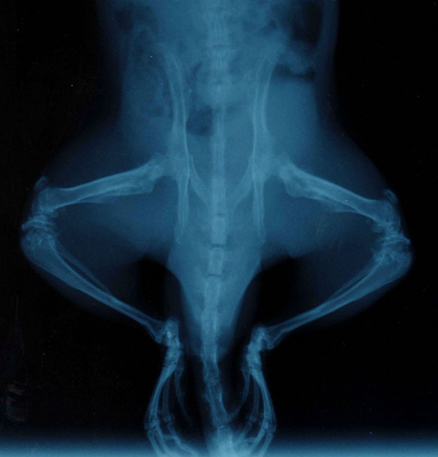
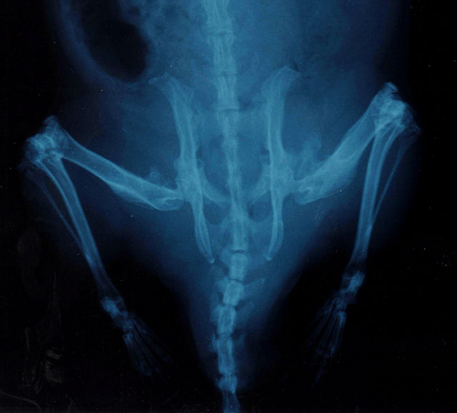
Gross Description:
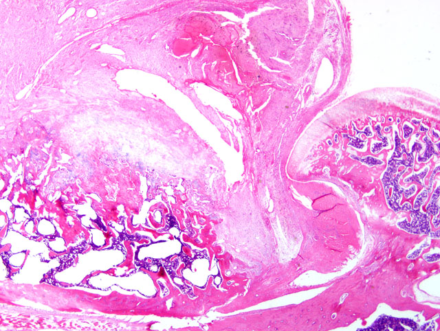
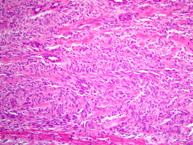
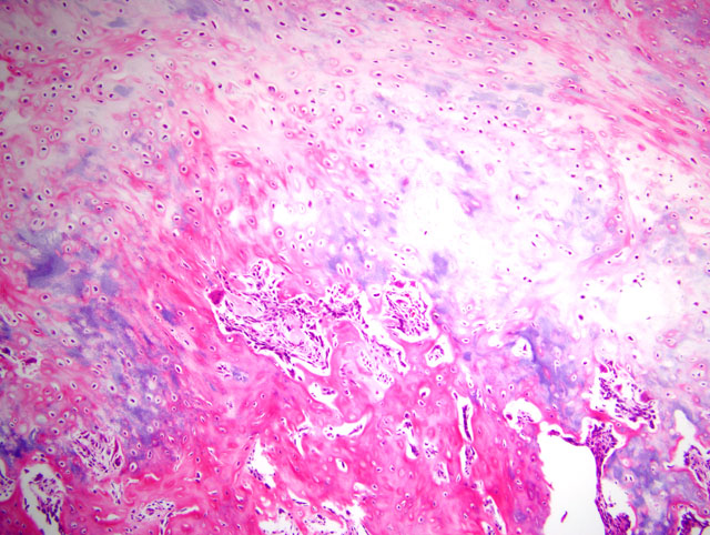
Histopathologic Description:
Morphologic Diagnosis:
Condition:
Contributor Comment:
Yeager and Hamre provide excellent descriptions of the evolution of the lesions of osteolathyrism.(7) Briefly, the exostoses develop at the site of the insertion of the tendons of the pectineus and adductor longus muscles on the periosteum of the femur through two distinct phases. In the preliminary proliferative stage, the inner layer of the periosteum becomes a highly cellular and less organized mass of tissue resembling a fibroma or fibrosarcoma. The second osteogenic stage occurs as soon as 7 to 8 days after initiation of a sweet pea diet, and consists of intra-membranous ossification at the periphery of the mass. No distant metastases were noted in any osteolathyrism studies, despite the infiltrative growth pattern, high cell density, and immature cytologic characteristics of the fibroblasts. Lesions may also develop at the deltoid ridge of the humerus, the gluteal trochanter of the femur, and the lamboidal ridge of the skull.
Although Yeager and Hamre observed no gross or histologic evidence of trauma at the site of the exostoses (7), additional studies noted that muscle tension was required for lesion development, as transected or denervated muscles failed to produce lesions, while lesions did develop at the sites of insertion of muscles that provided functional compensation for transected muscles.(1) Ponseti suggests that lifting of the periosteum is required for exostosis formation.(4)
JPC Diagnosis:
Conference Comment:
In addition to the detailed description of the lesion provided by the contributor, the moderator and attendees noted foci of new bone formation in the area of the trochanteric fossa and the lateral aspect of the proximal femur. In agreement with the contributors description, the Armed Forces Institute of Pathologys Department of Musculoskeletal and Soft Tissue Pathology summarized the lesion as a broad-based periosteal proliferation of new bone with a poorly-formed fibrocartilagenous cap and an overlying area composed of a large cellular proliferation of fibroblastic and myofibroblastic type cells that in one area blend with tendon and muscle cells; the possibility of an exuberant myofibroblastic reaction to an avulsive injury at a tendon insertion site was considered.Â
Ectopic ossification is the neoformation of non-neoplastic trabecular bone at extraosseous sites, and is distinct from ectopic or heterotopic mineralization, which lacks bone formation. Most occurrences of ectopic ossification are common, clinically insignificant findings, e.g. dural ossification (ossifying pachymeningitis) in aged dogs and ectopic bone formation in the lungs of dogs and cattle; or occur in the supporting stroma of certain neoplasms, such as mammary carcinoma in dogs. Two specific diseases associated with ossification of soft tissues are fibrodysplasia ossificans progressiva and myositis ossificans; the former is characterized by progressive, symmetrical ossification of the subcuticular and epimysial connective tissue of the neck, dorsum, and limbs. In contrast, myositis ossificans is characterized by localized and asymmetric lesions containing a peripheral zone of orderly maturation from fibrous tissue to mineralized osteoid, which is replaced by lamellar bone.(6)
The contributor provides a useful description of the effects of BAPN on lysyl oxidase and its similarity to the pathologic effects of copper deficiency. During the conference, the effects of BAPN and similar toxins produced by members of the genus Lathyrus on domestic animals and humans were discussed. The Lathyrus genus is composed of variety of species (e.g. Lathyrus sylvestris, L. sativus); toxins produced by this genus include BAPN (L. odoratus), diamino-butyric acid (L. sylvestris), and beta-oxalyl-diamino-propionic acid (L. sativus). In limited amounts, Lathyrus species of legumes are a nutritious source of protein for domestic animals and humans; however, consumption of large quantities of these legumes over prolonged periods (weeks to months) results in the disease condition referred to as lathyrism. Because of the plants ability to survive in poor soils, flood and drought, outbreaks of lathyrism often occur in impoverished areas of the world during prolonged drought conditions.(3)
In contrast to rats, which develop skeletal deformities referred to as osteolathyrism, ingestion of Lathyrus spp. in humans and most domestic animals results in neurologic disorders termed neurolathyrism. Neurolathyrism is characterized by degeneration and loss of neurons in the spinal cord, resulting in gradual paralysis of the posterior limbs in cattle, horses, pigs, humans, and other species. Peripheral nerves, such as the vagus and recurrent laryngeal, are also affected. In cattle, toxicosis also leads to blindness, torticollis, and skin anesthesia. In horses, paralysis of the recurrent laryngeal nerve results in roaring. Horses and pigs appear to be more susceptible than cattle. Death usually results from respiratory paralysis. Teratogenic effects in sheep and other species and aortic aneurysm in rats and turkeys are other pathologic effects of Lathyrus intoxication.(3)
References:
2. Hashimoto N, Handa H, Nagata I, Hazama F: Saccular cerebral aneurysms in rats. Am J Pathol 110:397-399, 1983
3. Jones TC, Hunt RD, King NW: Veterinary Pathology, 6th ed., p. 757. Williams and Wilkins, Baltimore, MD 1997
4. Ponseti IV: Lesions of the skeleton and of other mesodermal tissues in rats fed sweet-pea (Lathyrus odoratus) seeds. J Bone Joint Surg Am 36-A:1031-1058, 1954
5. Tinker D, Rucker RB: Role of selected nutrients in synthesis, accumulation, and chemical modification of connective tissue proteins. Physiol Rev 65:607-657, 1985
6. Thompson K: Bones and joints. In: Jubb, Kennedy, and Palmers Pathology of Domestic Animals, ed. Maxie MG, 5th ed., vol. 1, pp. 107-108, 127-129. Elsevier Saunders, Philadelphia, PA, 2007
7. Yeager VL, Hamre CJ: Histology of lathyrus-induced exostoses of rats; the initial changes at tendon-bone junction. AMA Arch Pathol 64:171-185