Signalment:
Term fetus female Alpine dairy goat,
Capra hircus.Twin female American Alpine dairy goat fetuses were submitted for determination of the cause of abortion. These were from a newly assembled dairy goat herd which had experienced multiple abortions over the last week.
Gross Description:
On physical exam, the fetuses were moderately autolytic, and contained serosanguinous fluid within their pleural cavities. No other gross lesions were detected in the fetuses or their accompanying placentas. All samples were taken from the larger of the two fetuses.
Histopathologic Description:
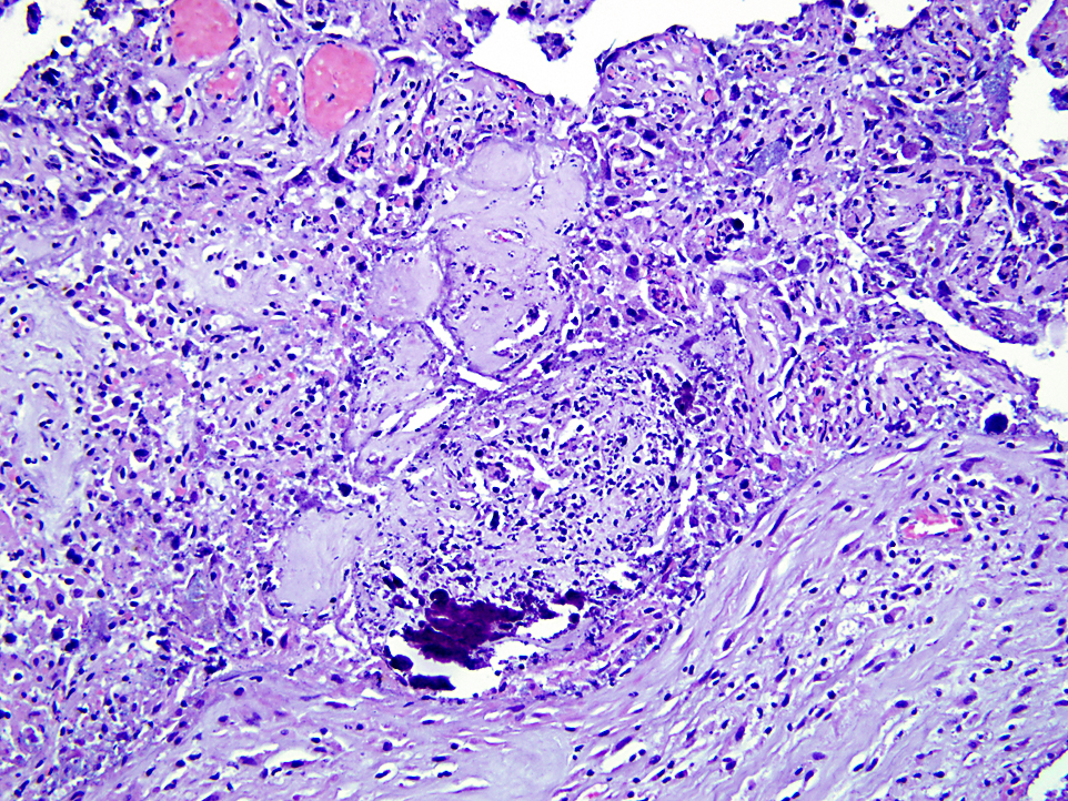
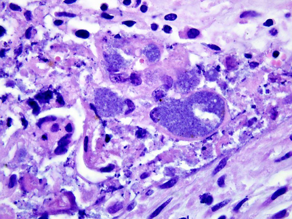
Slides contain sections of chorioallantois. Chorionic villi are necrotic, and the surface has adherent necrotic cellular debris and degenerate neutrophils. A mixed infiltrate of neutrophils, lymphocytes, plasma cells, and histiocytes extends throughout the interstitium of the chorioallantois. Numerous trophoblasts are distended by intracellular colonies of palely basophilic bacteria. Gram stains of the chorionallantois demonstrate these colonies consist of gram-negative short bacilli. Depending on the section, there is variable partial mineralization of the epithelium.
Immunohistochemistry using a generic antibody against
Coxiella burnetii was strongly immunopositive for the intracytoplasmic bacterial colonies.
Morphologic Diagnosis:
Chorioallantois: Severe acute necrotizing placentitis with intracellular bacterial colonies.
Lab Results:
PCR for
Coxiella burnetii was positive on placenta; PCR for
Chlamydophila sp. was negative. Mixed coliforms and
Streptococcus sp. were isolated from the placenta. Cultures of lung, liver, and stomach contents were negative.
Condition:
Coxiella burnetti
Contributor Comment:
Coxiella burnetii is a member of the Rickettsiaceae family, which are small, non-motile gram-negative bacteria that replicate only within host cells. Infection with
Coxiella burnetii is relatively common in domestic cattle, sheep and goats, which serve as the reservoir hosts. Infection is persistent, and shedding of the organism through urine, feces, milk and placental fluids contaminates the environment. Acute infection in domestic ruminants may manifest itself as late third trimester abortions, stillbirths, delivery of weak neonates, retained placentas, endometritis and infertility. When the placenta is infected, the obligate intracellular bacteria are encountered within the cytoplasm of trophoblasts. Gross lesions in the placenta (not noted in this case) include thickened and leathery appearance, and off-white exudates which are most prominent in the intercotyledonary regions of the chorioallantois. However, histologically, the predominantly necrotizing and suppurative placentitis frequently extends into cotyledonary regions as well. The principal differential for
C. burnetii in the placenta is
Chlamydophila infection, which produces a similar placentitis with intracytoplasmic colonies.
In humans,
C. burnetii results in the zoonotic condition known as Q fever. Acute infection may include mild flu-like symptoms, pneumonia, or hepatitis. The organism infects monocytes and macrophages which internalize the organism into phagolysosomes. In the environment, the organism survives as the highly resistant extracellular small cell variant. But once inside monocytes or macrophages, the organism switches to the large cell variant. The more debilitating chronic form of the disease in humans is characterized by endocarditis, hepatitis, and chronic fatigue syndrome. Due to
C. burnetiis resistance to environmental conditions of heat and pressure, and its highly infectious nature, it is classified as a Category B biological warfare agent.
JPC Diagnosis:
Chorioallantois: Placentitis, necrotizing, multifocal to coalescing, marked, with numerous intratrophoblastic bacilli.
Conference Comment:
Coxiella burnetii is transmitted by most tick species and can be acquired by aerosolization and direct contact. Although infection occasionally causes abortions in cattle, abortion is a more common result in small ruminants and typically presents as isolated abortions rather than as an abortion storm. Unless infection is overwhelming, as in this case, the organisms may not be visible with hematoxylin and eosin (H&E) staining, but silver stains and immunohistochemistry are useful.(3) Because of concerns for safety of laboratory personnel, the organism should only be cultured within a biosafety level-3 laboratory.Â
Brucella abortus, another differential for this case, also causes cotyledonary and intercotyledonary necrosis, but the fetus usually has bronchopneumonia, which helps differentiate infection with this organism from
Coxiella. Other differential causes for necrotizing placentitis are
Chlamydophila abortus, which commonly also causes vasculitis, and
Toxoplasma gondii, which primarily affects the cotyledons and spares the intercotyledonary areas.(3) Multifocal epithelial mineralization around blood vessels in small ruminant placentas can be a normal physiologic process and contributes to ossification of the fetal skeleton.Â
References:
1. Kazar J.Â
Coxiella burnetii infection. Ann NY Acad Sci. 2005;1063:105-114.Â
2. Sanchez J, Souriau A, Buendia AJ, et al. Experimental
Coxiella burnetii infection in pregnant goats: a histopathological and immunohistochemical study.Â
J Comp Path. 2006;135:108-115.
3. Schlafer DH, Miller RB. Female genital system. In: Maxie MG, ed.Â
Jubb, Kennedy, and Palmers Pathology of Domestic Animals. 5th ed. Vol 3. Philadelphia, PA: Saunders Elsevier; 2007:484, 502, 505, 513.