Signalment:
Gross Description:
Histopathologic Description:
Morphologic Diagnosis:
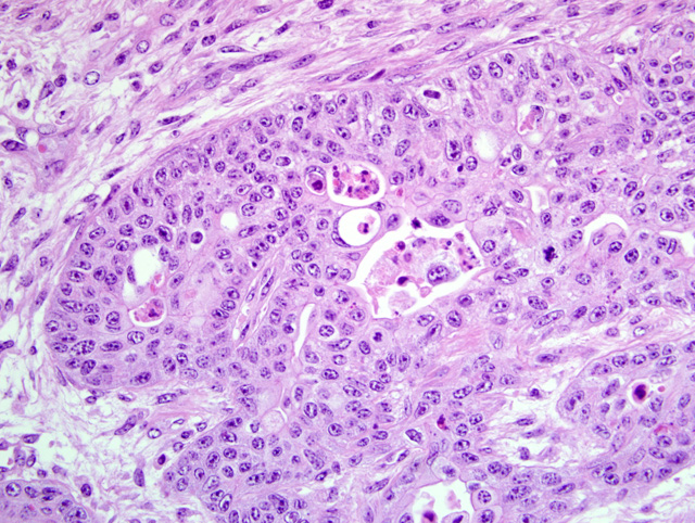
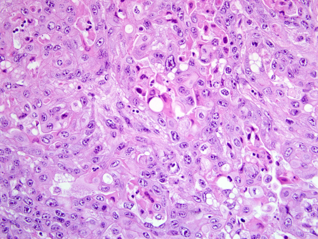
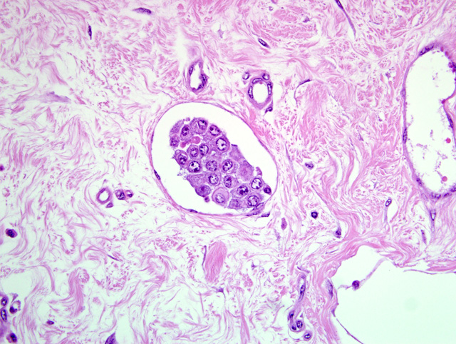
1. Urinary bladder, nonpapillary and infiltrating transitional cell carcinoma; Etiology: chronic ingestion of Pteridium aquilinum.
2. Kidney, pyelonephritis, chronic, suppurative, severe (slides not included).
3. Kidney, diffuse cortical atrophy (slides not included).
Condition:
Contributor Comment:
Chronic forms of disease are characterized by development of neoplasms: 1) squamous cell carcinomas in the upper digestive tract (base of tongue, esophagus and entrance of the rumen) (15) (please see also WSC 2004-2005, Conference 12, case IV) and (2) several types of the neoplasms in the urinary bladder.(5,12) This latter condition, the one of this report, is associated with bleeding from the urinary bladder and is universally known as bovine enzootic hematuria (BEH). Ptaquiloside is reportedly the active toxic principle in bracken fern; however, as a complexing factor, bovine papillomavirus type 4 (BPV-4) is frequently associated with bracken-induced tumors of upper digestive system (4) and bovine papillomavirus type 2 (BPV-2) is frequently associated with bracken-induced tumors of the urinary bladder.(3,5)
The prevalence of BEH varies depending on the region. Morbidity rates could be up to 10% and lethality rates are 100%.(8) BEH is observed in adult cattle and the most frequent clinical signs include intermittent or continuous hematuria, weight loss, pale mucous membranes, and anemia. In lactating cows there is a severe drop in milk yield.(16) Less frequently urinary incontinency, polyuria and dysuria are seen. The amount of daily loss of blood with urine varies from 0.01 to 1.0 L (5) and results in chronic anemia; in some cases a mass or thickening of the wall can be felt in the urinary bladder by rectal palpation. Cases of BEH can go through intervals of weeks and even months in which clinical signs become inapparent, but reccurrences are the rule and death of affected animals will occur months or even up to one year after the onset of clinical signs.(7)
Due to the ureteral obstruction caused by tumors, hydronephrosis, hydroureter and pyelonephritis are common complications observed in BEH. Laboratory findings in cattle affected by BEH include lymphocytosis, neutropenia, progressive anemia, decreased albuminemia, increased globulinemia, decreased calcium and phosphorous serum levels and increased serum activities of creatinine kinase and alkaline phosphatase.(7)
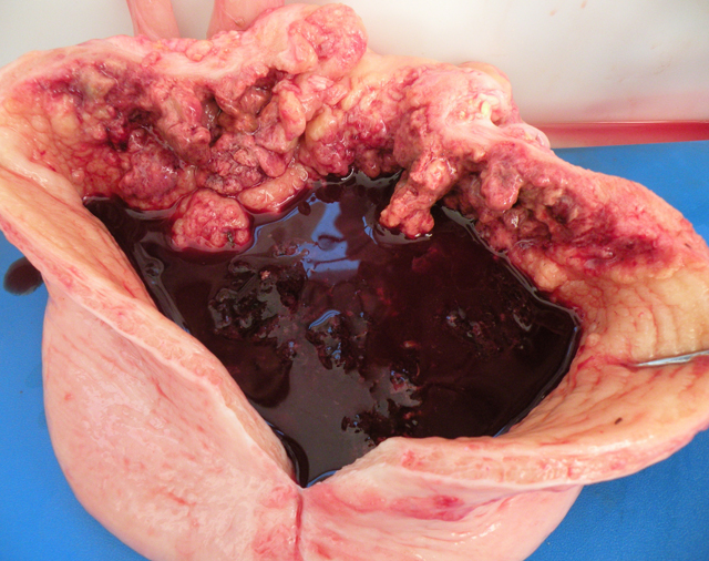
Gross findings in the urinary bladder of BEH affected cattle include dilatation of the bladder, with thickening of the wall and urine admixed with blood and blood clots. In the bladder mucosa there is congestion, markedly conspicuous vascularization, hemorrhagic foci, vascular growths, cauliflower-resembling nodules, red, white or yellow masses, pedunculated or sessile, frequently multilobulated and ulcerated and multiple translucent nodules arranged as bunches of grapes.(7,16)
Histologic examination of the bladder tumors associated with BEH reveals a large spectrum of tumor types, both of epithelial and mesencyhmal origin (7,12) and although mestastases are rare they are known to occur mainly to the lumboaortic lymph node.(5) In some cases the hematuria is not associated with the presence of neoplastic lesions, and the only lesions observed in the bladder mucosa are congestion and blood vessel ectasia.(7)
Other species, like sheep and horses, might be poisoned by Pteridium spp. Sheep, although much less frequently, are also affected by enzootic hematuria. In Australia the disease in sheep was attributed to the ingestion of P.esculentum. Changes in the bladder epithelium were metaplastic and neoplastic (carcinomas).(9) The acute hemorrhagic form of the disease, similarly to the one described in cattle, is also known to occur occasionally in sheep.(1) Three other conditions, namely intestinal tumors, the so called bright blindness and polioencephalomalacia are described in sheep that consume P. aquilinum.(10) Bright blindness is a form of retinal degeneration in which rods and cones are almost completely destroyed. Polioencephalomalacia is reportedly caused by the high thiaminase I contained in the plant.(10) Polioencephalomalacia has been reproduced in sheep by the feeding of bracken fern. A form of neurological disease due to the thiaminase action is also described in horses but the evidences for this disease are less convincing than the other forms of intoxication associated to this plant. Reportedly neurointoxication of the horse occurs more frequently in stabled horses receiving fodder contaminated with bracken fern, with clinical signs occurring 3-6 weeks after continuous exposure to the plant. Although the disease has been termed polioencephalomalacia, noteworthy pathological changes have not been described associated to presumptive bracken poisoning in horses. Horses fed for more than two months with dry bracken fern had no clinical or pathological findings in southern Brazil (unpublished data).
JPC Diagnosis:
Conference Comment:
Roperto et al (14) recently classified 400 bovine urothelial tumors in accordance with the World Health Organization morphological classification for human urothelial tumors, which is based on four distinct patterns of growth: flat, exophytic or papillary, endophytic, and invasive. In this series, urothelial tumors were usually multiple, and among flat urothelial lesions, carcinoma in situ (CIS) was the most common, accounting for 4% of the series, and occurring adjacent to papillary and invasive neoplasms in 80-90% of examined cases. The authors preferred the use of the term urothelial CIS to such synonyms as non-papillary and non-infiltrating TCC; urothelial CIS is characterized by marked local aggressive behavior, formation of von Brunns nests, pleomorphism, and frequent microinvasion into the lamina propria.(14) In the present case, participants noted CIS adjacent to the invasive neoplasm in many sections. In the same series, low-grade papillary urothelial carcinoma, consisting of papillary fronds lined by variably orderly urothelium, was the most common urothelial tumor recorded. Invasive urothelial tumors, most of which were high-grade carcinomas as with the present case, were less common than papillary urothelial neoplasms.(14)
Ptaquiloside is the major carcinogen in bracken fern. In alkaline environments, such as the urinary bladder or ileum of cattle, ptaquiloside is activated to its unstable reactive dienone form, which induces DNA alkylation.(14) The plant also contains thiaminase, tannins, phenolic acid, and several other carcinogens (e.g. quercetin, ptaquiloside Z, aquilide A, shikimic acid, and prunasin).(2,14) Bracken fern is also of significant public health interest in certain geographic locations due to epidemiological studies linking bracken fern consumption and cancer, either directly or indirectly through the milk of animals grazing on the plant or contaminated water.(14)
The contributor mentioned the role of BPV-2 and BPV-4 in association with bracken fern-induced tumors of the urinary bladder and upper digestive system, respectively. BPV-2 also is associated with cutaneous papillomas and fibropapillomas of the skin, esophagus, esophageal groove, and rumen.(2) The only BPVs known to infect the bladder urothelium, BPV-1 and BPV-2 are capable of infecting mesenchymal tissues and exhibiting cross-species transmission, and encode three oncoproteins (i.e. E5, E6, and E7) that initiate cellular transformation by a variety of mechanisms.(14)
References:
2. Brown CC, Baker DC, Barker IK: Alimentary system. In: Jubb, Kennedy and Palmers Pathology of Domestic Animals, ed. Maxie MG, 5th ed., vol. 2, p. 51. Saunders, Elsevier, Philadelphia, PA, 2007
3. Campo MS, Jarrett WF, Barron R, ONeil BW, Smith KT: Association of bovine papillomavirus type 2 and bracken fern with bladder cancer in cattle. Cancer Res 52:68986904, 1992
4. Campo MS: Bovine papillomavirus and cancer. Vet J 154:175188, 1997
5. Carvalho T, Naydan D, Nunes T, Pinto C, Peleteiro MC: Immunohistochemical evaluation of vascular urinary bladder tumors from cows with enzootic hematuria. Vet Pathol 46:211-221, 2009
6. Dawra RK, Sharma OP, Somvanshi R: Experience with enzootic hematuria in India Bracken Fern conference. In: International Bracken Fern Group Conference Bracken Fern: Toxicity, Biology and Control, ed. Taylor JA, Smith RT, Special Publication number 4, p. 150-154. International Bracken Group, Manchester, England, 2000
7. Fran+�-�a TN, Tokarnia CH, Peixoto PV: Diseases caused by the radiomimetic principle of Pteridium aquilinum (Polypodiaceae): a review [Enfermidades determinadas pelo princ+�-�pio radiomim+�-�tico de Pteridium aquilinum (Polypodiaceae). Pesq Vet Bras 22:85-96, 2002 (In Portuguese, Abstract in English)
8. Gava A, Neves DS, Saliba TM, Schild Al, Riet-Correa F. Bracken fern (Pteridium aquilinum) poisoning in cattle in southern Brazil. Vet Human Toxicol 44:362-365, 2002
9. Harbutt PR & Leaver DD: Carcinoma of the bladder of sheep. Aust Vet J 45:473-475, 1969.
10. Kellerman TS, Coetzer JAW, Naud+�-� TW, Botha CJ. Plant poisonings and mycotoxicoses of livestock in Southern Africa. 2nd ed., p 84, Oxford University Press, Oxford, 2005
11. Maxie MG, Newman SJ: Urinary system. In: Jubb, Kennedy, and Palmers Pathology of Domestic Animals, ed. Maxie MG, 5th ed., vol. 2, pp. 518-521. Elsevier Saunders, Philadelphia, PA, 2007
12. Peixoto PV, Fran+�-�a TN, Barros CSL, Tokarnia CH: Histopathological aspects of bovine enzootic hematuria in Brazil. Pesq Vet Bras 23:6581, 2003
13. Rissi DR, Rech RR, Pierezan F, Gabriel AL, Trost ME, Brum JS, Kommers GD, Barros CSL: Plant and plant-associated mycotoxins poisoning in cattle in Rio Grande do Sul, Brazil: 461 cases [Intoxica+�-�+�-�o por plantas e micotoxinas associadas a plantas em bovinos no Rio Grande do Sul]. Pesq Vet Bras 27:261-268, 2007 (In Portuguese, Abstract in English)
14. Roperto S, Borzacchiello G, Brun R, Leonardi L, Maiolino P, Martano M, Paciello O, Papparella S, Restucci B, Russo V, Salvatore G, Urraro C, Roperto F: A review of bovine urothelial tumours and tumour-like lesions of the urinary bladder. J Comp Pathol (in press), 2009
15. Souto MAM, Kommers GD, Barros CSL, Piazer JVM, Rech RR, Riet-Correa F, Schild AL: Neoplasms of the upper digestive tract of cattle associated with spontaneous ingestion of bracken fern (Pteridium aquilinum) [Neoplasias do trato alimentar superior de bovinos associadas ao consumo espont+�-�neo de samambaia (Pteridium aquilinum)]. Pesq Vet Bras 26:112-122, 2006 (In Portuguese, Abstract in English)
16. Souto MAM, Kommers GD, Barros CSL, Rech RR, Piazer JVM: Urinary bladder neoplasms associated with bovine enzootic [Neoplasmas da bexiga associados +� hematuria enzo³tica bovina]. Ci+�-�ncia Rural 36:1647-1650, 2006 (In Portuguese, Abstract in English)