CASE III:
Signalment:
Cat (Felis catus), breed: European shorthair
10 years of age
Male (castrated)
History:
Anorexia and losing weight since two months. Some teeth were extracted. In the following days the cat developed corneal edema in the right eye. Clinic diagnosed a glaucoma. CT was performed to identify cause of glaucoma and check for dental problems. During induction the cat developed high heartrate. Thoracic imaging shows bilateral pneumothorax with aberrant lung tissue. Due to poor prognosis the cat was euthanized.
Gross Pathology:
The right eye is very firm compared to the left eye. The right eye has a red, diffusely opaque cornea. The lungs contain bilaterally, multifocal, white, shiny, firm, round noduli. Both kidneys contain multiple white, shiny, firm, expansile noduli in the cortices.
Laboratory Results:
None.
Microscopic Description:
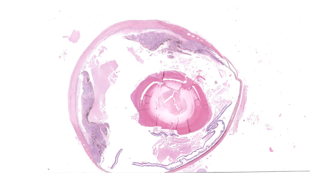
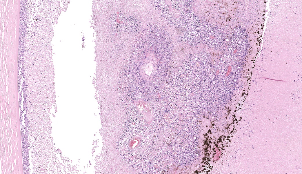
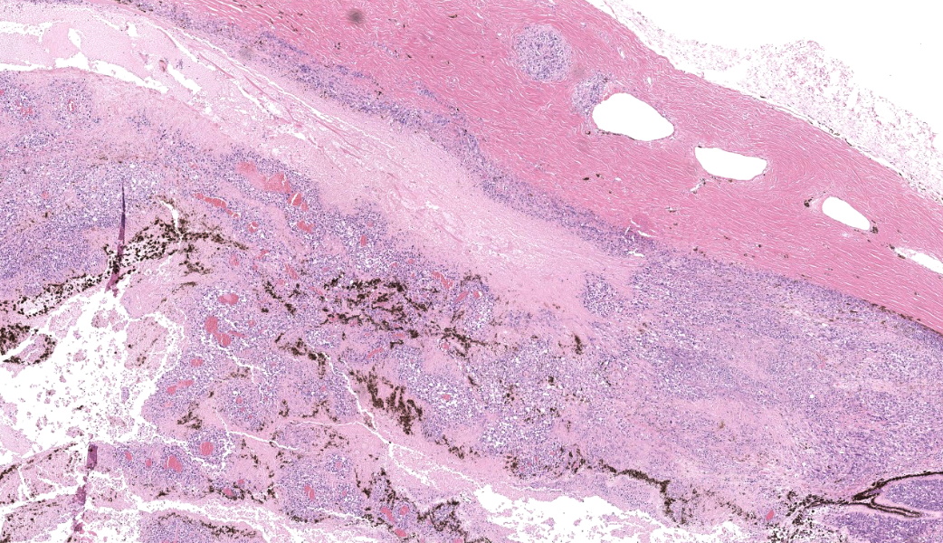
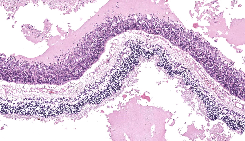
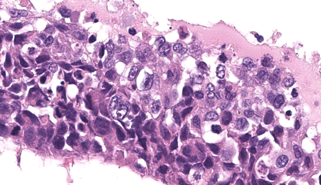
Right eye: The iris is almost diffusely irregularly thickened with nodular protrusions in the vitreous. This thickening is due to a development of neoplastic cells that appear in the ciliary body and infiltrate the filtration angle. The neoplastic mass is moderately cellular, poorly demarcated, growing infiltratively and not encapsulated. The cells lie in contiguous fields, regularly surrounding a newly created vascular structure, supported by a varying amount of fibrovascular stroma. Cells are polygonic, varying in size but averaging about 10 µm in diameter, with poorly visible cell borders, abundant eosinophilic, fibrillar cytoplasm and a high nucleus:cytoplasm ratio. Some cells contain a compact dark brown to fine granular brown pigment. The nucleus is round, oval, to elongated, centrally located with finely stippled chromatin, and 1 to 3 eosinophilic nucleoli. There are sometimes cells with multiple nuclei, there is significant anisokaryosis and anisocytosis, including karyomegaly. Mitoses are 1 to 3 per 400x HPF. Multifocally there are large areas of eosinophilic amorphous material, bordered by neutrophilic granulocytes, cells with karyorrhexis and cell debris (necrosis). The sclera contains a vascular structure with a tumor embolism. The cornea contains multifocal formation of vascular structures (neovascularization) surrounded by some neutrophilic granulocytes. Some vascular structures in the sclera are bordered by many lymphocytes and plasma cells (lymph aggregates). Around the corpus ciliary there are many macrophages with brown pigment granules (melanomacrophages).
The lens multifocally contains many “bladder cells” (cataract). There is a detached lens apsule, but no reaction (cutting artefact). In the optic nerve some dilated myelin sheaths with foamy macrophages are present (digestion chambers, indicating degeneration).
Other organs (not submitted): similar neoplastic cells in the lung and kidney.
Contributor’s Morphologic Diagnoses:
Right eye: ocular amelanotic melanoma; mild perivascular neutrophilic keratitis; cataract of the lens; multifocal, mild axonal degeneration of the optic nerve.
Contributor’s Comment:
Almost all intraocular melanocytic tumors in cats represent a specific histologic and behavioral entity known as diffuse iris melanoma.7 Moreover, Feline diffuse iris melanoma (FDIM) is by far the most common form of ocular melanocytic neoplasia.5
A wide variety in morphology is displayed including round or polygonal cell, spindle cell, balloon cell, anaplastic variants and giant cell types. The occurrence of amelanotic variants is low and can be clinically mistaken for inflammatory lesions.5 “FDIM progressively expands through the iris, iridocorneal drainage angle and ciliary body, eventually penetrating the sclera”.5 Due to the neoplasm infiltrating the iridocorneal angle dyscoria, reduced pupil motility, thickening of the iris and secondary glaucoma may be observed.5 Exfoliation of tumor cells into the anterior chamber and dissemination via the aqueous humor can result in metastasis as well as hematogenous spread via the intraocular vasculature.3 The lung and liver are considered the most common sites of metastasis although metastases in the spleen, bones and regional lymph nodes have been reported.6
Extent of the tumor has a significant impact on survival times, invasion of neoplastic melanocytes into the ciliary body and animals with secondary glaucoma have reported lower survival rates compared to those without.4
Due to the wide variety of morphologic and pigmentation characteristics, a definitive diagnosis based on H&E alone is difficult. Additional immunohistochemical stains used for diagnostics of melanocytic neoplasia’s include Melan-A, HMSA-5, S-100 and tyrosinase.2,5
Contributing Institution:
Veterinair Pathologisch Diagnostisch centrum – University of Utrecht
https://www.uu.nl/onderzoek/veterinair-pathologisch-diagnostisch-centrum/informatie-voor-dierenartsensen - Veterinair Pathologisch Diagnostisch Centrum - Universiteit Utrecht (uu.nl)
JPC Diagnoses:
- Eye: Metastatic carcinoma.
- Eye, lens: Cataract.
JPC Comment:
The contributor has provided an outstanding specimen of a pleomorphic, largely undifferentiated neoplasm in the feline globe. While melanoma must always be considered in the feline eye, it is not the only possibility, as post-traumatic ocular sarcomas may differentiate along a wide variety of morphologies. The feline uvea may also be a site of metastasis for poorly differentiated carcinomas.
This thorny issue was addressed by Gran et al. in 20162 in their retrospective review of 75 neoplasms of the feline globe. In this excellent review, immunohistochemical labeling of eight of the 75 cases ultimately conflicted with the initial morphologic diagnosis rendered on the H&E section. Discordant diagnoses included four tumors which were initially diagnosed as lymphoma and four as ocular sarcoma.2 Immunohistochemical testing amended the diagnosis of three of the lymphomas to melanoma, and the final case to ciliary body adenocarcinoma2. Two of the cases originally diagnosed as sarcoma ultimately demonstrated an immunohistochemical profile for melanoma, one for metastatic carcinoma, and the final case remained undifferentiated.2
In this case, the diagnosis of melanoma, particularly amelanotic melanoma, in the absence of any additional histochemical or immunohistochemical staining is somewhat problematic, especially in light of the findings of Grahn et al. discussed above.
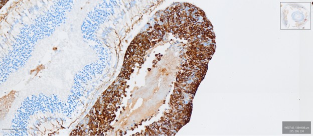
To confirm the contributor’s diagnosis of amelanotic melanoma, additional stains were run at the Joint Pathology Center, including Fontana-Masson, tyrosinase, MelanA, PNL2, and S-100. Neoplastic cells were immunonegative for all of these markers. Neoplastic cells demonstrate strong cytoplasmic immunopositivity for cytokeratin and approximately 10% of the neoplastic cells also demonstrate moderate immunoreactivity for vimentin.
Based on the strong immunopositivity for cytokeratin, a diagnosis of metastatic carcinoma was made in this case. An additional histologic finding which supports this finding is the “carpeting” of the inside of the eye by the neoplastic cells, which has been identified as a histologic feature of carcinomas metastatic to the eye in the dog and cat 1,8, and in a previous WSC case of feline pulmonary carcinoma metastatic to the eye (WSC 2014-2014 Conference 24, Case 1).
The JPC morphologic diagnosis is a simple one and to the eyes of some, may not reflect all of the histologic changes that may be found in this eye. However, in traditional JPC (and AFIP before it) fashion, the morphologic diagnosis of a neoplasm reflect the tumor, and not all of the secondary changes that appear over time because of it. We did not consider the cataract secondary to the tumor in this case, so we provided a second morphologic diagnosis for a second, likely independent pathologic process.
References:
- Dubielzig RR, Ketring KL, McLellan GJ. Veterinary Ocular Pathology: a comparative review. 2010.
- Grahn BH, Peiffer RL, Cullen CL. Classification of feline intraocular neoplasms based on morphology, histochemical staining, and immunohistochemical labeling. Vet Ophthalmol. 2016: 9(6), 395-403.
- Jajou S. Uveal amelanotic melanoma in a ragdoll cat. Can Vet J. 2020: Jun 61(6):645-647.
- Kalishman JB, Chappell R, Flood LA. A matched observational study of survival in cats with enucleation due to diffuse iris melanoma. Vet Ophthalmol. 1998 1(1), 25-29.
- Kayes D & Blacklock B. Feline Uveal Melanoma Review: Our Current Understanding and Recent Research Advances. Vet Sciences. 2022: 9(2), 46.
- Patnaik AK & Mooney S. Feline melanoma: a comparative study of ocular, oral, and dermal neoplasms. Vet Path. 1988: 25(2), 105-112.
- Wilcock BP, Bradley Njaa L. Special senses. In: Jubb, Kennedy, and Palmer’s Pathology of Domestic Animals. Sixth ed., vol 1, Philadelphia, PA: Elsevier Saunders; 2016: 407
- Stocklein H, Taylor RP, Shaw GC, Clarke LL. Histomorphologic and immunohistochemical characteristics of intraocular metastatic carcinomas of presumed pulmonary origin in cats: A retrospective case series from 2019 to 2025. Veterinary Pathology. 2026;63(5):820-824. doi:1177/03009858261445996