CASE I:
Signalment:
3 month old male Han Wistar rat
History:
Low dose rat in a 6 week duration oral toxicity study. Rats were 6-7 weeks old at study start.
Gross Pathology:
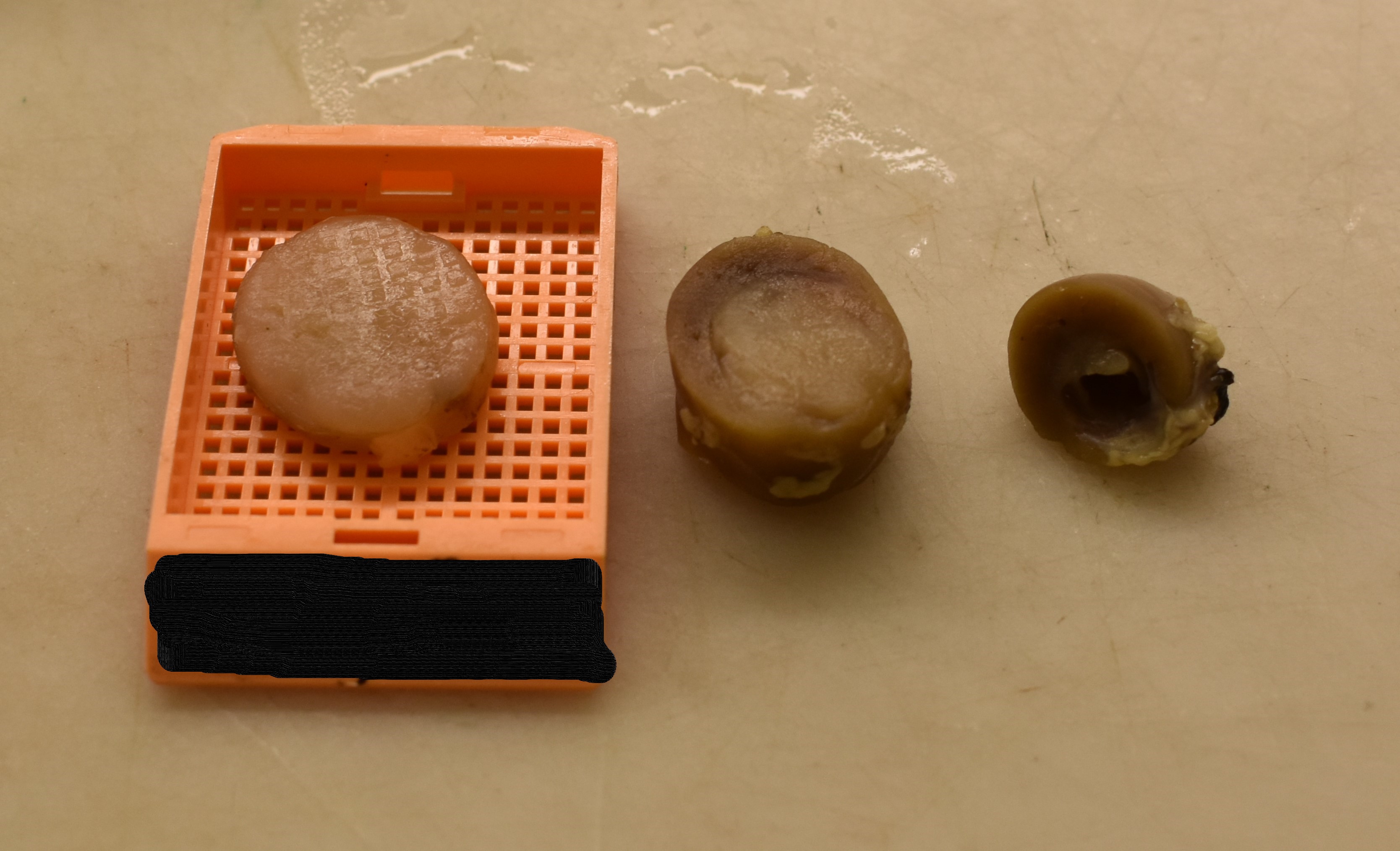
At scheduled terminal necropsy, a 2.5 x 1.5 cm, pale cylindrical mass emanating from the outer medulla of one kidney and proceeding caudally was noted. The mass clearly compressed the adjacent renal parenchyma. No other masses were noted during the full necropsy of this rat.
Laboratory Results:
None provided.
Microscopic Description:
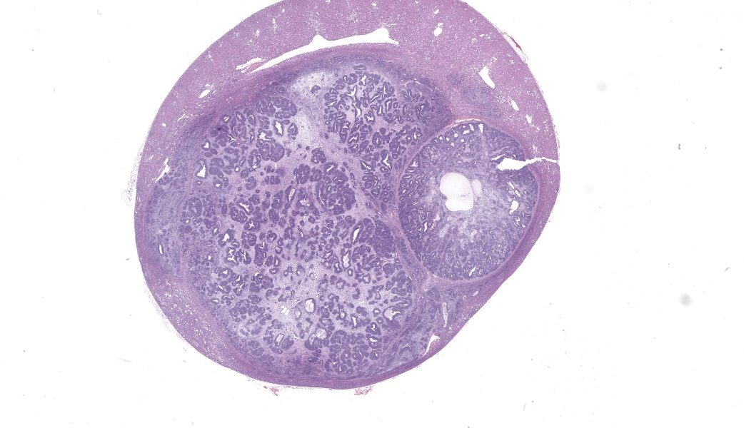
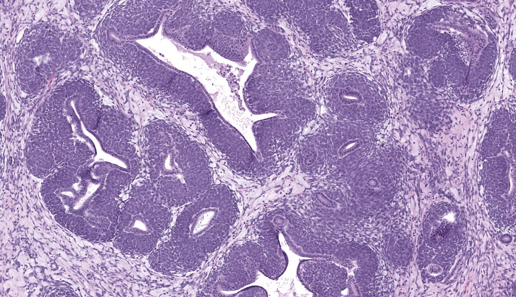
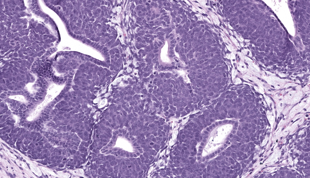
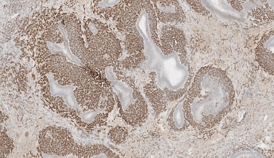
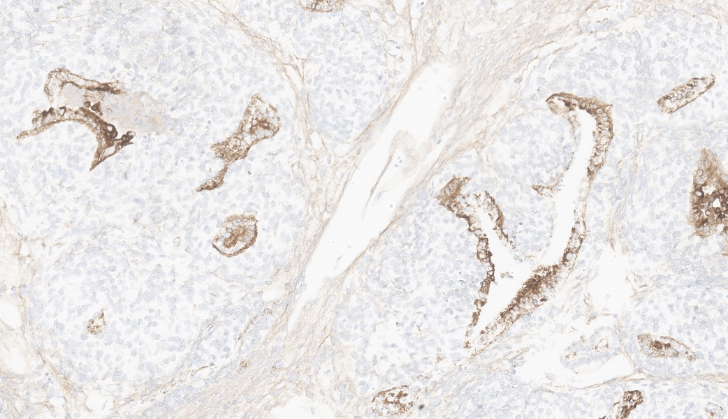
An expansile mass emanated from the corticomedullary junction of one kidney, obliterating the renal pelvis and nearly all of the medulla while compressing the remaining renal cortex. The mass was composed of biphasic islands of tubular structures (lined by darkly basophilic cuboidal to columnar epithelium when primitive, and lined with more eosinophilic, columunar epithelium when more mature) surrounded by darkly basophilic, crowded blastemal cells with occasional mitotic figures. These biphasic islands often touched and were either grouped into lobular structures separated from other portions of the mass by denser connective tissue or were scattered amongst a more primitive connective tissue stroma. The islands were often crowded at the periphery of the lobules with the centers of the larger lobules having scattered islands, loose stroma, and in most slides, cystic structures. There was degeneration of some tubular epithelium in some islands with lumenal cellular debris and an amorphous eosinophilic substance. Near the outer edges of the main mass where it abutted with the remaining renal parenchyma were a few primitive glomerular structures (characterized by foci of small dark mononuclear cells). Local invasion of the mass into the remaining renal parenchyma was characterized by variously sized aggregates of blastemal cell associated with either loose or condensed stroma similar to that in the main mass. Due to the compression by the mass, mild tubular dilatation was present in the remaining renal parenchyma; a few dilated tubules in many slides contained cell debris and inflammatory cells or hyaline casts. There was minimal interstitial infiltrate in the renal parenchyma with little to no inflammatory response within the mass.
Contributor's Morphologic Diagnoses:
Nephroblastoma, unilateral (Incidental)
Tubular dilatation, multifocal, mild
Interstitial infiltrate, lymphocytic, minimal
Contributor's Comment:
The topographic origin (corticomedullary junction), contents (biphasic structures of blastemal cells surrounding primitive renal tubules and occasional primitive glomerular structures), and primitive loose connective tissue were all consistent with nephroblastoma.2 Differential diagnoses were nephroblastemosis and renal mesenchymal tumor.1,3 The extent of the mass alone was sufficient to exclude nephroblastemosis, which is a much smaller, typically interstitial, aggregate of primitive renal structures.1 The remaining differential diagnosis, renal mesenchymal tumor,
is composed of several types of mesenchymally-derived structures, ranging from fibrous connective tissue to cartilage or bone and also contains entrapped but otherwise normal renal tubular elements.2 None of these features were present in the mass evaluated.
Tumors in young adult rats can be an interpretive challenge in toxicity studies as they are uncommon and must be distinguished from an actual test article-related effect. Recent retrospectives and case reports have been vital in de-risking these rare spontaneous tumors from potential early carcinogenic signals.3 The random placement of this particular rat into the low dose group was also helpful in the de-risking assessment in this case.
Contributing Institution:
Drug Safety Research & Development
Pfizer Inc.
www.Pfizer.com
JPC Diagnosis:
Kidney: Nephroblastoma
JPC Comment:
This week's moderator is our very own MAJ Sam Medlin, JPC Class of 2025 (that was a good year), Chief of the Necropsy Service at the Walter Reed Army Institute of Research, and a devotee of all things tumor. This week we covered some very interesting neoplasms from the stacks at the JPC, and found some surprising things in the process.
A nephroblastoma, also known as a Wilms tumor (WT), is an embryonal, mesodermal tumor with multiple lines of differentiation that is thought to arise from the primitive renal stem cells.4 It is the most common renal tumor in childhood.4 In human pediatric patients, it is associated with germline and/or somatic mutations of the WT1 gene, but other genes are thought to be involved as well.3
Histologically, WT presents with three distinct cell types: epithelial, stromal, and blastemal. All three elements are not required for diagnosis, but when all are present, the term triphasic
is used. Like the renal mesenchymal tumor mentioned by the contributor, these tumors may also contain heterologous components, including smooth muscle, skeletal muscle, cartilage, and bone. All of the components of these tumors, including the heterologous components, have been shown to demonstrate similar genetic changes, indicating that all of these components are neoplastic.6
Nephroblastomas have been documented in a wide variety of domestic, laboratory, and exotic species, including dogs, cats, pigs, chickens, non-human primates, ruminants, camelids, rabbits, parakeets, and fish.7,8,9,10 They are considered the most common renal neoplasm in pigs, chickens, and fish.7 In the dog, the spinal nephroblastoma (previously known by a wide range of names, including the early nomenclature of thoracolumbar spinal tumor of young dogs
) is an unusual manifestation of this tumor, which has previously been seen multiple times in the Wednesday Slide Conference (WSC 2013-2014 Conference 22, Case 3 and WSC 2019-2020, Conference 5, Case 4).11 While the origin remains controversial, leading theories are that these tumors arise from either ectopic metanephric blastema or mesonephric rest tissue, either of which becomes entrapped between the dura mater and the spinal cord during embryogenesis.12, 13
In laboratory animals, nephroblastomas are most commonly reported in rats14, and are less commonly seen in mice and non-human primates. In rats, nephroblastomas occur both experimentally and spontaneously.14
Experimentally, they have historically been induced by chemical administration of N-ethyl-N-nitrosourea or N-methyl-N-nitrosourea, both alkylating agents.15 Spontaneously, they have been reported in Sprague-Dawley and F344 rats, with reports of metastases to the lymph nodes and lungs in these strains.15,16
References:
- Kalaiselvan P, Mathur KY, Pande VV, Madheswaran R, Bhelonde JJ, Shelar PD, Udupa V, and Shingatgeri VM. Intralobar Nephroblastomatosis in a nine-week-old Wistar Rat. Toxicol Pathol 2009; 37:819-825.
- Seely JC. Renal mesenchymal tumor vs nephroblastoma: revisited. J Toxicol Pathol 2004; 17:131-136.
- Weber K. Differences in types and incidence of neoplasms in Wistar Han and Sprague-Dawley rats. Toxicol Pathol 2017; 45(1): 64-75.
- Pritchard-Jones K. Malignant origin of the stromal component of Wilms' tumor. J Natl Cancer Inst. 1997; 89:1089-1091.
- Beckwith J, Bruce NB, Bonadio K. Nephrogenic rests, nephroblastomatosis, and the pathogenesis of Wilms' tumor. Pediatr Pathol. 1990; 10:1-36
- Zhuang Z, Merino MJ, Vortmeyer AO. Identical genetic changes in different histologic components of Wilms' tumors. J Natl Cancer Inst. 1997; 89:1148-52.
- Meuten DJ, Meuten TLK. Tumors of the urinary system. In: Meuten DJ, ed. Tumors in Domestic Animals.5th ed. Ames, Iowa: Iowa State Press; 2017:646-649.
- Delaney MA, Treuting PM, Rothenburger JL. Lagamorpha. In: Terio KA, McAloose D, St. Leger J, eds. Pathology of Wildlife and Zoo Animals.San Diego, CA: Elsevier. 2018; 486.
- Frasca Jr S, Wolf JC, Kinsel MJ, Camus AC, Lombardini ED. Osteichthyes. In: Terio KA, McAloose D, St. Leger J, eds. Pathology of Wildlife and Zoo Animals.San Diego, CA: Elsevier. 2018; 961.
- Reavill DR, Dorrenstein G. Psittacines, Coliiformes, Musophagiformes, Cuculiformes. In: Terio KA, McAloose D, St. Leger J, eds. Pathology of Wildlife 11. and Zoo Animals.San Diego, CA: Elsevier. 2018; 784.
- Brewer DM, Cerda-Gonzalez S, Dewey CW, Diep AN, Van Horne K, McDonough SP. Spinal cord nephroblastoma in dogs: 11 cases (1985-2007). J Am Vet Med Assoc. 2011; 238(5):618-24.
- Gasser, AM., Bush, WW., Smith, S., Walton, R. Extradural spinal, bone marrow, and renal nephroblastoma. J Am Anim Hosp Assoc. 2003; 39: 80-85.
- Petit A, Rubio A, Durand C, Piolat C, Perret C, Pagnier A, Plantaz D, Sartelet H. A Wilms' Tumor with Spinal Cord Compression: An Extrarenal Origin? Case Rep Pediatr. 2018 Sep 3;2018:1709271.
- Chandra M, Carlton WW. Incidence, histopathologic and electron microscopic features of spontaneous nephroblastomas in rats. Toxico Lett. 1992; 62: 179-190.
- Yoshizawa K, Kinoshita Y, Emoto Y. N-Methyl-N-nitrosourea-induced Renal Tumors in Rats: Immunohistochemical Comparison to Human Wilms Tumors. J Toxicol Pathol. 2014; 26:141-148.
- Tanaka, N., Takeshi, I., Jyoji, Y. Spontaneous nephroblastoma with striated muscle differentiation in an F344 rat. J Toxicol Pathol. 2017; 30:231-234.