Signalment:
7yr old, castrated male,
Mustela putorius furo, ferretIn January of 2000 Eucchus presented with bilaterally symmetric hair loss over the dorsum, at that time both adrenals were at the upper range of normal size and nodular by ultrasound. Hormonal panels supported a clinical diagnosis of adrenal disease (Estradiol 174pmol/L (normal 30-180pmol/L); 17-OH-progesterone 0.85nmol/L (normal 0-0.8 nmol/L); androstenedione 30.7nmol/L (normal 0-15nmol/L). At that time the owner elected medical management with repeated injections of Depo-lupron. Depo-lupron is a GnRH analogue which inhibits production of LH and FSH. In September of 2000, the owner reported that Eucchus belly would be soaked in urine after he urinated. Eucchus also repeatedly presented with alopecia and flaky skin. Urinary symptoms and alopecia resolved following increasing doses of Depo-lupron. In December of 2001 and again in June of 2002 Eucchus presented with difficulty urinating and was found to have an enlarged bladder. A urinary tract infection and enlarged prostate were diagnosed and Eucchus was started on antibiotics. At this time an insulinoma was also suspected clinically. Several weeks after discontinuing the antibiotics (August 2002), Eucchus again presented with straining to urinate and antibiotics were resumed. While still on antiobiotics, Eucchus presented in October of 2002, for straining to urinate. At this time the left adrenal gland was markedly enlarged (1cm) by ultrasound and surgery was elected. At surgery, the left adrenal gland was removed and 2 periprostatic cysts were identified which communicated with the urinary bladder. Additionally, 2 discrete nodules were noted in the pancreas and were removed. Following surgery, Eucchus became lethargic, dehydrated and anuric and was euthanized.
Biopsy results of the adrenal and pancreas were consistent with an adrenocortical adenocarcinoma and islet cell tumors (presumptive insulinomas), respectively.
Gross Description:
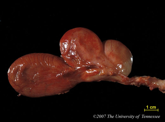
At necropsy, Eucchus was found to be obese and the abdomen contained 50ml of serosanguinous fluid. Two 3cm diameter cysts were found surrounding the prostate, just caudal to the
urinary bladder (fig. 1-1).
Histopathologic Description:
The prostate is markedly expanded by a single large cyst and multiple smaller cysts lined by keratinizing stratified squamous epithelium, and containing variable amounts of keratin. In some sections, there is focal loss of the epithelial lining of one of the cysts with free keratin in the surrounding stroma. This free keratin is surrounded by a variable infiltrate of neutrophils and macrophages. Throughout the prostate there are decreased amounts of glandular tissue. Remaining glandular structures are lined by a low cuboidal epithelium and rarely contain an eosinophilic secretory product. Scattered clusters of lymphocytes and rarely eosinophils are present in the surrounding fibrous connective tissue stroma.
Morphologic Diagnosis:
Prostate: Severe glandular atrophy and squamous metaplasia with cyst formation.
Condition:
Cystic prostatic disease
Contributor Comment:
Adrenal cortical lesions are the second most common neoplasm of ferrets, after pancreatic islet cell tumors. An increased incidence of proliferative adrenal lesions occurs in ferrets neutered at an early age (2-4months), and is likely due to chronic stimulation of the cells of the zona reticularis by luteinizing hormone.
4 Adrenal gland-associated endocrinopathy (AAE) is associated with the presence of hyperplastic or neoplastic adrenal lesions which produce high levels of estrogenic compounds (estradiol-17b, androstenedione, dehydroepiandrosterone sulfate, 17-hydroxyprogesterone, progesterone). Lesions associated with AAE include bone marrow toxicity
4 and Cushingoid features (thin skin, muscular atrophy, pot-bellied appearance)
1, bilateral symmetrical truncal alopecia, vulvar swelling in spayed females, reversion to sexual behavior in neutered animals, mammary gland hyperplasia in castrated males, and dysuria in males associated with squamous metaplasia of the prostate and prostatitis.Â
The squamous metaplasia in the prostate of this ferret is likely due to increased levels of circulating estrogenic compounds. Six cases of prostatic squamous metaplasia with concurrent prostatitis have been reported in male ferrets with proliferative adrenocortical lesions. As in dogs, the prostatitis has been attributed to the presence of keratin.
2 The absence of significant prostatitis in this case may be unusual. The observed prostatic atrophy is likely a result of castration at a young age and failure of the prostate to develop normally.
3
In dogs, squamous metaplasia of the prostatic glandular epithelium has been associated with estrogen-producing Sertoli cell tumors, or exogenous administration of estrogens. In such cases, squamous metaplasia affects the prostatic urethra, uterus masculinus and prostatic ducts. Similar changes have been reported in swine. In cats, exogenous estrogen results in prostatic enlargement due to epithelial hyperplasia and cystic dilation of the glands; squamous metaplasia and cornification, however, only occur in the urethral epithelium.Â
3 Enlargement of the prostate is most commonly associated with constipation, and less commonly stranguria.
3
JPC Diagnosis:
1. Prostate: Prostatic cysts, multiple,
ferret (Mustela putorius furo), carnivore.
2. Prostate, glands: Squamous metaplasia, multifocal,
mild, with prostatitis and keratinizing cysts.
Conference Comment:
Squamous metaplasia of the
prostate with keratinizing prostatic cysts is a common
sequel in male ferrets diagnosed with adrenal-associated
endocrinopathy.
1,8 Chronic elevation of circulating luteinizing
hormone (LH), resulting from early neutering,
is required for metaplastic transformation.
1 Elevated
circulating LH acts on the zona reticularis
4, stimulating
cellular proliferation as well as the production of high
levels of circulating estrogenic compounds, including
estradiol-17b, androstenedione, dehydroepiandrosterone,
17-hydroxyprogesterone, and progesterone.
2
Luteinizing hormone receptors (LHRs) are usually present
on ovarian thecal cells, granulosa cells, luteal cells,
and testicular Leydig cells.
1 LHRs have also been identified
in the adrenal gland of fetal (but not adult) mice, and
low levels of LHR mRNA has been detected in the adrenal
cortex of normal intact ferrets, indicating the presence
of non-functional receptors.
1
Tumors in the ferret adrenal gland include nodular hyperplasia,
adrenocortical adenoma, and adrenocortical carcinoma.
1,8
In the case of the latter, metastasis usually occurs
late in the disease, and early complete removal of
neoplastic adrenals carries a fair prognosis.
8 In contrast to
other species, plasma concentrations of cortisol are only
infrequently elevated in ferrets with AAE.
1,8
Squamous metaplasia of glandular epithelium due to hyperestrogenism,
has been documented in men, mice,
dogs, and sheep.
1,2,3,6 Experimental induction of prostatic
squamous metaplasia in the mouse model has revealed
proliferation of basal cells with keratinization following
injections with estrogen. In affected ferrets, squamous
metaplasia of prostatic epithelium is followed by cyst
formation and purulent inflammation as a result of keratin
production
2 and may ultimately result in dysuria and
post-renal azotemia.
References:
1. Bielinska M, Kiiveri S, Parviainen H, Mannisto S, Heikinheimo M, Wilson DB: Gonadectomy-induced adrenocortical neoplasia in the domestic ferret (
Mustela putorius furo) and laboratory mouse. Vet Pathol
43:97-117, 2006.
2. Coleman GD, Chavez MA, Williams BH: Cystic prostatic disease associated with adrenocortical lesions in the ferret (
Mustela putorius furo). Vet Pathol
35:547-549, 1998.
3. Foster RA and Ladds PW. Jubb, Kennedy, and Palmers Pathology of Domestic Animals. 5
th ed., vol. 3, pp. 605-611. Saunders Elsevier, Endinburgh, 2007.
4. Peterson RA, Kiupel M, Capen CC: Adrenal cortical
carcinomas with myxoid differentiation in the domestic
ferret (Mustela putorius furo). Vet Pathol 40:136-142,
2003
5. Pollock CG: Urogenital diseases. In: Ferrets, Rabbits,
and Rodents Clinical Medicine and Surgery, eds. Quesenberry
KE, Carpenter JW, 2nd ed., pp. 45-46. Saunders,
St. Louis, Missouri, 2004
6. Risbridger GP, Wang H, Frydenberg M, Cunha G:
The metaplastic effects of estrogen on mouse prostate
epithelium: proliferation of cells with basal cell phenotype.
Endocrinology 142:2443-2450, 2001
7. Quesenberry KE, Rosenthal KL: Endocrine diseases.
In: Ferrets, Rabbits, and Rodents Clinical Medicine and
Surgery, eds. Quesenberry KE, Carpenter JW, 2nd ed.,
pp. 83-87. Saunders, St. Louis, Missouri, 2004
8. Williams B: Pathology of the Domestic Ferret. At:
http://www.afip.org/ferrets/ferret.path.html