Signalment:
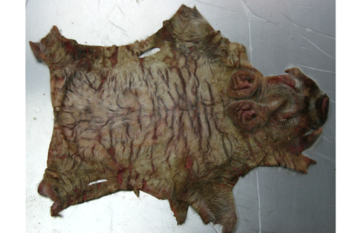
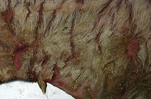
Gross Description:
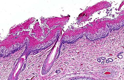
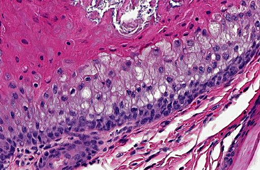
Histopathologic Description:
Morphologic Diagnosis:
Lab Results:
Condition:
Contributor Comment:
- Ichthyosis vulgaris: Orthokeratotic hyperkeratosis and a decreased or absent granular layer.
- X-linked ichthyosis: Orthokeratotic hyperkeratosis with normal or hyperplastic granular layer.
- Epidermolytic hyperkeratosis: Orthokeratotic and parakeratotic hyperkeratosis, vacuolation of keratinocytes in the upper stratum spinosum and stratum granulosum and a markedly thickened granular layer.
- Lamellar ichthyosis: Compact orthokeratotic hyperkeratosis and mild acanthosis.
- Harlequin ichthyosis: Thick compact stratum corneum with follicular hyperkeratosis and variable appearance of the stratum granulosum.
In cattle, two forms of ichthyosis are described: ichthyosis fetalis and ichthyosis congenita.(4) In ichthyosis fetalis morphological findings are similar to harlequin ichthyosis. Ichthyosis congenita has a prominent laminated orthokeratotic hyperkeratosis of the epidermis and superficial portion of hair follicles. In dogs, nonepidermolytic and epidermolytic classifications are used.(5) It has been concluded that well characterized cases of ichthyosis in animals do not correlate well with the human classification system.(4)
In this case, the lesions are somewhat similar to epidermolytic hyperkeratosis with orthokeratotic and parakeratotic hyperkeratosis and vacuolation of keratinocytes in the upper stratum spinosum and stratum granulosum, but lack the markedly thickened granular layer that is present in epidermolytic hyperkeratosis. Also lesions consistent with ichthyosis vulgaris (orthokeratotic hyperkeratosis and decreased or absent granular layer) are present. Furthermore, similarities with ichthyosis congenita are seen; however, there is no involvement of the hair follicles in the present case.
Due to the different morphologic findings in the present case and the current human classification systems our conclusion is that the disease is best regarded as ichthyosis with no further classification.
JPC Diagnosis:
Conference Comment:
The contributor outlines ichthyosis and its variable presentation among domestic animals as the cause of hyperkeratosis in this case. Another condition associated with hyperkeratosis in swine is zinc-responsive dermatosis, which occurs in 2-4 month-old piglets and is a secondary zinc deficiency due to the presence of phytic acid in plant protein rations that affects its availability. Zinc-responsive dermatoses are more commonly identified in dogs as one of three distinct varieties: reduced absorption in Arctic breeds, generic dog food, and lethal acrodermatitis of Bull Terriers. Thallium toxicity is another historical cause in many species and vitamin A-responsive dermatoses have been described in Cocker Spaniels as causing hyperkeratosis. Superficial necrolytic dermatitis in dogs and people characteristically exhibits parakeratosis along with laminar epidermal edema and basilar hyperplasia. This is also called hepatocutaneous syndrome due to its correlation with liver dysfunction and subsequent deranged glucose and amino acid metabolism inducing hypoaminoacidemia. Canine morbillivirus and pemphigus foliaceous both induce hyperkeratosis of the footpads in dogs.(4)
Congenital ichthyosis occurs in domestic animals as a heterogenous syndrome of autosomal recessive inheritance. Collectively, they are called autosomal recessive congenital ichthyoses (ARCIs) and their pathophysiology involves abnormal synthesis and/or secretion of lipid within the stratum corneum. Recently, specific genetic mutations have been linked to certain cornification disorders affecting particular breeds of dogs. ARCI in Golden Retrievers is associated with PNPLA1 mutations, in Jack Russell terriers an insertion in TGM1 has been described, and most recently an upstream insertion of NIPAL4 (ICHTHYIN) was identified in American bulldogs.(6)
References:
1. Baker JR, Ward WR. Ichthyosis in domestic animals: a review of the literature and a case report. Br Vet J. 1985;141:1-8.
2. Chittick EJ, Olivry T, Dalldorf F, et al. Harlequin Ichthyosis in two Greater Kudu (Tragelaphus strepsiceros). Vet Pathol. 2002;39:751-756.
3. Cho JK, Son JM, Lee DS, et al. Harlequin Ichthyosis in a Han Woo Calf. J Vet Med Sci 2007;69(5):553-555.
4. Ginn PE, Mansell JL, Rakich PM. Skin and appendages. In: Maxie MG, ed. Jubb, Kennedy and Palmers Pathology of Domestic Animals. 5th ed. Vol 1. Phildadelphia, PA: Elsevier Saunders; 2007:576-578, 628-648.
5. Gross TL, Ihrke PJ, Walder EJ, Affolter VK. Diseases with abnormal cornification. In: Skin Diseases of the Dog and Cat: Clinical and Histopathologic Diagnosis. 2nd ed. Oxford: Blackwell Publishing; 2005;174-179.
6. Mauldin EA, Wang P, Evans E, et al. Autosomal recessive congenital ichthyosis in American bulldogs is associated with NIPAL4 (ICHTHYIN) deficiency. Vet Pathol. 2014 Oct 16. pii: 0300985814551425. [Epub ahead of print]
7. Molteni L, Dardano S, Parma P, et al. Ichthyosis in Chianina cattle. Vet Rec. 2006;158:412-414.
8. Oji V, Traupe H. Ichthyosis: differential diagnosis and molecular genetics. Eur J Dermatol 2006;16(4):349-59.
9. Testoni S, Zappulli V, Gentile A. Ichthyosis in two Cianina calves. Dtsch Tierarztl Wochenschr. 2006;113(9):351-4.