Signalment:
Adult female white-tailed deer (Odocoileus virginianus).
The deer had no fear of humans or vehicles. He had human contact including petting and hand feeding. The animal was euthanized by gunshot wounds to the chest. The head was removed and send intact.
Gross Description:
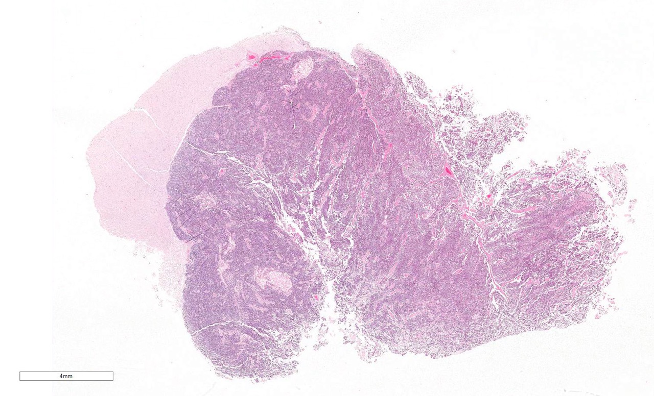
The head from the deer was submitted as part of routine Rabies surveillance by New York State Department of Health. A soft, white gelatinous mass effaced the right caudal nasal passage, cribriform plate and extended to involve the olfactory bulb of the brain.
Histopathologic Description:
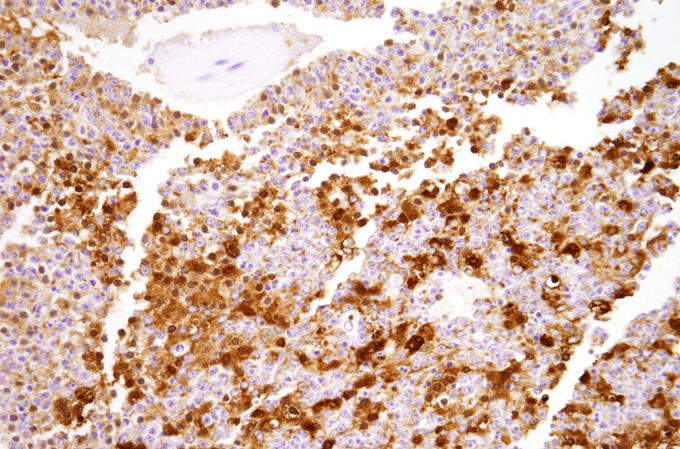
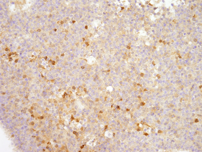
Immunohistochemistry was performed on a section of the brain
mass.Diffusely, neoplastic cells exhibited moderate to strong cytoplasmic
immunoreactivity for neuron specific enolase (NSE) (Fig. 4) and a subpopulation
of the cells showed strong cytokeratin immunoreactivity (Fig. 5).
Morphologic Diagnosis:
Lab Results:
- Fluorescent antibody test was negative for rabies
- Immunohistochemistry for chronic wasting disease was negative
- The brain mass was sent for aerobic
bacterial culture and many Aeromonas sp. were isolated.
Neuroendocrine carcinoma, deer
Condition:
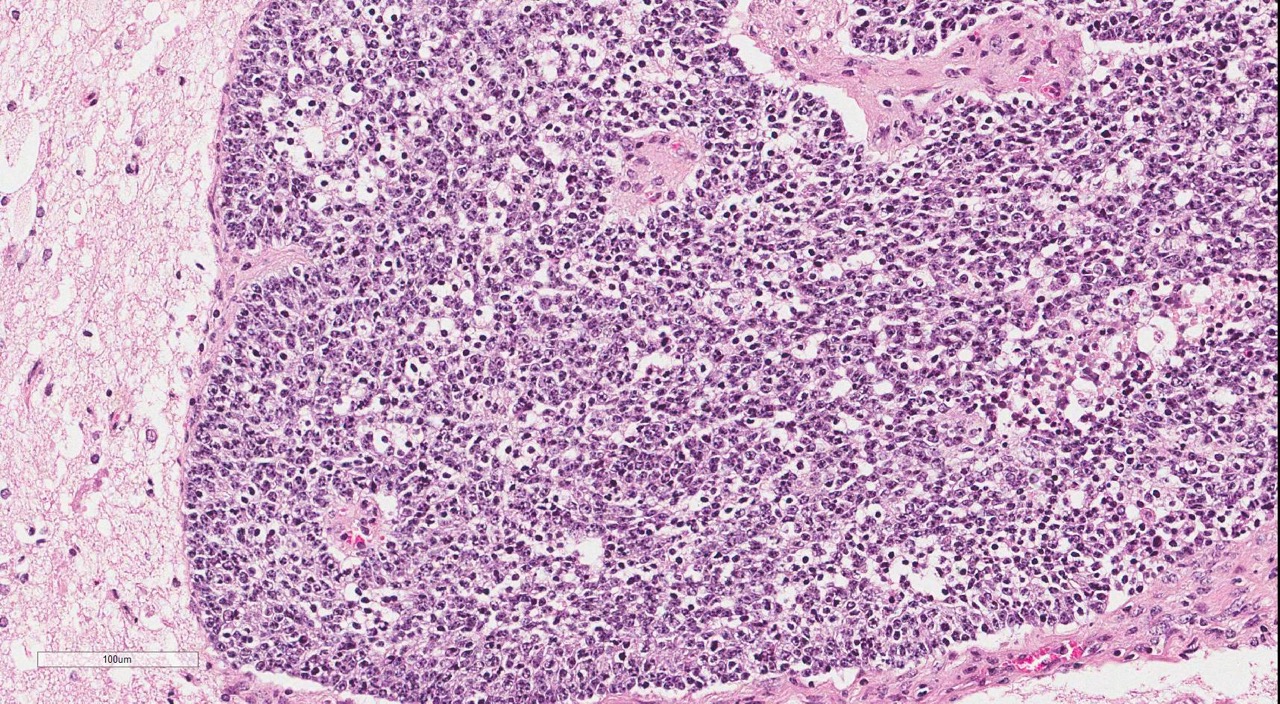
The two primary differential diagnoses for this tumor are neuroendocrine carcinoma and olfactory neuroblastoma.These two tumor types have similar histologic features and diff-erentiation often requires ultrastructural analysis and/or immunohistochemistry. Neuroendocrine tumors are a diverse group of neoplasms derived from cells which (A) produce a neurotransmitter, neuromodulator or neuropeptide hormone, (B) contain dense core granules on electron microscopy and (C) do not have axons or form synapses.10- Neuroendocrine tumors have been described in the almost every organ and are characterized by a typical pattern of small to medium sized cells with granular eosinophilic chromatin arranged in nests, cords and packets separated by fine fibrovascular stroma.Rosette formation and peripheral palisading is variably present.Ultrastructurally, tumor cells contain round, membrane bound dense core granules and have distinct intercellular junctions.Cells are argyrophilic (Grimelius positive) and show positive immunoreactivity for neuron specific enolase, chromogranin, cytokeratin and neuropeptides.Nasal neuroendocrine carcinomas have been reported in dogs14 and horses.16 In dogs, nasal neuroendocrine tumors have been reported to invade the underlying bone and adjacent sinuses, but not the brain.13
Contributor Comment:Olfactory neuroblastomas (ONB) or esthesioneuroblastomas are rare tumors, believed to be derived from the olfactory neuroepithelium. These tumors arise primarily in the upper nasal cavity and adjacent paranasal sinuses and frequently invade the cribriform plate and the intracranial cavity. Histological features of well-differentiated ONBs include growth in circumscribed lobules separated by richly vascularized fibrous stroma, or less commonly, a diffuse growth pattern. Pseudorosettes and true rosettes can be found. The neoplastic cells are surrounded by neurofibrillary matrix and have sparse cytoplasm and round to oval nuclei with inconspicuous nucleoli. Nuclear pleo-morphism, high mitotic activity and necrosis are frequently observed. Ultrastructural ch-aracteristics include dense core neur-osecretory granules and neurite-like cell pro-cesses with neurofilaments and neur-otubules.Like neuroendocrine carcinomas, these tumors are argyrophilic and often show positive immunoreactivity for neuron specific enolase.Synaptophysin, neur-ofilament protein, class III beta-tubulin and microtubule-associated protein-2 are less frequently detected. Cells are typically not cytokeratin immunoreactive. Olfactory neuroblastomas have been described previously in dogs, cats,2, 4, 13 horses5 and a cow.1
Other than cutaneous fibromas, neoplasms are uncommonly reported in white tailed deer. Central nervous tumors described in white tailed deer include astrocytomas,7 ependymomas10 and a mixed primitive neuroectodermal and rhabdomyoblastic tumor.6 Nasal tumors have not been previously reported in white tailed deer, but nasal adenocarcinomas have been described in Persian fallow deer and Elds deer.3,9A tentative diagnosis of neuroendocrine carcinoma was made in this case based on cytokeratin immunopositivity, which is a consistent feature of neuroendocrine carcinomas and uncommon in olfactory neuroblastomas; however, invasion through the cribriform plate and the fibril background of this tumor are more consistent with a diagnosis of olfactory neuroblastoma.A definitive diagnosis would require electron microscopy.
Cerebrum:- Neuroendocrine carcinoma.
JPC Diagnosis:
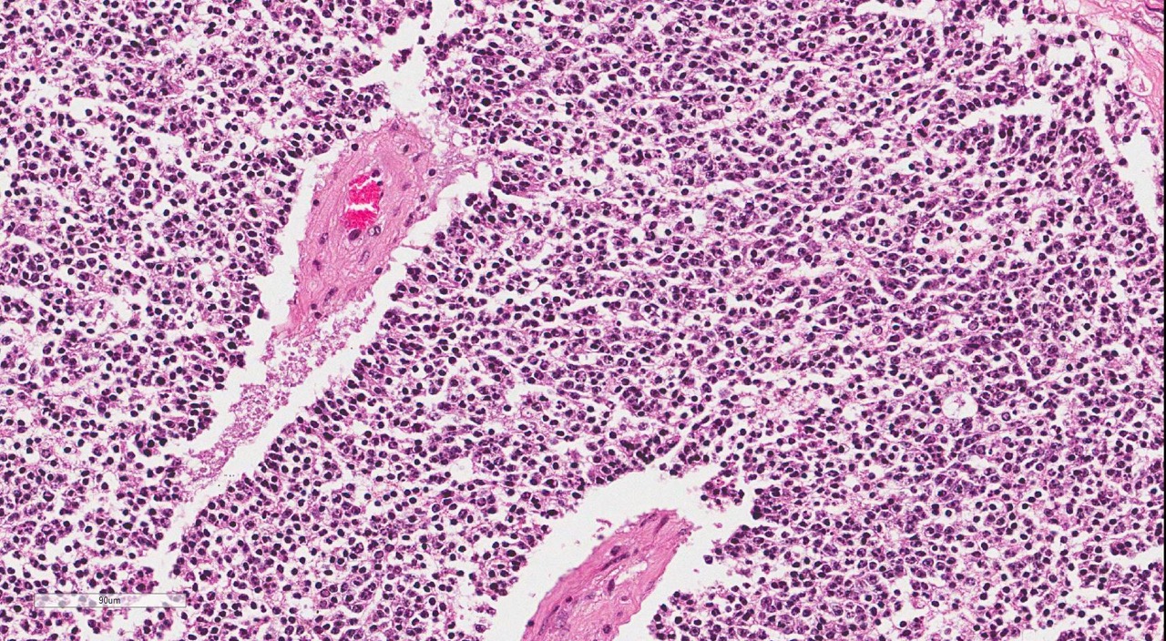
The conference description was closely aligned with the contributors description above; however, there was considerable discussion regarding the presence of and types of rosettes, with agreement that the poor fixation in this case gave a false appearance of Homer-Wright and Flexner-Wintersteiner rosettes. The moderator commented on the marked degree of vascular hyalinization and expansion of the perivascular space by dense, hyalinized, bright eosinophilic material.The di-fferential diagnosis list for this lesion included oligodendroglioma in addition to olfactory neuroblastoma discussed above.
Conference Comment:Immunohistochemical stains that can be useful in diagnosing oligodendroglioma include cyclic nucleotide phosphatase (CNPase) and oligodendrocyte transcription factor 2 (olig2); the absence of glial fibrillary acidic protein (GFAP) may also aid in the diagnosis.8 Other differentials discussed included primitive neu-roectodermal tumor and ependymoma.In general, neoplasms associated with the cribiform plate often extend from outside the CNS into the olfactory bulb / frontal lobe as opposed to extending from the brain outward through the cribiform plate.
Primitive neuroectodermal tumors include neuroblastoma, medulloblastoma and neu-resthesioblastoma and are composed of -Ç-ÿneuroblasts and generally arranged in sheets with the presence of Homer-Wright rosettes. Immunohistochemical markers used in the diagnosis of this type of tumor in dogs and cats include neuron-specific enolase (NSE), synaptophysin, a neuron-specific nuclear protein known as neuronal nuclei (NeuN) and neurofilament protein (NFP).8- These tumors may have a heterogenous or irregular staining pattern due to the presence of both neural and glial differentiation, which may aid in the diagnosis.Other stains which are reported as positive in some subsets of PNETs include vimentin and S100 among others.Stem cell markers which may be useful include nestin, beta III tubulin and doublecortin.Ependymomas are most often associated with the ventricle in some fashion and classically have a papillary pattern with ependymal rosette formation, although these features are not present in all cases. These tumors are generally GFAP and vimentin positive.8Neuroendocrine carcinomas are uncommon neoplasms which derive from neu-roendocrine cells distributed throughout the body.Other locations for neuroendocrine neoplasms include the gastrointestinal tract, lungs and integument.The most germane differential diagnosis in this case is olfactory neuroblastoma as discussed above, due in large part to overlapping histologic features with neuroendocrine carcinoma.-- Features which can aid in ultrastructural diff-erentiation include the lack of microtubule containing neural processes in neu-roendocrine carcinoma,9 as well as the IHC profiles discussed above. A pancytokeratin immunohistochemical (IHC) stain performed at the JPC showed diffuse, strong, cytoplasmic immunoreactivity and a neurofilament IHC was negative, supporting the contributors diagnosis of neuroendocrine carcinoma.
References:
1. Anderson BC, Cordy DR. Olfactory neuroblastoma in a heifer. Vet Pathol. 1981; 18: 536.
2. Brosinski K, Janik D, Polkinghorne A, Von Bomhard W, Schmahl W. Olfactory neuroblastoma in dogs and cats--a histological and immunohistochemical analysis. J Comp Pathol. 2012; 146: 152-9.
3. Brownstein DG, Montali RJ, Bush M, James AE. Nasal carcinoma in a captive Eld's deer. J Am Vet Med Assoc. 1975; 167: 569-71.
4. Cox NR, Powers RD. Olfactory neuroblastomas in two cats. Vet Pathol. 1989; 26: 341-3.
5. Do-¿pke C, Gro-¿ne A, Borstel Mv, et al. Metastatic esthesioneuroblastoma in a horse. J Comp Path. 2005; 132: 218222.
6. Holscher MA, Page DL, Powell HS, Kord CE. Mixed primitive neuroectodermal and rhabdomyoblastic tumor in the brain of a wild deer. J Wildl Dis. 1974; 10: 201.
7. Howard DR, Drehbiel JD, Fay LD, Whitenack DL. Visual defects in white-tailed deer from Michigan: six case reports. J Wildl Dis. 1976; 12: 143-7.
8. Johnson GC, Coates JR, Wininger F. Diagnostic immunohistochemistry of canine and feline intracalvarial tumors in the age of brain biopsies. Vet Pathol. 2014; 51(1):146-160.
9. Kubo M, Matsuo Y, Okano T, Sakai H, et al. Nasal neuroendocrine carcinoma in a free-living Japanese Raccoon dog (Nyctereutes procyonoides viverrinus). J. Comp Path. 2009;140:67-71.
10. Langley K. The neuroendocrine concept today. Annals New York Academy Sci. 1994; 733: 117.
11. Movassaghi AR, Davazdah Emami MR. Ethmoturbinate adenocarcinoma in a Persian fallow deer (Cervus dama mesopotamica). Vet Rec. 2001; 149: 493-4.
12. Nettles VF, Vandevelde M. Thalamic ependymoma in a white-tailed deer. Vet Pathol. 1978; 15:133-5.
13. Parker VJ, Morrison JA, Yaeger MJ. Olfactory neuroblastoma in a cat. J Feline Med Surg. 2010;12: 867-71.
14. Patnaik AK, Ludwig LL, Erlandson RA. Neuroendocrine carcinoma of the nasopharynx in a dog. Vet Pathol. 2002; 39: 496.
15. Sako T, Shimoyama Y, Akihara Y, Ohmachi T, Yamashita K, Kadosawa T, Nakade T, Uchida E, Okamoto M, Hirayama K, Taniyama H. Neuroendocrine Carcinoma in the Nasal Cavity of Ten Dogs. J Comp Pathol. 2005; 133: 155-163.
16. van Maanen C, Klein WR, Dik KJ, van den Ingh TS. Three cases of carcinoid in the equine nasal cavity and maxillary sinuses: histologic and immunohistochemical features. Vet Pathol. 1996; 33(1): 92-5.