Signalment:
Gross Description:
Histopathologic Description:
Morphologic Diagnosis:
Lab Results:
Condition:
Contributor Comment:
The multifocal and widespread pneumonia, and the skin lesions observed in this pig are compatible with a septicemia caused by Actinobacillus suis. Clinical cases of A. suis occur more frequently in high-health-status herds (6). The most common manifestation of the infection is septicemia and sudden death in suckling and recently weaned pigs (6). A disease resembling pleuropneumonia caused by A. pleuropneumonia, and skin lesions similar to those caused by Erysipelothrix rhusiopathiae are reported in older pigs (6).
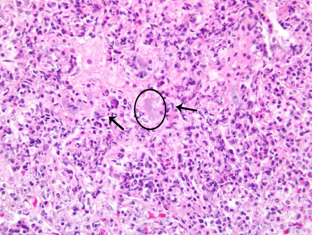
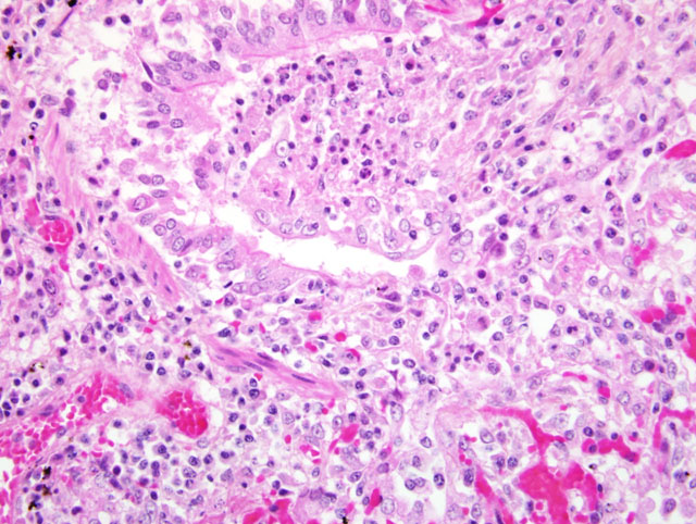
The pneumonic lesions caused by A. suis can have two patterns. One of them is a focal locally extensive fibrinohemorrhagic, fibrinoleukocytic and necrotizing pneumonia or pleuropneumonia affecting the middle or the caudal lung lobes, which may be unilateral or bilateral (2). These lesions are very similar to those caused by A. pleuropneumonia, and are probably originating from an airborne entry of the organism (2). The other pattern is a generalized multifocal pneumonia indicating hematogenous origin. This multifocal widespread pneumonia is a common finding in cases of A. suis septicemia. Other lesions observed in septicemic cases are petechial hemorrhages in serosa and other organs, multifocal necrosis and inflammation in the liver, spleen, kidney and skin, splenomegaly, serofibrinous pericarditis, pleuritis and peritonitis, polyarthritis, valvular endocarditis, and rhomboid skin lesions similar to those observed in cases of erysipelas (6).
The fibrinous pneumonia with many necrotic leukocytes appearing as oat cells is characteristic of A. pleuropneumonia and A. suis in swine (2). Different serotypes of A. pleuropneumonia produce RTX-toxins (ApxI, II and III) which are cytotoxic for the porcine neutrophils and macrophages (2, 4). Some strains of A. suis produce a RTX-toxin (Apx I) (6). Oat cells are also present in the fibrinous pneumonia caused by Mannheimia haemolytica in cattle, sheep and goat (2). All serotypes of M. haemolytica produce a leukotoxin being a member of the RTX family of bacterial toxins (2). These necrotic leukocytes appearing as oat cells are also present in the inflammatory lesions of other organs in cases of A. Suis septicemia.
JPC Diagnosis:
Conference Comment:
| Actinobacillus pleuropneumonia | Pigs | Serofibrinous pleuritis and necrotizing hemorrhagic pneumonia; caudodorsal distribution |
| Actinobacillus equuli | Horses | Common cause of suppurative embolic nephritis in foals |
| Actinobacillus lignieresii | Cattle | Glossitis and stomatitis in cattle (wooden tongue) |
| Actinobacillus seminis | Sheep | Common cause of bilateral epididymitis in rams |
References:
2. Caswell JL, Williams KJ: Respiratory system. In: Jubb, Kennedy and Palmers Pathology of Domestic Animals, ed. Maxie MG, 5th ed., vol. 2 pp. 587-589, 601-606. Elsevier Limited, St. Louis, MO 2007
3. Foster RA, Ladds PW: Male genital system. In: Jubb, Kennedy and Palmers Pathology of Domestic Animals, ed. Maxie MG, 5th ed., vol. 3 pg. 590. Elsevier Limited, St. Louis, MO 2007
4. Gottschalk M, Taylor DJ : Actinobacillus pleuropneumonia. In: Diseases of Swine, ed. Straw BE, Zimmerman JJ, DAllaire S, Taylor DJ, 9th ed., pp. 563-576. Blackwell Publishing, 2006.
5. Maxie MG, Newman SJ: Urinary system. In: Jubb, Kennedy and Palmers Pathology of Domestic Animals, ed. Maxie MG, 5th ed., vol. 2, pg. 480. Elsevier Limited, St. Louis, MO, 2007
6. Taylor DJ: Actinobacillus suis. In: Diseases of Swine, ed. Straw BE, Zimmerman JJ, DAllaire S, Taylor DJ, 9th ed., pp. 827-829. Blackwell Publishing, 2006.