Signalment:
Approximately 2-3 year old adult, male, Northern leopard frog (
Rana pipiens)The frog was euthanized and submitted for necropsy after presenting with a history of lethargy,
distended abdomen and possible abdominal mass on palpation.
Gross Description:
On external examination, the abdomen was severely distended and mild hyperemia and
erythema were noted on the distal extremities. On incision, the ventral abdomen contained a large,
approximately 4cm, space-occupying multilobulated, cauliflower-shaped pale pink soft tissue mass. The
testes were positioned ventral to and in contact with the mass. The mass was not adhering to any viscera
and caused displacement of the abdominal organs.
Histopathologic Description:
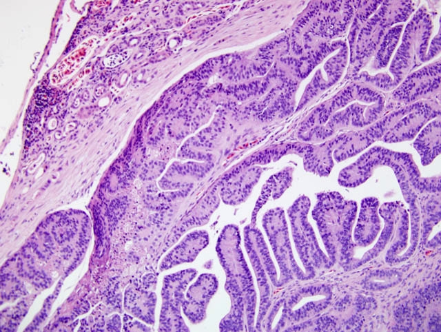
Abdominal Mass: The examined section is composed of part of the
abdominal mass, small segment of renal parenchyma and testes. The partially encapsulated, multilobulated
and moderately cellular neoplastic mass is well-differentiated and composed of a proliferation of closely
packed cells arranged in irregularly-shaped tubules or papillary projections that are supported by a fine
fibro-vascular stroma
(Fig. 1-1). Neoplastic cells are variably sized, mostly large, cuboidal to columnar
with distinct cell borders and contain moderate amounts of eosinophilic, granular cytoplasm that often
contains eosinophilic to mucinous globular droplets. The cells frequently form piles of 4-12 cell-layers
deep. The nuclei of the cells are round to oval, central to basally positioned, with coarsely stippled
chromatin, and contain one or more prominent basophilic nucleoli. Rarely, the nucleus contains 2-4 μm
diameter eosinophilic inclusion-like material with a clear halo and marginated chromatin. Mitotic figures
are 22 per 10 high-powered fields. There is mild anisocytosis and anisokaryosis. In multiple foci,
individual cells to aggregates of necrotic/ghost cells are present. Many tubules contain ectatic lumen filled
with necrotic cells, few lymphocytes and eosinophilic proteinaceous material.
Kidney: Within the submitted small remnant renal tissue, islands of dysplastic convoluted tubules mostly in
the renal pelvis are also lined by epithelium with morphological features similar to those observed in the
adjacent neoplasm. The neoplastic cells variably contain faintly visible micro-villi.
Morphologic Diagnosis:
Renal mass (presumed): Adenocarcinoma, well-differentiated,
tubulo-papillary with rare eosinophilic intranuclear inclusion-like material
Condition:
Adenocarcinoma (Lucke's tumor)
Contributor Comment:
This fairly large abdominal mass is suspected to be of renal origin, though no
remnants of renal parenchyma were present within the actual mass. However, the presence of islands of
tubules with features similar to those observed in the mass is highly indicative of renal origin along with
the massive growth of the tumor effacing the normal renal parenchyma. Rarely, indistinct eosinophilic
inclusion-like material was observed within the nucleus and rarely in the cytoplasm. The inclusions,
though not of typical size, are considered to be herpes viral inclusions. Additional electron microscopic
evaluation may be needed for confirmation. The morphological features are most consistent with that of
Ranid herpesvirus 1 (RaHV-1) induced adenocarcinoma of leopard frogs.
RaHV-1 is the etiologic agent of Luck+�-� renal adenocarcinoma and occurs spontaneously in
Rana pipiens
typically in frogs aged 2 years or older.(1) Tumor incidence can be as high as 50% in laboratory
populations living at 25°C.(2) The viral replication and growth kinetics of the tumor are dependent on
temperature and season. High environmental temperature during summer is permissive for viral invasion
and rapid growth of tumor with very few inclusion bodies. During cooler, winter temperatures invasion is
restricted but viral replication occurs in the convoluted tubules of the kidney with dormant phase of tumor
growth.(1) When frogs are hibernating or maintained at low temperature, tumor cells contain intranuclear
inclusions whereas frogs in summer months or maintained at 25°C do not contain virus or inclusions in
tumor cells.(2) Frogs may not show clinical signs of lethargy, emaciation and ascites until the disease is
well advanced.(1) Whitish tumors can be seen at necropsy on the kidneys, though tumors can grow very
large and metastasize.(1) There is no treatment and affected animals should be euthanized.(1) The other
well-known spontaneous amphibian tumor is lymphosarcoma, occurring in
Xenopus laevis.(3)
JPC Diagnosis:
Kidney: Adenocarcinoma, tubulopapillary
Conference Comment:
Luckes tumor, or renal adenocarcinoma of frogs, is commonly found in the
northern and northeastern United States. It can be found in up to 10% of frogs captured in the wild.(2) The
Lucke tumor herpesvirus, (LTHV), is the causative agent of the tumor, and ultrastructurally the virions are
icosahedral and 95-100nm.
Frogs shed this virus in the colder months of the year, and it travels via water to infect frog eggs during
spawning season. Mortality due to renal adenocarcinoma usually occurs after spawning when temperatures
are warmer and the tumor has grown considerably within the coelomic cavity. Frogs often seem clinically
normal until just prior to death. Neoplastic cells can often be isolated from ascitic fluid to aid in an antemortem
diagnosis.(1)
References:
1. Fox JG, Anderson LC, Loew FM, Quimby FW: Laboratory Animal Medicine, 2nd ed.,
pp. 817-818. Academic Press, London, England, 2002
2. Granoff A: Herpesvirus and the Luck+�-� tumor. Cancer Res
33:1431-1433, 1973
3. Ruben LN, Clothier RH, Balls M: Cancer resistance in amphibians. Altern Lab Anim
35:463-470, 2007