Signalment:  

12-month-old crossbred calf (Bos taurus).Out of a group of 1200 from Canada, this was the 3rd calf to become ill and die after treatment. Calves were reported to be depressed and off feed and water. This calf presented to the field veterinarian terminally ill in lateral recumbency and was euthanized.


Gross Description:  

Necropsy findings noted by the field veterinarian included: esophagus contained non-hemorrhagic erosions. Small and large intestine contained multiple hemorrhagic ulcers.


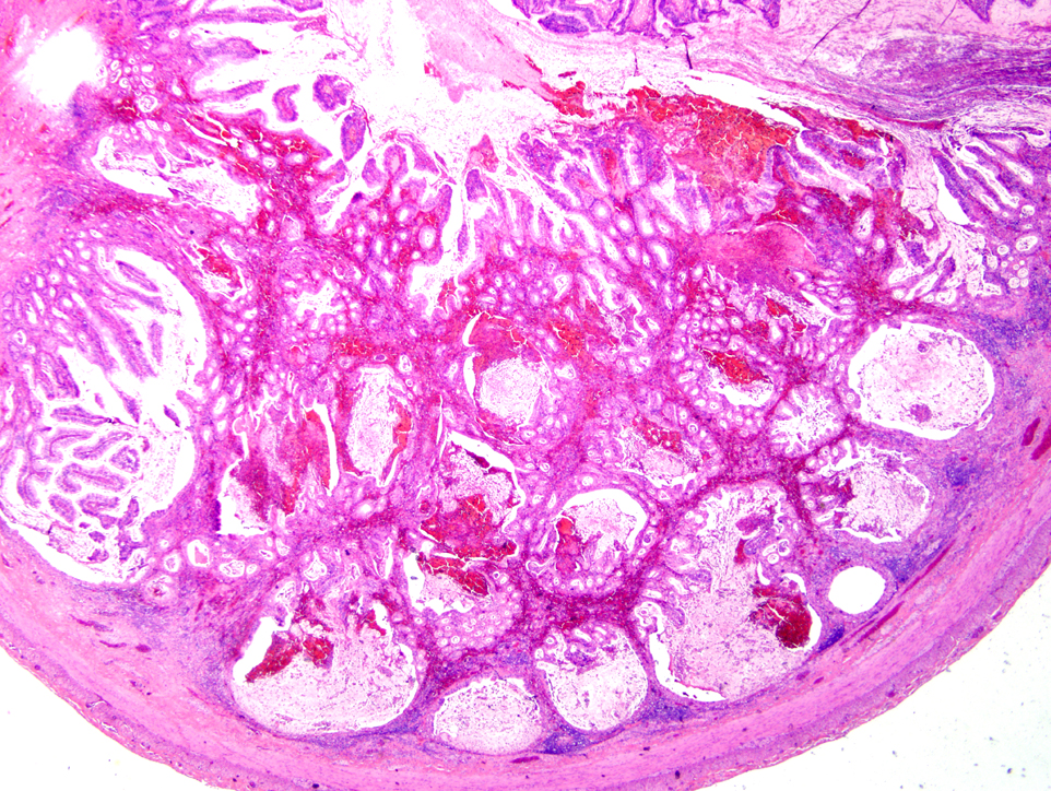
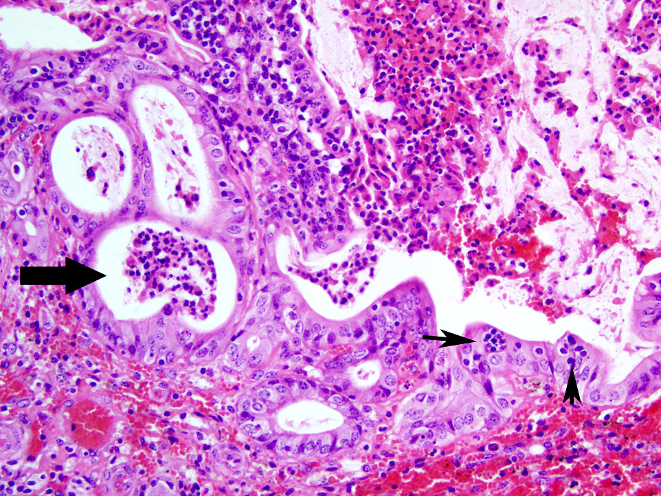
Histopathologic Description:

Within sections of small intestine, there is multifocal moderate to severe suppurative, mucohemorrhagic enteritis with attenuation and necrosis of crypt epithelium primarily confined to areas overlying Peyers patches. Peyers patches are severely depleted and their normal architecture has been destroyed; dilated crypts containing degenerate epithelial cell, neutrophils, mucous and hemorrhage are herniated into the submucosa previously occupied by Peyers patches. Moderate infiltration of the lamina propria by lymphocytes and plasma cells is also present. Within mesenteric adipose tissue, there is mild accumulation of perivascular lymphocytes and plasma cells; vasculitis is not present. 


Morphologic Diagnosis:  

Small intestine: Enteritis, mucohemorrhagic, neutrophilic, acute to subacute, moderate to severe, with crypt epithelial necrosis and destruction of Peyers patches due to BVD virus infection.


Lab Results:  

Florescent antibody tests on lung, esophagus, and small intestine were positive for BVD virus. Cytopathic-BVD virus was isolated from lung and kidney. BVD ear notch ELISA test was positive on a fresh ear notch. Fecal flotation was negative for parasites.


Condition:  

BVD/Mucosal disease


Contributor Comment:  

BVD virus is an RNA virus belonging to the genus Pestivirus. Infection of pregnant cattle can result in fetal abortion or birth of persistently-infected (PI) calves. PI calves typically are unthrifty and do not live beyond 2 years of age. PI calves can succumb to fatal mucosal disease if infected with cytopathic BVD virus (mutation of non-cytopathic BVD virus can also cause mucosal disease in these PI calves).(1)

Infection of healthy, immunocompetent, non-pregnant cattle by BVD virus usually results in only subclinical to mild clinical disease; however, primary infection by very virulent strains of BVD virus can result in severe clinical disease with mortality which cannot be differentiated from mucosal disease by clinical signs and necropsy findings.(1)

In this particular case, it is uncertain whether this calf was persistently infected and died as a result of mucosal disease, or if this was a healthy calf which became infected by a particularly virulent strain of BVD virus. The low morbidity, high mortality of the group suggests this was a PI calf, which died of fatal mucosal disease. Molecular characterization of the isolated virus may have aided in the differentiation of these two syndromes.


JPC Diagnosis:  

Small intestine: Enteritis, necrotizing, diffuse, moderate with focally extensive Peyers patch necrosis and crypt herniation.


Conference Comment:  

Bovine viral diarrhea (BVD) is an acute, highly contagious disease of cattle with worldwide distribution that results in enteric disease, respiratory disease and reproductive loss. BVD virus can also infect sheep, goats and pigs and has been isolated in many wild and captive African species. BVD is separated into two biotypes, cytopathic (CP) and noncytopathic (NCP), based on cytopathic effects in vitro, and separated into two genotypes based on antigenic variation. NCP strains are associated with acute bovine virus diarrhea, BVD-induced thrombocytopenia, abortions, and are teratogenic, and CP strains produce mucosal disease. BVD is immunosuppressive and predisposes animals to secondary infections.(1,2)

Virus is shed in body fluids, and primary replication occurs in the tonsils and oropharyngeal lymphoid tissues. Virus then enters circulating monocytes and is transported to lymphoid tissues and the subepithelial connective tissue of the dermis and GI tract, where it spreads locally to overlying epithelial cells. In addition to the subclinical form in immunocompetent adults as mentioned by the contributor, BVD manifests in two other forms. Transplacental infections during days 50-100 of gestation result in fetal death, abortion, or mummification; infection at day 100-150 of gestation results in congenital defects such as microencephaly, cerebellar hypoplasia, hydranencephaly, hydrocephalus, microphthalmia, thymic aplasia, hypotrichosis, alopecia, brachygnathism, growth retardation, and pulmonary hypoplasia. If a calf survives infection prior to 125 days of gestation, it may develop immunotolerance and lifelong, subclinical infection, which is the most pervasive source of infection of other cattle. A second form is mucosal disease, which occurs when a calf is infected with a noncytopathic genotype prior to 125 days of gestation and becomes immunotolerant, and is then infected with a cytopathic strain. Mortality in calves with mucosal disease approaches 100%.(1,2,3)

Gross lesions with subclinical BVD are seen as mild erosions or shallow ulcerations of the oral cavity. Mucosal disease manifests with erosions and ulcerations of mouth, tongue, esophagus, oral and ruminal papillae, abomasum, cecum, and colon; linear esophageal ulcerations (tiger-stripe); swollen, necrohemorrhagic Peyer's patches with diphtheritic membranes; and erosive or ulcerative interdigital dermatitis and coronitis. The virus causes widespread vasculitis, epithelial necrosis, and lymphocytolysis.(1,2,3)


References:

1. Brown CC, Baker DC, Barker IK. Alimentary system. In: Maxie MG, ed. Jubb, Kennedy, and Palmers Pathology of Domestic Animals. 5th ed. Edinburgh, Scotland: Saunders Elsevier; 2007:140-8. 
2. Gelberg HB. Alimentary system and the peritoneum, omentum, mesentery, and peritoneal cavity. In: McGavin MD, Zachary JF, eds. Pathologic Basis of Veterinary Disease. 5th ed. St. Louis, MO: Mosby Elsevier; 2011:384.
3. Zachary JF. Mechanisms of microbial infections. In: McGavin MD, Zachary JF, eds. Pathologic Basis of Veterinary Disease. 5th ed. St. Louis, MO: Mosby Elsevier; 2011:206-7.


Click the slide to view.


1-1. Small intestine


1-2. Small intestine


Back | VP Home | Contact Us |